Skip to main page content
U.S. flag

An official website of the United States government

Dot gov

The . gov means it’s official. Federal government websites often end in . gov or . mil. Before sharing sensitive information, make sure you’re on a federal government site. VSports app下载.

Https

The site is secure. The https:// ensures that you are connecting to the official website and that any information you provide is encrypted and transmitted securely V体育官网. .

. 2012 May-Jun;3(3):250-60.
doi: 10.4161/gmic.20529. Epub 2012 May 1.

Activated fluid transport regulates bacterial-epithelial interactions and significantly shifts the murine colonic microbiome

Affiliations

Activated fluid transport regulates bacterial-epithelial interactions and significantly shifts the murine colonic microbiome (V体育安卓版)

Simon Keely (V体育平台登录) et al. Gut Microbes. 2012 May-Jun.

Abstract

Within the intestinal mucosa, epithelial cells serve multiple functions to partition the lumen from the lamina propria. As part of their natural function, intestinal epithelial cells actively transport electrolytes with passive water movement as a mechanism for mucosal hydration. Here, we hypothesized that electrogenic Cl(-) secretion, and associated mucosal hydration, influences bacterial-epithelial interactions and significantly influences the composition of the intestinal microbiota. An initial screen of different epithelial secretagogues identified lubiprostone as the most potent agonist for which to define these principles. In in vitro studies using cultured T84 cells, lubiprostone decreased E VSports手机版. coli translocation in a concentration-dependent manner (p < 0. 001) and decreased S. typhimurium internalization and translocation by as much as 71 ± 6% (p < 0. 01). Such decreases in bacterial translocation were abolished by inhibition of electrogenic Cl(-) secretion and water transport using the Na/K/Cl(-) antagonist bumetanide (p < 0. 01). Extensions of these findings to microbiome analysis in vivo revealed that lubiprostone delivered orally to mice fundamentally shifted the intestinal microbiota, with notable changes within the Firmicutes and Bacteroidetes phyla of resident colonic bacteria. Such findings document a previously unappreciated role for epithelial Cl(-) secretion and water transport in influencing bacterial-epithelial interactions and suggest that active mucosal hydration functions as a primitive innate epithelial defense mechanism. .


Figures

Figure 1.
Figure 1.
Screen of epithelial chloride channel activators and resultant water transport. (A) Epithelial electrogenic Cl- secretion was used to define responses to the secretagogues forskolin (1 μM, Fsk), adenosine (10 μM, Ado), prostaglandin E2 (1 μM, PGE2) and lubiprostone (100 nM, Lub) relative to Hank’s buffer (HBSS) control. Cells were exposed to each agonist for 1 h and Isc was monitored every 5 min. Results are pooled from eight monolayers in each condition and results are expressed as the mean ± SEM where * indicates p < 0.01. (B) Influence of individual secretagogues on water transport. Monolayers of T84 cells were incubated with individual secretagogues for 24 h and examined for vectorial basolateral-to-apical fluid transport. T84 cells alone served as a negative control. Data are pooled from eight monolayers in each condition and results are expressed as the mean ± SEM fluid movement over 24 h where * indicates p < 0.01.
Figure 2.
Figure 2.
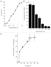
Influence of epithelial electrogenic Cl- secretion on E. coli translocation. (A) Epithelial electrogenic Cl- secretion was used to define the dose response to indicated concentrations of lubiprostone (0.001–10 μM) relative to Hank’s buffer (HBSS) control. Results are pooled from 6–8 monolayers in each condition and results are expressed as the mean ± SEM. (B) Translocation of E. coli across T84 cells (120 min time point) in the presence and absence of lubiprostone stimulation at indicated concentrations. Results are pooled from 6–8 monolayers in each condition and results are expressed as the mean ± SEM. (C) Correlation of electrogenic Cl- secretion (X-axis) with E. coli translocation (Y-axis, plotted as percent inhibition of translocation).
Figure 3.
Figure 3.
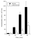
Influence of lubiprostone on E. coli translocation. Time course translocation of E. coli across T84 cells in the presence and absence of lubiprostone stimulation (100 nM). Results are pooled from 6–8 monolayers in each condition and results are expressed as the mean ± SEM where * indicates p < 0.01.
Figure 4.
Figure 4.
Influence of lubiprostone on S. typhimurium internalization and translocation: (A) Time course of lubiprostone (100 nM) on S. typhimurium internalization relative to vehicle control (p < 0.01 by ANOVA). (B) Time course of lubiprostone (100 nM) on S. typhimurium translocation across T84 epithelia relative to vehicle control. Results are pooled from 8 monolayers in each condition and results are expressed as the mean ± SEM where * indicates p < 0.01.
Figure 5.
Figure 5.
Influence of blocking electrogenic Cl- secretion on lubiprostone-mediated inhibition of E. coli translocation: (A) Epithelial electrogenic Cl- secretion was used to define responses to lubiprostone (100 nM, Lub) in the presence and absence of the NKCC1 inhibitor bumetanide (1 μM) relative to Hank’s buffer (HBSS) control. Results are pooled from eight monolayers in each condition and results are expressed as the mean ± SEM (p < 0.001 by ANOVA). (B) Influence of bumetanide on lubiprostone-mediated on water transport in the presence and absence of the NKCC1 inhibitor bumetanide (1 μM) relative to Hank’s buffer (HBSS) control or Fsk control (where * indicates p < 0.01 in the comparison of lubiprostone vs. lubiprostone + 
bumetanide). (C) Influence of lubiprostone on E. coli translocation in the presence and absence of bumetanide (p < 0.01 by ANOVA in the comparison of lubiprostone vs. lubiprostone + bumetanide).
Figure 6.
Figure 6.
Influence of lubiprostone on murine colonic bacterial microbiota: C57Bl/6 mice (n = 4 per group) were administered lubiprostone (1 mg/kg/day) by oral gavage for 7 d. Mice were sacrificed on day 7 and colonic stool was harvested. Bacterial genomic DNA was isolated and samples were amplified using 16S rDNA-specific primers. Samples were resolved by denaturing gradient gel electrophoresis (DGGE). (A) is an example of stool DGGE from animals exposed to vehicle or lubiprostone for 7 d. Indicated bands (A–E) signify bands over-represented in vehicle exposed animals and bands (A’–D’) signify bands over-represented following 7 d of lubiprostone treatment. (B) represents densitometric analysis of indicated bands relative to the control band depicted in (A), where * over closed bars indicates significantly over-represented in control group (p < 0.05) and * over open bars indicates significantly over-represented in lubiprostone-treated group (p < 0.05).
Figure 7.
Figure 7.
Analysis of lubiprostone-mediated changes on murine colonic stool microbiota: C57Bl/6 mice (n = 13 per group) were administered lubiprostone (1 mg/kg/day) by oral gavage for 7 d. Stool was harvested on days 0 and 7 following treatment. Bacterial genomic DNA was isolated and samples were amplified using 16S rDNA-specific primers. Shown here a comparison of genus level groups identified in day 0 and day 7 samples of lubiprostone treated mice. In (A), the Manhattan plot displays results of statistical significance testing (p-value plotted on the y-axis) for the 230 genera identified in the two groups. Numbers corresponding to individual bacterial genera are depicted in (B) as the change in relative abundance between the two groups, plotted as the median of non-zero sequencing counts. Values > 1 indicate enrichment with lubiprostone treatment and values < 1 indicate enrichment with vehicle treatment, where # indicates p = 0.02, @ indicates p = 0.03 and * indicates p = 0.05.
Figure 8.
Figure 8.
Comparison of mucosal and stool microbiota in lubiprostone-treated mice: C57Bl/6 mice (n = 13 per group) were administered lubiprostone (1 mg/kg/day) by oral gavage for 7 d. Mice were sacrificed on day 7 and colonic stool and mucosal scrapings were harvested. Bacterial genomic DNA was isolated and samples were amplified using 16S rDNA-specific primers. Samples were analyzed by next-generation sequencing (230 genus level taxa identified). Shown here a comparison of differences in the microbiota between the mucosa and the stool of lubiprostone treated mice on day 7. In (A), the Manhattan plot displays results of statistical significance testing (p-value plotted on the y-axis) for the 230 genera identified in the two groups. Numbers corresponding to individual bacterial genera are depicted in (B) as the change in relative abundance between the two groups, plotted as the median of non-zero sequencing counts. Values > 1 indicate enrichment in the stool of mice with lubiprostone treatment and values < 1 indicate enrichment in the mucosa of mice with lubiprostone treatment, where # indicates p = 0.02, @ indicates p = 0.03 and indicates p = 0.04, and * indicates 
p = 0.05.

References

    1. Gamba G. Molecular physiology and pathophysiology of electroneutral cation-chloride cotransporters. Physiol Rev. 2005;85:423–93. doi: 10.1152/physrev.00011.2004. - DOI - PubMed
    1. Barrett KE, Keely SJ. Chloride secretion by the intestinal epithelium: molecular basis and regulatory aspects. Annu Rev Physiol. 2000;62:535–72. doi: 10.1146/annurev.physiol.62.1.535. - DOI (VSports注册入口) - PubMed
    1. Campbell EL, Serhan CN, Colgan SP. Antimicrobial aspects of inflammatory resolution in the mucosa: a role for proresolving mediators. J Immunol. 2011;187:3475–81. doi: 10.4049/jimmunol.1100150. - DOI - PMC - PubMed
    1. Ivanov AI, Parkos CA, Nusrat A. Cytoskeletal regulation of epithelial barrier function during inflammation. Am J Pathol. 2010;177:512–24. doi: 10.2353/ajpath.2010.100168. - DOI - PMC - PubMed
    1. Turner JR. Intestinal mucosal barrier function in health and disease. Nat Rev Immunol. 2009;9:799–809. doi: 10.1038/nri2653. - DOI - PubMed

VSports - Publication types

MeSH terms

LinkOut - more resources