Skip to main page content
U.S. flag

An official website of the United States government

Dot gov

The . gov means it’s official. Federal government websites often end in . gov or VSports app下载. mil. Before sharing sensitive information, make sure you’re on a federal government site. .

Https

The site is secure V体育官网. The https:// ensures that you are connecting to the official website and that any information you provide is encrypted and transmitted securely. .

. 2012 Jun 22;287(26):21673-85.
doi: 10.1074/jbc.M111.336537. Epub 2012 May 8.

Comparative processing and function of human and ferret cystic fibrosis transmembrane conductance regulator

Affiliations

Comparative processing and function of human and ferret cystic fibrosis transmembrane conductance regulator

John T Fisher et al. J Biol Chem. .

VSports注册入口 - Abstract

The most common cystic fibrosis transmembrane conductance regulator (CFTR) gene mutation is ΔF508, and this causes cystic fibrosis (CF). New CF models in the pig and ferret have been generated that develop lung, pancreatic, liver, and intestinal pathologies that reflect disease in CF patients. Species-specific biology in the processing of CFTR has demonstrated that pig and mouse ΔF508-CFTR proteins are more effectively processed to the apical membrane of airway epithelia than human ΔF508-CFTR. The processing behavior of ferret WT- and ΔF508-CFTR proteins remains unknown, and such information is important to predicting the utility of a ΔF508-CFTR ferret. To this end, we sought to compare processing, membrane stability, and function of human and ferret WT- and ΔF508-CFTR proteins in a heterologous expression system using HT1080, HEK293T, BHK21, and Cos7 cells as well as human and ferret CF polarized airway epithelia. Analysis of the protein processing and stability by metabolic pulse-chase and surface On-Cell Western blots revealed that WT-fCFTR half-life and membrane stability were increased relative to WT-hCFTR. Furthermore, in BHK21, Cos7, and CuFi cells, human and ferret ΔF508-CFTR processing was negligible, whereas low levels of processing of ΔF508-fCFTR could be seen in HT1080 and HEK293T cells. Only the WT-fCFTR, but not ΔF508-fCFTR, produced functional cAMP-inducible chloride currents in both CF human and ferret airway epithelia VSports手机版. Further elucidation of the mechanism responsible for elevated fCFTR protein stability may lead to new therapeutic approaches to augment CFTR function. These findings also suggest that generation of a ferret CFTR(ΔF508/ΔF508) animal model may be useful. .


Figures

FIGURE 1.
FIGURE 1.
Expression of HA-tagged and non-tagged ferret and human CFTR. A and B, human fibrosarcoma cells (HT1080) were transiently transfected by electroporation with expression plasmids containing either the HA- and non-tagged (A) human and (B) ferret WT- and ΔF508-CFTR cDNAs. The cells were harvested 48 h post-transfection, and the recombinant proteins were immunoprecipitated with a combination of both anti-CFTR and anti-HA antibodies. The protein was then in vitro phosphorylated by PKA in the presence of [γ-32P]ATP and resolved by SDS-PAGE. The gels were dried and exposed to a phosphoscreen, and the autoradiographs were developed using phosphorimaging. HA-tagged and untagged bands B and C are represented by empty and solid arrows, respectively. C and D, cell lysate from the HA-tagged samples (A and B) was subjected to the IP kinase assay. However, before SDS-PAGE analysis, the samples were treated with endoglycosidase H (Endo H) or peptide N-glycosidase (PNGase F) for 2 h at 37 °C. Bands A, B, and C are shown in gray, empty, and black, respectively. Note that the peptide N-glycosidase F-sensitive band (Band A) runs slightly slower than band B in the other lanes as seen in other reports (19, 25).
FIGURE 2.
FIGURE 2.
Multiple cell lines have increased steady state expression of ferret WT-CFTR band C, whereas only certain cell lines process ferret ΔF508-CFTR to band C. A–C, shown are representative autoradiographs from immunoprecipitated and in vitro phosphorylated CFTR protein derived from human embryonic kidney (HEK293T) (A), baby hamster kidney (BHK21) (B), and monkey kidney fibroblast (Cos7) cells (C) transiently overexpressing HA-tagged human and ferret WT- and ΔF508-CFTR proteins. D, Northern blot analysis was performed on HT1080 cells transiently expressing HA-tagged ferret and human WT-CFTR to evaluate the relative abundance of the CFTR mRNAs relative to GAPDH. The mRNA was harvested at 48 h after transfection, and a Northern blot was performed using 25 μg of total RNA. The human and ferret blots were probed with homologous regions to their respective CFTR ortholog cDNA, and the specific activities of the probes varied less than 5%. After CFTR probing, the blots were stripped and reprobed for GAPDH. E, quantification of the CFTR mRNA levels relative to the GAPDH is shown. Data represent the mean ± S.E. (n = 3). Marked comparison (*) demonstrates a significant difference relative to fCFTR as determined by Student's t test (p < 0.0001).
FIGURE 3.
FIGURE 3.
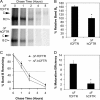
The half-life of complex-glycosylated ferret WT-CFTR is elevated relative to its human ortholog in HT1080 cells. A, metabolic [35S]methionine pulse-chase experiments were performed on HT1080 cells transiently expressing HA-tagged ferret and human WT-CFTR to evaluate the rates of protein synthesis, processing, maturation, and stability. Top and bottom panels are representative autoradiographs for ferret and human WT-CFTR, respectively. B, shown is quantification of the average intensity of band B at 0 h (Band B0) for ferret and human CFTR normalized to total protein. C, shown is quantification of the disappearance of band B (lower graph) over the course of the experiment relative to B and B0 (Band BT and B0). The upper graph represents quantification of the maturation efficiency as calculated by the percentage of band C4 h to band B0. D, shown is quantification of the stability of band C over the course of the experiment relative to B and C4 h (band CT/band C4 h). Data for initial protein labeling and maturation efficiency (B and C, inset, respectively) represent the mean ± S.E. (n = 9 from 3 independent experiments labeled in triplicate). Time course graphs in C and D represent the mean ± S.E. with n = 6. Marked comparisons demonstrate significant differences as determined by Student's t test (*, p < 0.03; †, p < 0.007).
FIGURE 4.
FIGURE 4.
Maturation of ferret ΔF508-CFTR band B to band C is more efficient than the human ortholog in HT1080 cells. A, metabolic [35S]methionine pulse-chase experiments were performed on HT1080 cells transiently expressing HA-tagged ferret and human ΔF508-CFTR to evaluate the rate of band B disappearance and maturation efficiency of the proteins as done in Fig. 3. Top and bottom panels are representative autoradiographs for ferret and human ΔF508-CFTR, respectively. B, shown is quantification of the average intensity of band B at 0 h (Band B0) for ferret and human ΔF508-CFTR normalized to total protein. C, shown is quantification of the disappearance of band B over the course of the experiment relative to Band B0 (Band BT/Band B0). D, shown is quantification of the maturation efficiency as calculated by the percentage of band C4 h relative to band B0. Data in all graphs represent the mean ± S.E. with n = 4 for panel C and n = 9 for panels B and D derived from three independent experiments done in triplicate. Marked comparisons (*) demonstrate significant differences relative to ferret ΔF508-CFTR as determined by Student's t test (p < 0.0001).
FIGURE 5.
FIGURE 5.
Ferret and human ΔF508-CFTR lack significant cell surface expression relative to the wild-type counterparts. A, BHK21 cells grown on coverslips were transfected with the indicated plasmid expression constructs. At 48 h post-transfection, the cells were brought to 4 °C, and a mouse anti-HA antibody was applied for 1 h to stain surface HA-tagged CFTR (red). The unbound antibody was washed away, and the cells were then fixed and permeabilized. The cells were then stained for total CFTR using a rat anti-CFTR antibody (green) and DAPI to mark nuclei (blue). Immunofluorescent images of ferret and human WT-CFTR (left panels) and ΔF508-CFTR (right panels) are shown with the single anti-HA channel shown to the right of the merged color images. Boxed insets show a second example to the larger image. Scale bars represent 10 μm. B–E, surface On Cell Western blots were performed by binding an anti-HA antibody at 4 °C to cell surface CFTR on live BHK21 (B and C) and HT1080 (D and E) cells that were transfected with the indicated constructs as in A and a non-HA tagged CFTR control construct. Excess anti-HA antibody was washed away at 4 °C followed by fixation and staining with an IR secondary antibody. Wells were then imaged using an Odyssey IR scanner. The resulting example images (B and D) were quantified using ImageJ software to give the data shown in (C and E). Only regions containing cells, as indicated by the hashed circles on the WT-fCFTR samples (B and D) were evaluated for the average intensity of staining (which is independent of the area of the field quantified). This was necessary because variable sloughing of cells occurred during the assay washings. Data in all graphs represent the control background subtracted mean ± S.E. with n = 16 from 4 independent experiments. All comparisons with the exception of human ΔF508-CFTR versus ferret ΔF508-CFTR were statistically significant as determined by the Student's t test (p < 0.001). Human ΔF508-CFTR versus ferret ΔF508-CFTR was not significant for BHK21 cells but was significant for HT1080 (p < 0.008). With the exception of ferret ΔF508-CFTR in HT1080 cells, all other ΔF508-CFTR analyses were not significantly above control background levels of cells transfected with a non-HA-tagged version of CFTR.
FIGURE 6.
FIGURE 6.
Cell surface ferret WT-CFTR is more stable than human WT-CFTR in HT1080 cells. A, HT1080 cells were transfected with human or ferret WT-CFTR HA-tagged expression constructs. After 48 h the medium was replaced with warm medium containing an anti-HA antibody and incubated for 1 h at 37 °C, thus labeling any CFTR that made it to the cell surface during the labeling time. The unbound antibody was removed by washing, and the cells were returned to 37 °C for 0, 4, 24, and 48 h. The cells were then quickly washed at room temperature, fixed in 4% PFA, permeabilized with 0.1% Triton X-100, stained with an infrared secondary antibody, and imaged using an Odyssey IR imager. Permeabilization allowed access of the secondary antibody to all CFTR that was labeled during the 1-h labeling period including the endocytosed intracellular pools. B, shown is quantification of data from multiple experiments as shown in A using ImageJ software. Data represent the mean ± S.E. with n = 12 individual wells from three independent experiments; the marked comparison (*) demonstrates a significant difference as determined by Student's t test of the comparison (p < 0.012). C and D, surface ferret or human WT-CFTR protein on HT1080 cells was labeled and stained as in A but under non-permeabilization conditions (i.e. no Triton X-100). The quantified surface CFTR data contained within these two graphs are identical but are depicted on a linear (C) or logarithmic (D) scale with ferret and human WT-CFTR represented by closed and open circles, respectively. These data represent the mean ± S.E. with each time point represented by 6–8 individual wells; marked comparisons demonstrate significant differences as determined by the Student's t test (**, p < 0.022; † p < 0.001).
FIGURE 7.
FIGURE 7.
Ferret and human ΔF508-CFTR lack significant apical surface CFTR in human CF polarized airway cells. Polarized human CF airway epithelial cells (CuFi) grown on Millicell membrane inserts were infected with human and ferret HA-tagged WT- and ΔF508-CFTR adenoviral vectors. At 48 h post-infection, the cells were brought to 4 °C, and a mouse anti-HA antibody was applied for 1 h to stain the surface HA-tagged CFTR (red). The unbound antibody was removed, and the cells were then fixed and permeabilized. Total CFTR (green) was stained using rat anti-CFTR (3G11) and rat anti-HA antibodies. The tight junctions (white) were stained using a rabbit anti-ZO-1 antibody. The membranes were excised and mounted with VectaShield containing DAPI (blue). Confocal immunofluorescent images of ferret and human WT-CFTR and ΔF508-CFTR are shown in the XY, XZ, and YZ planes with the single anti-HA channel shown to the right of the merged color images. The dotted lines represent the location of the Millicell membrane. Scale bars represent 10 μm.
FIGURE 8.
FIGURE 8.
Ferret and human ΔF508-CFTR lacks the ability to correct chloride transport and permeability in CF human and ferret polarized airway epithelia. A, polarized human CF airway epithelial cells (CuFi) grown under air liquid interface (ALI) on Millicell inserts were infected with the indicated adenoviral vectors. At 48 h post infection, the inserts were mounted in Ussing chambers, and short circuit current (ISC) measurements were made. Representative tracings are shown for the indicated samples in response to serial addition of 3-isobutyl-2-methylxanthine (IBMX, 100 μm)/forskolin (Forsk, 10 μm), Bumet (100 μm), and GlyH101 (50 μm). B, shown is quantification of the ΔISC (A). Data in this graph represent plateau value (not peak) mean ± S.E. with n = 12 Millicells for WT-CFTR and n = 10 for ΔF508-CFTR. Mock refers to uninfected controls. No significant differences between human and ferret ΔF508-CFTR values were observed by Student's t test. However, human and ferret WT-CFTR values in response to 3-isobutyl-2-methylxanthine/forskolin were significantly greater for ferret (p < 0.017); this difference and its significance was greater when peak currents were evaluated (mean data not shown). C, ferret CFTR-knock-out tracheal xenografts were infected with either ferret WT- or ΔF508-CFTR adenoviruses overnight. Transepithelial potential difference measurements were made as described under “Experimental Procedures.” Representative TEPD tracings before and after infection are shown. D, quantification of the average cAMP/forskolin and GlyH101 responses is shown. Data in this graph represent the mean ± S.E. (n = 7 measurements from 5 WT-fCFTR infected xenografts and n = 8 from 6 ΔF508-fCFTR infected xenografts). Significant differences between WT- and ΔF508-CFTR responses to both cAMP/forskolin and GlyH101 were observed by Student's t test (p < 0.0055).

References

    1. Rosenstein B. J., Zeitlin P. L. (1998) Cystic fibrosis. Lancet 351, 277–282 - PubMed
    1. Rowe S. M., Miller S., Sorscher E. J. (2005) Cystic fibrosis. N. Engl. J. Med. 352, 1992–2001 - "VSports app下载" PubMed
    1. Egan M. E. (2009) How useful are cystic fibrosis mouse models? Drug Discov. Today 6, 35–41
    1. Fisher J. T., Zhang Y., Engelhardt J. F. (2011) Comparative biology of cystic fibrosis animal models. Methods Mol. Biol. 742, 311–334 - PMC - PubMed
    1. Grubb B. R., Boucher R. C. (1999) Pathophysiology of gene-targeted mouse models for cystic fibrosis. Physiol. Rev. 79, S193–S214 - PubMed

Publication types

MeSH terms

"V体育官网" Substances