Skip to main page content
U.S. flag

An official website of the United States government

Dot gov

The . gov means it’s official. Federal government websites often end in . gov or . mil. Before sharing sensitive information, make sure you’re on a federal government site VSports app下载. .

Https

The site is secure V体育官网. The https:// ensures that you are connecting to the official website and that any information you provide is encrypted and transmitted securely. .

. 2012;7(4):e36019.
doi: 10.1371/journal.pone.0036019. Epub 2012 Apr 26.

YopJ-induced caspase-1 activation in Yersinia-infected macrophages: independent of apoptosis, linked to necrosis, dispensable for innate host defense

Affiliations

YopJ-induced caspase-1 activation in Yersinia-infected macrophages: independent of apoptosis, linked to necrosis, dispensable for innate host defense (VSports)

Ying Zheng et al. PLoS One. 2012.

Abstract

Yersinia outer protein J (YopJ) is a type III secretion system (T3SS) effector of pathogenic Yersinia (Yersinia pestis, Yersinia enterocolitica and Yersinia pseudotuberculosis) that is secreted into host cells. YopJ inhibits survival response pathways in macrophages, causing cell death. Allelic variation of YopJ is responsible for differential cytotoxicity in Yersinia strains. YopJ isoforms in Y. enterocolitica O:8 (YopP) and Y. pestis KIM (YopJ(KIM)) strains have high cytotoxic activity. In addition, YopJ(KIM)-induced macrophage death is associated with caspase-1 activation and interleukin-1β (IL-1β secretion VSports手机版. Here, the mechanism of YopJ(KIM)-induced cell death, caspase-1 activation, and IL-1β secretion in primary murine macrophages was examined. Caspase-3/7 activity was low and the caspase-3 substrate poly (ADP-ribose) polymerase (PARP) was not cleaved in Y. pestis KIM5-infected macrophages. In addition, cytotoxicity and IL-1β secretion were not reduced in the presence of a caspase-8 inhibitor, or in B-cell lymphoma 2 (Bcl-2)-associated X protein (Bax)/Bcl-2 homologous antagonist/killer (Bak) knockout macrophages, showing that YopJ(KIM)-mediated cell death and caspase-1 activation occur independent of mitochondrial-directed apoptosis. KIM5-infected macrophages released high mobility group protein B1 (HMGB1), a marker of necrosis, and microscopic analysis revealed that necrotic cells contained active caspase-1, indicating that caspase-1 activation is associated with necrosis. Inhibitor studies showed that receptor interacting protein 1 (RIP1) kinase and reactive oxygen species (ROS) were not required for cytotoxicity or IL-β release in KIM5-infected macrophages. IL-1β secretion was reduced in the presence of cathepsin B inhibitors, suggesting that activation of caspase-1 requires cathepsin B activity. Ectopically-expressed YopP caused higher cytotoxicity and secretion of IL-1β in Y. pseudotuberculosis-infected macrophages than YopJ(KIM). Wild-type and congenic caspase 1 knockout C57BL/6 mice were equally susceptible to lethal infection with Y. pseudotuberculosis ectopically expressing YopP. These data suggest that YopJ-induced caspase-1 activation in Yersinia-infected macrophages is a downstream consequence of necrotic cell death and is dispensable for innate host resistance to a strain with enhanced cytotoxicity. .


Conflict of interest statement

Competing Interests: The authors have declared that no competing interests exist.

"V体育平台登录" Figures

Figure 1
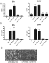
Figure 1. Caspase-3/7 activity is low in KIM5-infected macrophages.
(A) BMDMs were left uninfected (U) or infected with Y. pestis strains expressing YopJKIM or YopJC172A in 96-white walled tissue culture plates. Caspase-3/7 activity was measured 4, 8, 12 or 24 hr post-infection with fluorometer. The results from three independent experiments were averaged and are shown as fold change compared to uninfected cells. Error bars represent standard deviations. Differences in caspase-3/7 activities between uninfected and infected cells were not significant as determined by two way ANOVA (B) BMDMs were left uninfected (U) or infected with Y. pestis strains expressing YopJKIM or YopJC172A or treated with 1 µM of staurosporine (STS) for 16 hr. Macrophage lysates were collected and analyzed by PARP immunoblotting. Sizes of molecular weight standards (kDa) are shown on the left. Positions of full length PARP and cleaved PARP (c-PARP) are showed on right.
Figure 2
Figure 2. Mitochondrial-induced apoptosis is not required for KIM5-induced macrophage death and IL-1β secretion.
Figure 3
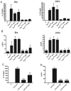
Figure 3. Caspase-8 activity is dispensable for KIM5-triggered macrophage death and IL-1β secretion.
BMDMs were treated with 40 µM caspase-8 inhibitor Z-IETD (IETD) or vehicle 1 hr prior to infection. The BMDMs were then infected with Y. pestis strains expressing YopJKIM or YopJC172A or left uninfected (U). Infected cells were maintained in the presence of Z-IETD or the vehicle for the remainder of the experiment. At 8 hr or 24 hr post-infection, supernatants were collected and LDH release (A) and IL-1β (B) were measured. Results shown are averages from three independent experiments. Error bars represent standard deviations. ★, P<0.05 as determined by one way ANOVA compared to the YopJKIM infection without inhibitor condition. (C) BMDMs were treated with 5 µM of MG-132 in the presence or absence of 40 µM Z-IETD for 30 min, followed by 1 µg/ml of LPS for 3 hrs. Representative phase images of the treated BMDMs were captured by digital photomicroscopy.
Figure 4
Figure 4. KIM5-infected macrophages have necrotic morphology as shown by Annexin V staining and PI uptake assay.
BMDMs were seeded on glass coverslips in a 24-well plate and infected with Y. pestis strains expressing YopJKIM or YopJC172A or left uninfected (U). Annexin V staining and PI uptake assay was performed at 4 hr, 8 hr or 12 hr post-infection. Representative images were captured by digital photomicroscopy. Average percentages of Annexin V positive or Annexin V/PI double positive cells as counted from three random fields in three independent experiments are shown. Error bars represent standard deviations. Difference in values of single or double positive cells were not significant as determined by one way ANOVA.
Figure 5
Figure 5. HMGB1 is released from KIM5-infected macrophages.
BMDMs were infected with Y. pestis strains expressing YopJKIM or YopJC172A or left uninfected (U). Medium from infected macrophages was collected at 24 hr post infection and immunoblotted for HMGB1. Total cell lysate (Lys) was used as a positive control. Position of molecular weight standard (kDa) is shown on the left.
Figure 6
Figure 6. KIM5-infected necrotic macrophages contain active caspase-1.
BMDMs were seeded on glass coverslips in a 24-well plate and left uninfected (U) or infected with Y. pestis strains expressing YopJKIM or YopJC172A. FAM-YVAD-FMK was added at 9 hr post infection to stain for active caspase-1 and PI uptake assay was performed immediately before microscopic analysis. (A) Representative images of phase, active caspase-1 (green) and PI uptake (red) signals captured by digital photomicroscopy are shown in a-c and e-g, respectively. Panels d and h show merged images of green and red signals. (B) Average percentages of BMDMs positive for active caspase-1, PI or both signals was calculated (∼100–300 cells per field) from three random fields in three independent experiments. Error bars represent standard deviations.
Figure 7
Figure 7. RIP1 is not required for YopJKIM-induced cell death or IL-1β secretion.
BMDMs were treated with 30 µM RIP1 inhibitor necrostatin-1 (Nec) or vehicle 1 hr prior to and during infection. BMDMs were infected with Y. pestis strains expressing YopJKIM or YopJC172A or left uninfected (U). Supernatants were collected and LDH release (A) and secreted IL-1β (B) were measured at 8 hr or 24 hr post-infection from three independent experiments. Results shown are averages and error bars represent standard deviations (★★, P<0.01 as determined by one way ANOVA as compared to YopJKIM no inhibitor).
Figure 8
Figure 8. ROS are not required for cytotoxicity or IL-1β secretion in macrophages infected with KIM5.
BMDMs were treated with 10 µM of DPI or 10 mM of NAC for 2 hours or left untreated. (A and B) BMDMs were infected with Y. pestis strains expressing YopJKIM or YopJC172A or left uninfected (U). Supernatants were collected at 8 hr and 24 hr post-infection and analyzed by IL-1β ELISA (A) or LDH release assay (B). (C and D) BMDMs treated or not with DPI or NAC as above were exposed to 50 ng/ml of LPS for 3 hr. The treated BMDMs were then exposed to 5 mM ATP for 1 hr to activate pyroptosis. Supernatants were tested by IL-1β ELISA (C) or LDH release assay (D). Results shown are the averages from three independent experiments. Error bars represent standard deviations (★★, P<0.01; ★★★, P<0.001, determined by one way ANOVA as compared to LPS+ATP no inhibitor).
Figure 9
Figure 9. Inhibitors of cathepsin B reduce caspase-1 activation in macrophages infected with KIM5.
BMDMs were left untreated or treated with 25 µM of E64d or CA-074 Me (CA) for 1 hr. Following infection with Y. pestis strains expressing YopJKIM in the absence or presence of the inhibitors, supernatants were collected (A, B) or microscopic assay was performed (C). IL-1β ELISA (A) and LDH release assay (B) was done on supernatants collected 24 hr post-infection. Results shown are the averages from three independent experiments. Error bars represent standard deviations. (★★, P<0.01 as determined by one way ANOVA as compared to infection in absence of inhibitor) (C) Infected BMDMs on coverslips were incubated with FAM-YVAD-FAM 9 hr post-infection stained for active caspase-1 (green) for 1 hr and PI uptake (red) immediately before observation. Representative images of phase, green and red signals were captured by digital photomicroscopy.
Figure 10
Figure 10. Enhanced YopP-mediated macrophage cell death is associated with elevated levels of IL-1β release.
Y. pseudotuberculosis IP26 (ΔyopJ) carrying the empty pACYC184 plasmid (pACYC) or pACYC184 encoding the indicated YopP or YopJ isoforms was used to infect BMDMs. Twenty four hours post-infection, medium was collected for IL-1β ELISA (A) and LDH release assay (B). Results shown are the averages from three independent experiments. Error bars represent standard deviations (★★, P<0.01; ★★★, P<0.001 as determined by one way ANOVA as compared to pACYC condition).
Figure 11
Figure 11. Caspase-1 is not required for innate host protection against Yersinia endowed with enhanced cytotoxicity.
(A) Six to eight-week old Casp1+/+ (wild type, WT) or Casp1−/− (Casp1-) C57BL/6J mice were infected orogastrically with 1×10⁹ CFU of Y. pseudotuberculosis IP26 carrying pACYC184 encoding YopP or YopJYPTB. Mouse survival was monitored for 21 days. Results shown are pooled from two independent experiments. Total numbers of mice infected are shown in parenthesis. (B) Data from (A) are reformatted by grouping mice according to infecting strain. Significant difference between survival curves was determined by log rank test.

References

    1. Dockrell DH. Apoptotic cell death in the pathogenesis of infectious diseases. J Infect. 2001;42:227–234. - "VSports" PubMed
    1. Fairbairn IP. Macrophage apoptosis in host immunity to mycobacterial infections. Biochem Soc Trans. 2004;32:496–498. - PubMed
    1. Jonas D, Walev I, Berger T, Liebetrau M, Palmer M, et al. Novel path to apoptosis: small transmembrane pores created by staphylococcal alpha-toxin in T lymphocytes evoke internucleosomal DNA degradation. Infect Immun. 1994;62:1304–1312. - PMC - PubMed
    1. Navarre WW, Zychlinsky A. Pathogen-induced apoptosis of macrophages: a common end for different pathogenic strategies. Cell Microbiol. 2000;2:265–273. - PubMed
    1. Porcelli SA, Jacobs WR., Jr Tuberculosis: unsealing the apoptotic envelope. Nat Immunol. 2008;9:1101–1102. - PubMed

Publication types (VSports)

MeSH terms

Substances

LinkOut - more resources (VSports app下载)