Skip to main page content (VSports)
U.S. flag

An official website of the United States government

Dot gov

The . gov means it’s official. Federal government websites often end in . gov or . mil VSports app下载. Before sharing sensitive information, make sure you’re on a federal government site. .

Https

The site is secure. The https:// ensures that you are connecting to the official website and that any information you provide is encrypted and transmitted securely V体育官网. .

. 2012 Jun 15;287(25):20986-95.
doi: 10.1074/jbc.M112.356709. Epub 2012 May 1.

"VSports在线直播" Target specificity of an autoreactive pathogenic human γδ-T cell receptor in myositis

Affiliations

Target specificity of an autoreactive pathogenic human γδ-T cell receptor in myositis

Jessica Bruder et al. J Biol Chem. .

Abstract

In polymyositis and inclusion body myositis, muscle fibers are surrounded and invaded by CD8-positive cytotoxic T cells expressing the αβ-T cell receptor (αβ-TCR) for antigen. In a rare variant of myositis, muscle fibers are similarly attacked by CD8-negative T cells expressing the γδ-TCR (γδ-T cell-mediated myositis). We investigated the antigen specificity of a human γδ-TCR previously identified in an autoimmune tissue lesion of γδ-T cell-mediated myositis. We show that this Vγ1. 3Vδ2-TCR, termed M88, recognizes various proteins from different species. Several of these proteins belong to the translational apparatus, including some bacterial and human aminoacyl-tRNA synthetases (AA-RS). Specifically, M88 recognizes histidyl-tRNA synthetase, an antigen known to be also targeted by autoantibodies called anti-Jo-1 VSports手机版. The M88 target epitope is strictly conformational, independent of post-translational modification, and exposed on the surface of the respective antigenic protein. Extensive mutagenesis of the translation initiation factor-1 from Escherichia coli (EcIF1), which served as a paradigm antigen with known structure, showed that a short α-helical loop around amino acids 39 to 42 of EcIF1 is a major part of the M88 epitope. Mutagenesis of M88 showed that the complementarity determining regions 3 of both γδ-TCR chains contribute to antigen recognition. M88 is the only known example of a molecularly characterized γδ-TCR expressed by autoaggressive T cells in tissue. The observation that AA-RS are targeted by a γδ-T cell and by autoantibodies reveals an unexpected link between T cell and antibody responses in autoimmune myositis. .


Figures

FIGURE 1.
FIGURE 1.
Screening of a cDNA library. A, growth curves of E. coli BL21-Star-DE3 cells transfected with the single-chain Fv γδ-TCR M88. Blue line and squares: bacterial growth in the absence of M88 expression. Red line and triangles, growth with induction of M88 expression. The time point of induction by isopropylthiogalactoside is indicated by an arrow. After induction of M88, bacterial growth was drastically inhibited. B, bacteria expressing the single-chain Fv M88 are rescued from its growth inhibitory effect by supertransfection with a cDNA library. Some components of the library rescued the bacteria, presumably by binding to and neutralizing M88. This yielded big colonies from which the inserts of the library could be cloned and sequenced. Many small colonies were observed in addition, in which the library components did not neutralize the growth-inhibiting effect of M88. C, control experiments in which the promotor driving M88 expression was shut down in the presence of 2 mm glucose. All colonies were of approximately the same size. To prevent formation of a confluent layer of bacteria, colonies were grown for a shorter period of time. D, activation of M88 by EcK-RS. EcK-RS was identified as one of the cDNA library components that could rescue M88-transfected E. coli BL21-Star-DE3. We tested recognition of purified EcK-RS in an ELISA experiment. EcK-RS and an anti-CD3 antibody (α-CD3-mAb) as positive control were coated to microtiter plates, M88-transfected T hybridoma cells were added, and secreted IL-2 was measured in the supernatant. EcK-RS and the α-CD3-mAb both activated M88, whereas medium alone had no effect.
FIGURE 2.
FIGURE 2.
Recognition of diverse bacterial proteins. A, size-exclusion HPLC chromatography of the recombinant E. coli proteins EcK-RS (green curve), EcD-RS (black curve), EcN-RS (blue curve), and EcIF1 (red curve). All proteins were produced identically in E. coli. Elution profiles were recorded at 210 nm. 12 fractions were collected in the range from 150 to 5 kDa. Purity of all proteins was tested by SDS-PAGE before and after chromatography (supplemental Fig. S1A). EcD-RS migrated faster than expected, presumably because it formed oligomers. B, activation of M88 transfectants by the HPLC fractions shown in A. All fractions from all proteins were coated to microtiter plates and tested for their capability to activate M88 transfectants. To this end, we measured secreted IL-2 in the supernatant. The color coding is identical to that described in A. Activation of M88 transfectants was coincident with the elution peaks for EcK-RS, EcN-RS, and EcIF1. EcD-RS did not activate M88.
FIGURE 3.
FIGURE 3.
Recognition of diverse human proteins. A, size-exclusion HPLC chromatography of the recombinant human proteins hH-RS (blue curve), hA-RS (green curve), hLC1 (red curve), and hPCNA (black curve). All proteins were produced identically in insect cells. Purity of all proteins was tested by SDS-PAGE before and after chromatography (supplemental Fig. S1B). hH-RS migrated with a higher molecular mass than expected, indicating oligomerization. hA-RS and hLC1 were retained presumably due to interaction with the column matrix. hT-RS denatured during chromatography and could not be detected in the eluate (data not shown). B, size-exclusion HPLC chromatography of the recombinant human proteins shown in A. IL-2 production was measured as detailed in Fig. 2B. Activation of M88 transfectants was coincident with the elution peaks for hH-RS, hA-RS, and hLC1. hPCNA was not recognized. Data represent two independent experiments.
FIGURE 4.
FIGURE 4.
Specific recognition of human aminoacyl-tRNA-synthetases by M88. The putative antigens hH-RS (blue bar), hT-RS (orange bar), and hA-RS (green bar) were tested for activation of M88. Candidate antigens were coated to microtiter plates, incubated with M88-transfected T hybridoma cells in the absence or presence of blocking antibodies, and secreted IL-2 was measured in the supernatant. Recognition of hH-RS by M88 could be blocked by two types of specific antibodies (Ab). We used polyclonal human serum (α-hH-RS serum) at two different dilutions (1:10 and 1:100, dark and light red bars, respectively), and a monoclonal antibody (α-hH-RS mAb, yellow bar). The isotype control antibody X40 (isotype control mAb, gray bar) did not block recognition. Recognition of hT-RS and hA-RS could only be blocked at the high concentration of polyclonal α-hH-RS serum, indicating some cross-reactivity. Recognition of hA-RS was lower than recognition of hH-RS or hT-RS. This may be explained by the presence of more contaminating proteins (supplemental Fig. S1B) that may compete for binding to the microtiter plate. However, M88 activation could also be blocked to some extent by high concentrations of α-hH-RS serum. The antibodies alone did not induce IL-2 secretion. Error bars represent ± S.E. Data represent four independent experiments.
FIGURE 5.
FIGURE 5.
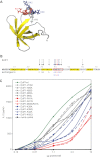
Amino acids of the target antigen EcIF1 recognized by M88. A, structural model of EcIF1 based on NMR spectroscopy (21). The entire molecule is shown in ribbon presentation. The five β-sheets that form a rigid β-barrel are shown in yellow. The amino acids Lys-39 (blue), Met-40 (gray), Arg-41 (red), and Lys-42 (blue) in the short helix, which is recognized by the γδ-TCR, are highlighted as stick models. We used Protein Data Bank code 1AH9 (no.11) and the program PyMOL for display. B, amino acid sequence of EcIF1 of E. coli. The structural elements shown in A are indicated: β-sheets are highlighted in yellow, and the α-helix is boxed in red. The 15 amino acids that were exchanged by site-directed mutagenesis are indicated by arrows. The most relevant amino acid, Arg-41, is indicated by a red arrow. The two other relevant amino acids, Lys-39 and Lys-42, are indicated by blue arrows. Other amino acids are indicated by gray arrows. Continuous arrows indicate amino acid exchanges where the data are shown in C (Asn-28, Val-31, His-35, Met-40). Dashed gray arrows indicate amino acids that were tested but without showing the data in C (His-30, Asn-43, Tyr-44, Tyr-60). C, activation of M88 transfectants by EcIF1 wild-type protein and by EcIF1 molecules carrying site-specific mutations. M88 transfectants were incubated with wild-type and mutated EcIF1 proteins, which were coated to microtiter plates at the indicated concentrations. TCR activation was determined by measuring the secreted IL-2 in the supernatant. EcIF1 wild-type data are shown in dark green. The color code for the different amino acid substitutions is given in the inset. It is identical to the color code used in B. Amino acid substitutions that are indicated with dashed gray arrows in B were also tested but are not shown here. They all showed no significant deviation from the data of the wild-type. We also include a mock control, i.e. bacteria that expressed myelin oligodendrocyte glycoprotein, which forms insoluble inclusion bodies. An identically prepared sample from these bacteria contains all bacterial contaminations. It did not activate M88 (black squares). Data were statistically significant with p values of < 0.05 according to a Student's two-tailed unpaired t test at protein concentrations of 1.1, 3.3, and 10 μg/well for the mutants R41A, R41Q, M40A/R41A, K42Q, and K39A, respectively. Data represent four independent experiments.
FIGURE 6.
FIGURE 6.
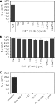
M88 recognizes a conformational epitope. A, the synthetic peptide EcIF1(33–46) did not activate M88 when coated to microtiter plate wells at concentrations between 5.0 × 10² and 5.0 × 10⁻⁵ μg/well. Wild-type EcIF1 in its native conformation was coated at 0.5 μg/well and served as positive control (left panel). B, EcIF1(33–46) did not compete with wild-type EcIF1 for activation of M88. EcIF1 was coated at 0.5 μg/well to microtiter plates, plates were washed, and then EcIF1(33–46) was added at concentrations between 6.6 × 10² and 3.3 × 10⁻⁴ μg/ml together with M88-transfected hybridoma cells. Recognition of EcIF1 was not influenced by the synthetic peptide EcIF1(33–46). C, wild-type EcIF1 was denatured by exposure to 6 m guanidinium thiocyante (Gua+ SCN), 2 m HCl, 5 m NaOH. Furthermore, EcIF1 was subjected to digestion with the proteases proteinase K and trypsin. All conditions abolished recognition by M88. We show untreated wild-type EcIF1 as positive control experiment. Error bars represent ± S.E.
FIGURE 7.
FIGURE 7.
Specific recognition of the paradigmatic antigen EcIF1 by the CDR loops of the γδ-TCR. We compared the recognition of EcIF1 by the wild-type M88 and M88 molecules that contained mutations in the variable (V) and/or CDR3 regions of either chain. CDR3 regions are composed of random nucleotides (N), diversity (D), and joining (J) elements. The first row lists the wild-type γδ-TCR M88 (Vγ1.3+Vδ2+), and the TCR transfectants with mutated γ- or δ-chains: two mutants had altered V(D)J regions of either chain (Vγ9*altVJγVδ2+, Vγ1.3+Vδ1*altVDJδ), three mutants had amino acid exchanges in the γ-N region (Vγ1.3*altγCDR3Vδ2+), the δ-NDN (Vγ1.3+Vδ2*altδCDR3), or δ-NDNJ regions (Vγ1.3+Vδ2*altδCDR3J), and one mutant had wild-type V(D)J regions, but an altered constant region (C) of the γ-chain (Vγ1.3*altγCVδ2+). The second and third rows illustrate these exchanges. We list the variable and joining families of the γ- and δ-chains (V-region nomenclature according to Arden et al. (50)), and show the amino acids of the N- or NDN regions in the single amino acid code. Wild-type regions and amino acids are highlighted in green, and exchanges of entire regions or amino acids are highlighted red. On the right panel, we show the activation of γδ-TCR transfectants by EcIF1. EcIF1 was adsorbed to microtiter plates, incubated with γδ-TCR transfected hybridoma cells, and secreted IL-2 was measured by ELISA. Only the wild-type Vγ1.3Vδ2-TCR M88 and the mutant with altered γ-chain constant region recognized EcIF1. All other mutants with altered V- and/or CDR3 regions were not activated. This provides evidence that EcIF1 is recognized specifically by the complementarity determining regions. Data represent two independent experiments.

"V体育安卓版" References

    1. Chien Y. H., Konigshofer Y. (2007) Antigen recognition by γδ T cells. Immunol. Rev. 215, 46–58 - PubMed
    1. Beetz S., Wesch D., Marischen L., Welte S., Oberg H. H., Kabelitz D. (2008) Innate immune functions of human γδ T cells. Immunobiology 213, 173–182 - V体育2025版 - PubMed
    1. Hayday A. C. (2009) γδ T cells and the lymphoid stress-surveillance response. Immunity 31, 184–196 - V体育官网 - PubMed
    1. Bonneville M., O'Brien R. L., Born W. K. (2010) γδ T cell effector functions: A blend of innate programming and acquired plasticity. Nat. Rev. Immunol. 10, 467–478 - PubMed
    1. Wucherpfennig K. W., Newcombe J., Li H., Keddy C., Cuzner M. L., Hafler D. A. (1992) γδ T-cell receptor repertoire in acute multiple sclerosis lesions. Proc. Natl. Acad. Sci. U.S.A. 89, 4588–4592 - PMC - PubMed

Publication types

V体育官网入口 - MeSH terms

Substances