Skip to main page content
U.S. flag

An official website of the United States government

Dot gov

The . gov means it’s official. Federal government websites often end in . gov or . mil. Before sharing sensitive information, make sure you’re on a federal government site VSports app下载. .

Https

The site is secure V体育官网. The https:// ensures that you are connecting to the official website and that any information you provide is encrypted and transmitted securely. .

. 2012 May 15;26(10):1055-69.
doi: 10.1101/gad.187252.112. Epub 2012 May 1.

VSports - Abrogation of BRAFV600E-induced senescence by PI3K pathway activation contributes to melanomagenesis

Affiliations

"V体育ios版" Abrogation of BRAFV600E-induced senescence by PI3K pathway activation contributes to melanomagenesis

Liesbeth C W Vredeveld et al. Genes Dev. .

"VSports" Abstract

Human melanocytic nevi (moles) are benign lesions harboring activated oncogenes, including BRAF. Although this oncogene initially acts mitogenically, eventually, oncogene-induced senescence (OIS) ensues. Nevi can infrequently progress to melanomas, but the mechanistic relationship with OIS is unclear. We show here that PTEN depletion abrogates BRAF(V600E)-induced senescence in human fibroblasts and melanocytes. Correspondingly, in established murine BRAF(V600E)-driven nevi, acute shRNA-mediated depletion of PTEN prompted tumor progression. Furthermore, genetic analysis of laser-guided microdissected human contiguous nevus-melanoma specimens recurrently revealed identical mutations in BRAF or NRAS in adjacent benign and malignant melanocytes VSports手机版. The PI3K pathway was often activated through either decreased PTEN or increased AKT3 expression in melanomas relative to their adjacent nevi. Pharmacologic PI3K inhibition in melanoma cells suppressed proliferation and induced the senescence-associated tumor suppressor p15(INK4B). This treatment also eliminated subpopulations resistant to targeted BRAF(V600E) inhibition. Our findings suggest that a significant proportion of melanomas arise from nevi. Furthermore, these results demonstrate that PI3K pathway activation serves as a rate-limiting event in this setting, acting at least in part by abrogating OIS. The reactivation of senescence features and elimination of cells refractory to BRAF(V600E) inhibition by PI3K inhibition warrants further investigation into the therapeutic potential of simultaneously targeting these pathways in melanoma. .


Figures

Figure 1.
Figure 1.
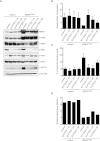
PTEN depletion abrogates BRAFV600E-induced senescence in cultured human fibroblasts. TIG3 HDF stably expressing one of two nonoverlapping PTEN shRNAs (#1 or #2) as well as hTERT (to avoid confounding effects due to replicative senescence; hTERT does not interfere with BRAFV600E-induced senescence) were transduced with BRAFV600E-encoding retrovirus and selected for integration of the construct. Empty vectors were used as controls. (A) Samples were analyzed by Western blot for the indicated proteins 8 d post-infection. β-Actin served as a loading control. (B) Samples were seeded at equal densities and fixed and stained 8 d post-infection. (C) Samples were analyzed for BrdU incorporation 3 d post-infection. Data are represented as mean with standard deviation (SD) (paired ratio two-tailed t-test; [*] P = 0.0001; [**] P = 0.0006).
Figure 2.
Figure 2.
PTEN depletion abrogates BRAFV600E-induced senescence in cultured human melanocytes. (A) Primary human melanocytes stably expressing sh-p16INK4A and either control or sh-PTEN (#1) were transduced with control or BRAFV600E-encoding lentivirus and pharmacologically selected for successful integration of the construct. Empty vectors were used as controls. Samples were seeded at equal densities and fixed and stained 15 d post-infection. (B) Samples were fixed and analyzed for SA-β-Gal activity 15 d post-infection. Data are represented as percentage of cells ± SD (paired two-tailed t-test; P = 0.0001 for the bottom panel). (C) Samples were analyzed for BrdU incorporation 15 d post-infection. Data are represented as mean ± SD (paired ratio two-tailed t-test; [*] P = 0.0001). (D) Samples were analyzed by Western blot for protein expression as indicated. β-Actin served as a loading control. (E) Melanocytes stably expressing sh-p16INK4A were first transduced with BRAFV600E-encoding lentivirus, subsequently transduced with either control or sh-PTEN, and analyzed for BrdU incorporation 8 d post-infection. The relative ratios of three independent experiments are illustrated in the graph. Statistical analysis was performed by a paired ratio two-tailed t-test; P = 0.025.
Figure 3.
Figure 3.
Abrogation of BRAFV600E-induced senescence by PTEN depletion is dose-dependent. Primary human melanocytes stably expressing sh-p16INK4A and either control or sh-PTEN (#1) were transduced with control or BRAFV600E-encoding lentivirus and selected for integration of the construct. Empty vectors were used as controls. (A) Samples were analyzed by Western blot for protein expression as indicated. β-Actin served as a loading control. (B) Samples were analyzed for BrdU incorporation 15 d post-infection. Data are represented as mean ± SD (one-way ANOVA/Bonferroni's multiple comparison test; P < 0.05). (C) Samples were fixed and analyzed for SA-β-Gal activity 15 d post-infection. Data are represented as percentage of cells ± SD (one-way ANOVA/Tukey's multiple comparison). For both B and C, BRAFV600E sh-PTEN1:10 and BRAFV600E sh-PTEN1:25 samples are significantly different from BRAFV600E controls (P < 0.05). (D) Samples were seeded at equal densities and fixed and stained 15 d post-infection. Crystal violet staining was extracted and quantified; similar results were obtained in duplicate experiments.
Figure 4.
Figure 4.
Acute in vivo PTEN depletion in BRAFV600E-expressing nevi drives tumor formation. (A) H&E staining of mouse skin injected with lentivirus carrying either control or shRNA constructs. The sh-PTEN-injected lesion is more cellular and larger; both lesions show focal melanotic pigmentation indicative of the melanocytic origin. (B) Photographs of mice bearing tumors 18.5 wk after intradermal injection of sh-PTEN-carrying lentivirus. Adult mice received a sublethal total body irradiation (5 Gy) 3 d prior to lentivirus application to reduce an immune response eradicating virus-infected cells. shRNA targeting PTEN (sh-PTEN#3) or empty vector control were delivered by intradermal injection of lentivirus into the dorsal skin of 4-OHT-treated Tyr::CreER; BRafCA mice. Arrows indicate sites of injections. (C) High-power micrograph of melanocytic tumor of a sh-PTEN-injected mouse showing a population of plump to elongated cells (red arrows), some of which are laden with melanin pigment, highlighting the characteristic melanocytic dendrites (black arrows).
Figure 5.
Figure 5.
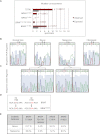
Significant conservation of BRAF and NRAS mutations during progression from nevi to melanomas. (A) Bar graph representing expected and observed frequencies of melanomas and their associated nevi harboring identical BRAF and NRAS mutations in nevi and contiguous melanomas. Seventeen specimens were suitable for laser capture microdissection, after which the mutational status of BRAF exon 15 and NRAS exon 3 (in which most activating mutations reside) was analyzed by genomic DNA PCR amplification and sequencing. Red bars (Observed) represent the mutational status observed for BRAF and NRAS in 17 nevi that are directly contiguous to melanomas. Gray bars (Expected) represent the calculated percentages of random (co-)occurrence. For statistical analysis, we carried out the “one-sample test” for proportions, with the null hypothesis being that the mutations occurred randomly. The “two-sided alternative test” was used to calculate P-values, which were <0.05 for the BRAFT1799A, BRAFTG1799–1800AA, and NRASA182G mutations, indicating that it is highly unlikely that the observed co-occurrence of these mutations (in red) has arisen due to chance. See Supplemental Table 1 for details. (B) A rare BRAF double mutation co-occurring in the nevus and the adjacent melanoma. Chromatograms of BRAF exon 15 PCR fragments amplified from genomic DNA of laser-microdissected normal skin, nevus, and melanoma and of a fibroblast cell line as a control. Arrowheads indicate nucleotide thymine 1799 in BRAF, which is often found mutated to an adenine, and asterisks indicate nucleotide guanine 1800, which is rarely found mutated to an adenine. (C) Chromatograms of cloned PCR fragments encompassing BRAF exon 15 show either the wild-type allele or the allele with the double mutation. (D) Schematic overview of the BRAFT1799A and BRAFTG1799–1800AA mutations, both encoding BRAFV600E. (E) To perform the statistical calculations, percentages of BRAFT1799A, BRAFTG1799–1800AA, NRASC181A, and NRASA182G mutations were extracted from the COSMIC database (http://www.sanger.ac.uk/genetics/CGP/cosmic) (Forbes et al. 2006). Melanomas and benign melanocytic nevi originating from skin were selected, excluding acral, mucosal, ocular, and genital origin; spitz; and blue nevi, as these types were not included in our specimen panel.
Figure 6.
Figure 6.
Decrease in PTEN and increase in AKT3 and P-AKT expression in melanomas relative to their contiguous nevi. Representative immunohistochemical stainings of consecutive sections of a contiguous nevus–melanoma specimen with PTEN, AKT3, and P-AKT antibodies. Both nevus and melanoma compartments harbor the BRAFT1799A mutation, as determined by laser capture microdissection and sequence analysis. The melanoma compartment (M) exhibits clearly weaker PTEN (A–C) and stronger AKT3 (D–F) and P-AKT (G–I) immunostaining compared with the nevus compartment (N). B, E, and H show higher magnifications of the nevus, and C, F, and I, show higher magnifications of the melanoma. The cell-rich area underneath the melanoma compartment comprises mainly infiltrating lymphocytes.
Figure 7.
Figure 7.
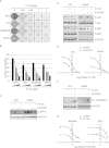
PI3K inhibition induces cell cycle arrest in melanoma cells and enhances cytotoxicity of BRAFV600E inhibition. Melanoma cell lines were treated with Pi-103 and/or PLX4720. (A) Samples were kept in medium containing Pi-103 for 7 d and fixed and stained with crystal violet. (B) Samples were analyzed for BrdU incorporation 4 d after treatment with increasing concentrations of Pi-103 (0.25, 0.5, 1, and 2 μM). Error bars represent SEM (standard error of the mean) of three independent experiments. For 453A0 melanoma cells, a representative experiment is shown. (C) Samples were analyzed by Western blotting for p15INK4B 2 d after treatment with 0.25 μM Pi103 and/or 1 μM PLX4720. β-Actin served as a loading control. (D) Melanoma cell lines were treated with 0.5 μM Pi-103 and/or 2 μM PLX4720 for 4 h. Samples were analyzed by Western blotting for the indicated proteins. β-Actin served as a loading control. (E) Melanoma cell lines were treated with a dilution series of Pi-103 either alone or in combination with the BRAFV600E inhibitor PLX4720 at a concentration of 3 μM (D10) or 1 μM (453A0) for 3 d. Total cell numbers were determined with a cell titer blue assay. The Y-axis represents the percentage of living cells. (F) Cells were treated with 0.25 μM Pi-103 and/or 1 μM PLX4720 for 1 d (D10) or 3 d (453A0). Samples were analyzed by Western blotting for cleaved caspase 3. Apoptotic cells in the supernatant were included in the analysis. β-Actin served as a loading control. (G) Cells were treated with a dilution series of PLX4720 either alone or in combination with 0.5 μM Pi-103. Total cell numbers were determined with a cell titer blue assay. The Y-axis represents the percentage of living cells.

References

    1. Alimonti A, Nardella C, Chen Z, Clohessy JG, Carracedo A, Trotman LC, Cheng K, Varmeh S, Kozma SC, Thomas G, et al. 2010. A novel type of cellular senescence that can be enhanced in mouse models and human tumor xenografts to suppress prostate tumorigenesis. J Clin Invest 120: 681–693 - PMC - PubMed
    1. Atefi M, von Euw E, Attar N, Ng C, Chu C, Guo D, Nazarian R, Chmielowski B, Glaspy JA, Comin-Anduix B, et al. 2011. Reversing melanoma cross-resistance to BRAF and MEK inhibitors by co-targeting the AKT/mTOR pathway. PLoS ONE 6: e28973 doi: 10.1371/journal.pone.0028973 - V体育ios版 - PMC - PubMed
    1. Bauer J, Curtin JA, Pinkel D, Bastian BC 2007. Congenital melanocytic nevi frequently harbor NRAS mutations but no BRAF mutations. J Invest Dermatol 127: 179–182 - PubMed
    1. Bennett DC 2003. Human melanocyte senescence and melanoma susceptibility genes. Oncogene 22: 3063–3069 - PubMed
    1. Bevona C, Goggins W, Quinn T, Fullerton J, Tsao H 2003. Cutaneous melanomas associated with nevi. Arch Dermatol 139: 1620–1624 - VSports最新版本 - PubMed

Publication types

V体育安卓版 - MeSH terms