<kbd date-time="wAkO4"></kbd><del id="tHD20iO"></del> Skip to main page content
U.S. flag

An official website of the United States government

Dot gov

The . gov means it’s official. Federal government websites often end in . gov or VSports app下载. mil. Before sharing sensitive information, make sure you’re on a federal government site. .

Https

The site is secure. The https:// ensures that you are connecting to the official website and that any information you provide is encrypted and transmitted securely. V体育官网.

. 2012 Jun 15;72(12):2970-9.
doi: 10.1158/0008-5472.CAN-11-3396. Epub 2012 Apr 13.

Cell-mediated autophagy promotes cancer cell survival

Affiliations

Cell-mediated autophagy promotes cancer cell survival

William J Buchser et al. Cancer Res. .

Abstract (V体育安卓版)

Immune effector cells integrate signals that define the nature and magnitude of the subsequent response. Experimental measures for immune cell-mediated lysis of tumors or virally infected targets rely on average responses of permeability or apoptotic changes within a population of targets. Here, we examined individual target cells following interaction with lymphoid effectors. We found that human peripheral blood lymphocytes not only provide lytic signals but also promote autophagy in the remaining cells. At high effector-to-target ratios, autophagy was induced in several human tumors, as assessed by induction of LC3 puncta and diminished p62 VSports手机版. Natural killer cells are a primary mediator of this process. In addition, target cell autophagy was enhanced by provision of interleukin (IL)-2, whereas IL-10 attenuated this effect, and cell-to-cell contact strongly enhanced lymphocyte-mediated autophagy. Although IFN-γ can induce autophagy in target cells, IFN-α acted directly on the targets or in concert with lymphocytes to diminish target autophagy in some cell types. Importantly, cell-mediated autophagy promoted resistance from treatment modalities designed to eradicate tumor cells. Our findings therefore show that the lymphocyte-induced cell-mediated autophagy promotes cancer cell survival and may represent an important target for development of novel therapies. .


"V体育2025版" Conflict of interest statement

Conflict of Interest: No Conflict of Interest

Figures

Figure 1
Figure 1. Human Peripheral Lymphocytes and NK cells Induce Autophagy in Human Tumors
A–D Fluorescence microscope images of CAKI-1 renal cancer cells cultured for 24h; untreated (A), treated with 500 IU/ml IL-2 (B), 100:1 effector:target (E:T) ratio of peripheral lymphocytes (C), or 100:1 lymphocytes concurrently treated with 500 IU/ml IL-2 (D) for 16h. 100:1 ratio was used to allow identification of lymphocyte-target interactions. Cancer cell nuclei (blue), PBL nuclei (false-color magenta), LC3 (green). E–F Line charts representing the mean +/−SD for the number of remaining cancer cells (targets) per field (E) and LC3 puncta area (F) of T-24 bladder cancer cells exposed to varying E:T ratios (on a log scale) of negatively selected NK cells (black markers) or unsorted lymphocytes (gray markers) for 24h (ANCOVA comparing NK vs. PBLs ** p<0.01, ***p<0.001). The effector series was in the presence of 6000 IU/ml IL-2 (left panels), or without IL-2 (right panels). All effector series were significant. G, Cytoplasmic p62 mean fluorescence intensity (MFI) in T-24 cells. H, Membrane-associated Caveolin-1 intensity. I, Western blot for LC3 I and II, p62, and actin in HCT116 cells untreated (UT), treated by 4 hour starvation (HBSS) or 30:1 E:T ratio of lymphocytes (PBL). 30:1 was used to limit the effector number required for protein analysis. J, P-values (Chi squared) for cell mediated lysis (remaining cells per field), and two measures of autophagy (LC3 puncta area and #LC3 puncta). Scale bar 50 μm.
Figure 2
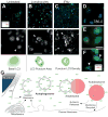
Figure 2. Autophagic Flux and Phenotype in Human Cancer Cell Lines
Epi-fluorescent imaging of human pancreatic and renal cell lines plated for 24h under various conditions. A–C, Panc2.03, top row; 20x images, Hoechst (blue), LC3 (green), and PBL (false-color magenta), yellow box is magnified in micrographs below. Second row, magnified fields, LC3 channel in grayscale for enhanced contrast; nuclei are outlined with dotted lines. Arrowheads indicate an illustrative cell which is cartooned below. Third row, cartoon of cell showing nuclear outline, diffuse LC3 (light green), and LC3 puncta (dark green). D, A renal cancer cell line (786-0) with resting LC3 staining. EF, Panc2.03 cells stained for Hoechst (blue), LC3 (green) and LAMP1 (red). Perinuclear puncta from both LC3 and LAMP1 are visible in treated condition (arrow heads). Cells in control conditions (A,D,E), lymphocyte coculture (B,G) and IFNγ (C). G, Overview of classical macroautophagy process. Cytoplasmic LC3 is microtubule associated while membrane LC3 is lipidated. Scale bars 50 μm.
Figure 3
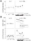
Figure 3. IL-2 Promotes and IL-10 Inhibits PBL Induced Autophagy in Cocultures
A, Human pancreatic cancer cells (Panc2.03) were cultured alone (left panel) or with lymphocytes (right panel) and treated with IL-2. Markers show LC3 puncta area. B, T-24 bladder cancer cells were treated with a dose series of IL-10 (left panels), or a dose series of IL-10 over a constant E:T ratio of 50:1 PBLs (right panels), with and without IL-2 (black or gray markers). Markers show MHC-I mean fluorescent intensity (upper panels) and LC3 puncta area (lower panels). Error bars are SEM of field replicates.
Figure 4
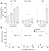
Figure 4. Cell-Cell Contact Enhances Autophagy
Lymphocytes were cocultured with hepatocellular, pancreatic, renal and bladder cancer cell lines to examine the extent of cell-mediated autophagy. A, induction of autophagy is measured in targets; untreated (UT), supernatant from 2 and 6 hour coculture, and direct coculture with lymphocytes (PBL). Bars show average with SD, ** p<0.01 by Dunnet’s posttests after ANOVA. B, Cancer cell lines were analyzed with lymphocytes in coculture (filled circles) or separated by a 0.4 μm transwell (open circles) for 24h. IL-2 was added concurrently at 6000 IU/ml under all conditions. Line chart shows averages with SEM, E:T ratio is plotted on a log scale, ANCOVAs were significant for HCT116 p=0.022, T-24 p<0.001.
Figure 5
Figure 5. IFNγ Is Sufficient To Induce Autophagy In Epithelial Cancer Cell Lines
Human colorectal (HCT116), pancreatic (Panc2.03), and bladder (T-24) cancer lines were cultured for 24h alone before the addition of primary lymphocytes or recombinant IFNγ. A–C Micrographs of Panc2.03 in the control condition (A), mixed with lymphocytes at 50:1 E:T ratio (B), or with 53,000 IU/ml IFNγ (C). Note the varied localization and distribution of the LC3 (red) when comparing PBLs with IFNγ treatment (blue Hoechst, green HMGB1). D–G Line charts summarizing the dose response of primary human lymphocytes (PBLs+IL-2, D,F), or IFNγ (E,G) showing Mean+/−StDev of MHC-I expression (D,E) or LC3 puncta area (F,G) on target cancer cells (x-axes log scale). Scale bar 50 μm.
Figure 6
Figure 6. Cell-Mediated Autophagy is inhibited by Alpha Interferon
Tumor cell lines were grown alone for 24h, then treated with combinations of IFNα and lymphocytes for 24h before measuring cytolysis, MHC-I expression, and autophagic puncta. Line charts showing MHC-I expression (A) and # LC3 puncta (B) with the five columns on each graph representing a dose response to IFNα, increasing E:T ratios of PBLs with no IFNα, a gradient of PBLs with a constant IFNα 70,000 IU/ml, Constant 50:1 E:T Ratio (PBLs) with increasing concentrations of IFNα, and a dual increase in both E:T ratio and IFNα. All x-axes are on a log scale. Micrographs of T-24 bladder cancer (C) and Panc2.03 (D) representing the various patterns of autophagy and MHC-I expression. C, Control growth of cells in Hoechst channel, LC3, MHC-I and merge (blue, red, green respectively). On the second row, 50:1 E:T ratio of lymphocyte coculture after 24h (arrowhead indicating lymphocyte nuclei). Bottom row showing IFN-gamma treatment for comparison. D, Panc2.03 control (top), 70,000 IU/ml IFNα (middle), and IFNγ (bottom) for comparison. Scale bar 50 μm.
Figure 7
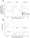
Figure 7. Coculture with Lymphocytes protects HCT116 and 786-0 against subsequent Gamma Irradiation
HCT116, 786-0 and Panc2.03 cells were cultured with human PBLs for 24h. Following the culture, the lymphocytes were rinsed off, and the cells were harvested and exposed to gamma irradiation. A, Remaining cell numbers, displayed as a fraction of the control (no gamma irradiation) for cultures previously exposed to 16:1 E:T ratio of PBL (black) and 50:1 E:T ratio of PBL, or no PBLs (red), all with 6000 IU/ml IL-2, are plotted as the mean+/−SD of well replicates (** p < 0.01, ** p < 0.05). B, Number of cytoplasmic LC3 puncta are plotted (mean+/− SD of well replicates), indicative of sustained autophagy at 24h post irradiation.

V体育安卓版 - References

    1. Vakkila J, Lotze MT. Inflammation and necrosis promote tumour growth. Nat Rev Immunol. 2004;4:641–8. - "VSports app下载" PubMed
    1. Coussens LM, Werb Z. Inflammation and cancer. Nature. 2002;420:860–7. - PMC - PubMed
    1. Balkwill F, Mantovani A. Inflammation and cancer: back to Virchow? Lancet. 2001;357:539–45. - PubMed
    1. Lotze MT, Deisseroth A, Rubartelli A. Damage associated molecular pattern molecules. Clin Immunol. 2007;124:1–4. - PMC - PubMed
    1. Rubartelli A, Lotze MT. Inside, outside, upside down: damage-associated molecular-pattern molecules (DAMPs) and redox. Trends Immunol. 2007;28:429–36. - "V体育ios版" PubMed

Publication types

VSports注册入口 - MeSH terms