Skip to main page content
U.S. flag

An official website of the United States government

Dot gov

The VSports app下载. gov means it’s official. Federal government websites often end in . gov or . mil. Before sharing sensitive information, make sure you’re on a federal government site. .

Https

The site is secure. The https:// ensures that you are connecting to the official website and that any information you provide is encrypted and transmitted securely. V体育官网.

. 2012;7(4):e34788.
doi: 10.1371/journal.pone.0034788. Epub 2012 Apr 6.

Growth hormone improves growth retardation induced by rapamycin without blocking its antiproliferative and antiangiogenic effects on rat growth plate

Affiliations

VSports在线直播 - Growth hormone improves growth retardation induced by rapamycin without blocking its antiproliferative and antiangiogenic effects on rat growth plate

Óscar Álvarez-García (V体育2025版) et al. PLoS One. 2012.

Abstract

Rapamycin, an immunosuppressant agent used in renal transplantation with antitumoral properties, has been reported to impair longitudinal growth in young individuals VSports手机版. As growth hormone (GH) can be used to treat growth retardation in transplanted children, we aimed this study to find out the effect of GH therapy in a model of young rat with growth retardation induced by rapamycin administration. Three groups of 4-week-old rats treated with vehicle (C), daily injections of rapamycin alone (RAPA) or in combination with GH (RGH) at pharmacological doses for 1 week were compared. GH treatment caused a 20% increase in both growth velocity and body length in RGH animals when compared with RAPA group. GH treatment did not increase circulating levels of insulin-like growth factor I, a systemic mediator of GH actions. Instead, GH promoted the maturation and hypertrophy of growth plate chondrocytes, an effect likely related to AKT and ERK1/2 mediated inactivation of GSK3β, increase of glycogen deposits and stabilization of β-catenin. Interestingly, GH did not interfere with the antiproliferative and antiangiogenic activities of rapamycin in the growth plate and did not cause changes in chondrocyte autophagy markers. In summary, these findings indicate that GH administration improves longitudinal growth in rapamycin-treated rats by specifically acting on the process of growth plate chondrocyte hypertrophy but not by counteracting the effects of rapamycin on proliferation and angiogenesis. .


Conflict of interest statement

Competing Interests: The authors have declared that no competing interests exist.

Figures

Figure 1
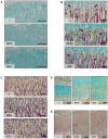
Figure 1. GH effects on longitudinal growth rate and growth plate morphology.
(A) Representative images showing the distance between the metaphyseal end of the growth cartilage and the fluorescent calcein front, which indicates longitudinal bone growth rate during the last 3 days of the study, in control rats (C), rats treated with rapamycin (RAPA) or rapamycin and GH (RGH). (B) Representative alcian blue/safranine stained sections showing tibial growth cartilage morphology of C, RAPA and RGH animals. (C) Representative Von Kossa stained sections showing the pattern of extracellular matrix mineralization in proximal tibial growth plate of C, RAPA and RGH animals. Mineralized transverse septa (Red arrows) were often found in RAPA and RGH animals and not in C group. Magnification bars  =  100 µm.
Figure 2
Figure 2. GH effects on growth plate cell proliferation and angiogenesis.
(A) Representative images of BrdU immunodetection in the proliferative zone of the epiphyseal cartilage of control rats (C), rats treated with rapamycin (RAPA) or rapamycin and GH (RGH). (B) Representative sections of proximal tibial growth plates stained with picrosirius red/alcian blue showing trabeculae and vascular sprouts arrangement in the primary spongiosa of C, RAPA and RGH animals. Transverse unresorbed septa are indicated with yellow arrows. Vascular sprouts are indicated with red arrows. (C) Representative sections of proximal tibial growth plates stained with tartrate-resistant acid phosphatase (TRAP) showing positive chondroclasts/osteoclasts in the chondro-osseous junction of C, RAPA and RGH animals. Representative images of immunohistochemistry (D) and in situ hybridization (E) experiments showing VEGF expression in growth plates of C, RAPA and RGH animals. Magnification bars  =  100 µm.
Figure 3
Figure 3. GH effects on chondrocyte autophagy.
(A) Immunofluorescent detection of LC3, a marker of autophagy, in the growth plates of rats treated with rapamycin (RAPA) or rapamycin and GH (RGH). Fluorescent signal was observed in prehypertrophic chondrocytes often displaying a punctuate distribution (white arrows in B). (C) Western blot of LC3-I (18 kDa) and LC3-II (16 kDa) in the growth cartilage of rats treated with rapamycin (RAPA) or rapamycin and GH (RGH). GAPDH was used as loading control. Image is representative of three blots giving similar results.
Figure 4
Figure 4. GH effects on chondrocyte metabolism.
(A) Periodic acid-Schiff (PAS) reaction in the proximal tibial growth plate of control rats (C), rats treated with rapamycin (RAPA) or rapamycin and GH (RGH). Magnification bars  =  100 µm. (B) Western blot of p-GSK3β (Ser9) and GSK3β in the growth cartilage of C, RAPA and RGH animals. (C) Western blot of β-catenin in the growth cartilage of C, RAPA and RGH animals. (D) Western blot of p-AKT (Thr308), AKT, p-ERK1/2 (Thr202/Tyr204) and ERK1/2 in the growth cartilage of C, RAPA and RGH animals. GAPDH was used as loading control. Images are representative of three blots giving similar results. (E) Densitometry analysis of the western blot experiments shown in B,C and D. At least four animals per group were used per experiment and each assay was repeated at least three times. aMeans statistically different from C group (P≤0.05). bMeans statistically different from RAPA group (P≤0.05).
Figure 5
Figure 5. Proposed mechanism of the stimulating effect of GH on the hypertrophy of growth cartilage chondrocytes.
GH would signal through PIP3/Akt and MAPK pathways to phosphorylate and inactivate GSK3β in growth plate chondrocytes. This would lead to an increase of glycogen synthesis and stabilization of β-catenin that, eventually, would enhance chondrocyte hypertrophy.

References

    1. Wullschleger S, Loewith R, Hall MN. TOR signaling in growth and metabolism. Cell. 2006;124:471–484. - "VSports最新版本" PubMed
    1. Geissler EK, Schlitt HJ. The potential benefits of rapamycin on renal function, tolerance, fibrosis, and malignancy following transplantation. Kidney Int. 2010;78:1075–1079. - PubMed (VSports)
    1. Koehl GE, Andrassy J, Guba M, Richter S, Kroemer A, et al. Rapamycin protects allografts from rejection while simultaneously attacking tumors in immunosuppressed mice. Transplantation. 2004;77:1319–1326. - PubMed
    1. Hudes G, Carducci M, Tomczak P, Dutcher J, Figlin R, et al. Temsirolimus, interferon alfa, or both for advanced renal-cell carcinoma. N Engl J Med. 2007;356:2271–2281. - PubMed
    1. Cravedi P, Ruggenenti P, Remuzzi G. Sirolimus to replace calcineurin inhibitors? Too early yet. Lancet. 2009;373:1235–1236. - PubMed

Publication types

MeSH terms