Skip to main page content
U.S. flag

An official website of the United States government

Dot gov

The . gov means it’s official. Federal government websites often end in . gov or . mil. Before sharing sensitive information, make sure you’re on a federal government site. VSports app下载.

Https

The site is secure. The https:// ensures that you are connecting to the official website and that any information you provide is encrypted and transmitted securely. V体育官网.

. 2012 Apr 15;188(8):3859-68.
doi: 10.4049/jimmunol.1103729.

"V体育官网" T cell factor-1 and β-catenin control the development of memory-like CD8 thymocytes

Affiliations

T cell factor-1 and β-catenin control the development of memory-like CD8 thymocytes (V体育安卓版)

Archna Sharma et al. J Immunol. .

Abstract

Innate memory-like CD8 thymocytes develop and acquire effector function during maturation in the absence of encounter with Ags. In this study, we demonstrate that enhanced function of transcription factors T cell factor (TCF)-1 and β-catenin regulate the frequency of promyelocytic leukemia zinc finger (PLZF)-expressing, IL-4-producing thymocytes that promote the generation of eomesodermin-expressing memory-like CD8 thymocytes in trans VSports手机版. In contrast, TCF1-deficient mice do not have PLZF-expressing thymocytes and eomesodermin-expressing memory-like CD8 thymocytes. Generation of TCF1 and β-catenin-dependent memory-like CD8 thymocytes is non-cell-intrinsic and requires the expression of IL-4 and IL-4R. CD8 memory-like thymocytes migrate to the peripheral lymphoid organs, and the memory-like CD8 T cells rapidly produce IFN-γ. Thus, TCF1 and β-catenin regulate the generation of PLZF-expressing thymocytes and thereby facilitate the generation of memory-like CD8 T cells in the thymus. .


Figures
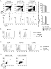
Figure 1
Figure 1
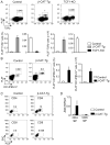
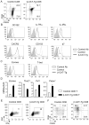
Memory-like CD8 T cell development in the thymus is regulated by β-catenin and TCF1. A) Flow-cytometric analysis of surface CD4, CD8 expression on total thymocytes and TCR-β expression on the gated CD8SP population from control, β-CAT-Tg and TCF1-deficient mice. Numbers show the percentage of cells. Data are representative of eight independent analyses. The graph on upper right panel shows total thymic cellularity and the graph on lower right panel shows absolute numbers of TCRβhi CD8SP thymocytes from control, β-CAT-Tg and TCF1-deficient mice. B) Flow-cytometric analysis of Eomes and IL-4Rα expression on the gated TCRβhi CD8SP thymocytes from control, β-CAT-Tg and TCF1-deficient mice. Data are representative of six independent analyses. C) Flow-cytometric analysis of cell surface memory markers expression on the gated CD8SP thymocytes from control and β-CAT-Tg mice. Data are representative of eight independent analyses. D) Flow-cytometric analysis of intracellular IFN-γ expression on gated CD8SP thymocytes after 5 h PMA plus ionomycin stimulation of total thymocytes from control and β-CAT-Tg mice is shown. Data are representative of three independent analyses with total five to six mice in each group.
Figure 2
Figure 2
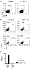
TCRβ+ and CD1dPBS-57+ PLZF-expressing thymic population is increased in β-CAT-Tg mice. A) Flow-cytometric analysis of expression of PLZF and TCRβ on total thymocytes from control, β-CAT-Tg and TCF1-deficient mice. Numbers adjacent to outlined areas (upper panels) and graphs (lower panels) show percentage and absolute numbers of PLZF+TCRβ+ cells as indicated. Data are representative of five independent analyses with total eight to ten mice per group. B) Flow-cytometric analysis of expression of PLZF and CD1dPBS-57 on total thymocytes from control and β-CAT-Tg mice. Numbers adjacent to outlined areas (left panels) and graphs (right panels) show percentage and absolute numbers of PLZF+CD1dPBS-57+ cells as indicated. Data are representative of five independent analyses with total eight to ten mice per group. C) Flow-cytometric analysis of expression of PLZF on gated CD4 and CD8SP populations of total thymocytes from control and β-CAT-Tg mice. Numbers adjacent to outlined areas show percent of PLZF+ CD4SP and PLZF+ CD8SP thymocytes as indicated. Data are representative of five independent analyses. D) Real-time PCR analysis for Zbtb16 mRNA in purified CD4SP and CD8SP thymocytes, presented relative to Hprt. Data are from four independent samples.
Figure 3
Figure 3
β-CAT-Tg PLZF+ CD4SP thymocytes over-produce IL-4. A) Flow-cytometric analysis of intracellular expression of IL-4 and PLZF in control and β-CAT-Tg thymocytes stimulated for 5 h with PMA and ionomycin. Numbers adjacent to outlined areas show percent IL-4+PLZF+ cells. Data are representative of four independent analyses. B) Flow-cytometric analysis of intracellular IL-4 expression on gated CD4 and CD8SP thymocytes after 5h PMA plus ionomycin stimulation of total thymocytes from control and β-CAT-Tg mice. Numbers adjacent to outlined areas show percent of IL-4+ CD4SP and IL-4+ CD8SP thymocytes as indicated. Data are representative of five independent analyses. C) Real-time PCR analysis for Il4 mRNA in purified CD4SP and CD8SP thymocytes, presented relative to Actb. Data are from four independent samples.
Figure 4
Figure 4
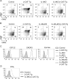
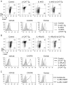
The memory-like CD8 phenotype in β-CAT-Tg mice is dependent on IL-4 and IL-4Rα. A) Flow-cytometric analysis of the cell surface memory markers CD44 and CD62L on the gated CD8SP population of total thymocytes from control, β-CAT-Tg, IL-4KO and IL-4KO-β-CAT-Tg mice. Numbers show the percentage of cells. Data are representative of four independent analyses. B) Flow-cytometric analysis of the cell surface memory markers CD44 and CD62L on the gated CD8SP population of total thymocytes from control, β-CAT-Tg, IL-4RαKO and IL-4RαKO-β-CAT-Tg mice. Numbers show the percentage of cells. Data are representative of four independent analyses. C) Flow-cytometric analysis of the surface CD122, CXCR expression and intracellular Eomes expression in the gated CD8SP thymocytes from control, β-CAT-Tg, IL-4KO, IL-4RαKO, IL-4KO-β-CAT-Tg and IL-4RαKO-β-CAT-Tg mice. Data are representative of four independent analyses. D) Flow cytometric analysis of intracellular IFN-γ expression on gated CD8SP thymocytes after 5 h PMA plus ionomycin stimulation of total thymocytes from control, β-CAT-Tg, and IL-4KO-β-CAT-Tg mice is shown. Data are representative of four independent analyses.
Figure 5
Figure 5
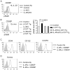
Enhanced IL-4 signaling and IL-4Rα expression regulates β-CAT-Tg CD8 memory-like phenotype. A) Flow-cytometric analysis of intracellular pSTAT6 in gated CD8SP thymocytes from ex vivo control, IL-4KO and IL-4RαKO total thymocytes transiently stimulated with IL-4 for 20 min. Data are representative of two independent analyses. B) Flow-cytometric analysis of intracellular pSTAT6 on gated CD8SP thymocytes from ex vivo total thymocytes from control, β-CAT-Tg, IL-4KO-β-CAT-Tg and IL-4RαKO-β-CAT-Tg mice. Data are representative of two independent analyses. Graph on right side represents pSTAT6 levels as mean fluorescence intensity (MFI). C) Flow-cytometric analysis of the cell surface memory markers on the gated IL-4Rα and IL-4Rα+ CD8SP population of total thymocytes from β-CAT-Tg and IL-4RαKO-β-CAT-Tg 1:1 bone marrow chimera. Data are representative of two independent analyses with total 5 chimera mice. D) Flow-cytometric analysis of Eomes expression on the gated IL-4Rα and IL-4Rα+ CD8SP thymocytes from β-CAT-Tg and IL-4RαKO-β-CAT-Tg 1:1 bone marrow chimera. Data are representative of two independent analyses with total 5 chimera mice.
Figure 6
Figure 6
Peripheral CD8 T cells in β-CAT-Tg also have memory-like features and function. A) Flow-cytometric analysis of the cell surface memory markers CD44 and CD62L on the gated CD8 T population of lymph node cells from control and β-CAT-Tg mice; Numbers show the percentage of cells. Data are representative of ten independent analyses. B) Flow-cytometric analysis of the cell surface memory markers CD122, IL-7Rα, IL-4Rα, CXCR3, CD103 and β7 expression on the gated CD8 T population of lymph node cells from control and β-CAT-Tg mice. Data are representative of ten independent analyses. C) Flow-cytometric analysis of Eomes and T-bet expression on the gated CD8 T population of lymph node cells from control and β-CAT-Tg mice. Data are representative of six independent analyses. D) Real-time PCR analysis for mRNA abundance of Eomes, Tbx21, Perforin and Prdm-1 in purified ex vivo CD8 T cells from lymph nodes of control and β-CAT-Tg mice, presented relative to Actb. Data are from four independent samples. E) Flow cytometric analysis of intracellular IFN-γ expression on gated CD8 T cells after 5 h PMA plus ionomycin stimulation of lymph node cells from control and β-CAT-Tg mice is shown. Data are representative of four independent analyses. F) Flow-cytometric analysis of the expression of cell surface memory markers CD44 and CD62L on the gated CD8 T population of IEL from small intestines of control and β-CAT-Tg mice. Numbers show the percentage of cells.
Figure 7
Figure 7
IL-4 or IL-4Rα chain deficiency results in loss of memory-like phenotype of β-CAT-Tg CD8 T cells. A) Flow-cytometric analysis of the cell surface memory markers and Eomes expression by the gated CD8T cells from lymph nodes from control, β-CAT-Tg, IL-4KO and IL-4KO-β-CAT-Tg mice; CD44 and CD62L expression (upper panel) and CD122, CXCR3 and Eomes expression (lower panel). Numbers show the percentage of cells. Data are representative of four independent experiments. B) Flow-cytometric analysis of the cell surface memory markers and Eomes expression by the gated CD8T cells from lymph nodes from control, β-CAT-Tg, IL-4Rα-KO and IL-4RαKO-β-CAT-Tg mice; CD44 and CD62L expression (upper panel) and CD122, CXCR3 and Eomes expression (lower panel). Numbers show the percentage of cells. Data are representative of four independent experiments. C) Flow-cytometric analysis of the cell surface memory markers on and Eomes expression in the gated IL-4Rα- and IL-4Rα+ CD8 T population of spleenocytes from β-CAT-Tg and IL-4RαKO-β-CAT-Tg 1:1 bone marrow chimera. Data are representative of two independent analyses with total 5 chimera mice.
Figure 8
Figure 8
IL-4 signals induce memory-like gene expression in naïve CD8 T cells. A) CD8 T cells were infected with control MSCV-human CD8 vector or MSCV-β-CAT-human-CD8 construct. Human CD8+ infected CD8 T cells were sorted and the relative abundance of mRNA for transcription factors and effector molecules was analyzed by real time RT-PCR. Data are presented relative to Actb and are from six independent samples. B) CD122CD44lo naïve CD8 T cells that were treated with various cytokines and the relative abundance of mRNA for transcription factors and effector molecules was analyzed by real time RT-PCR. Data are presented relative to Actb and are from four to six independent samples. C) Lymph node cells from control and TCF1-deficient mice were stimulated with anti-CD3 for 3 d and then rested in culture that contained IL-7 and IL-4 for 6 d. After this in vitro stimulation CD8 T cells were purified and transcription factors and effector molecules analyzed by real-time RT-PCR. Data are presented relative to Actb and are from three independent samples.

References

    1. Lee YJ, Jameson SC, Hogquist KA. Alternative memory in the CD8 T cell lineage. Trends Immunol. 2011;32:50–56. - PMC (VSports手机版) - PubMed
    1. Broussard C, Fleischacker C, Horai R, Chetana M, Venegas AM, Sharp LL, Hedrick SM, Fowlkes BJ, Schwartzberg PL. Altered development of CD8+ T cell lineages in mice deficient for the Tec kinases Itk and Rlk. Immunity. 2006;25:93–104. - PubMed
    1. Atherly LO, Lucas JA, Felices M, Yin CC, Reiner SL, Berg LJ. The Tec family tyrosine kinases Itk and Rlk regulate the development of conventional CD8+ T cells. Immunity. 2006;25:79–91. - PubMed
    1. Jordan MS, Smith JE, Burns JC, Austin JE, Nichols KE, Aschenbrenner AC, Koretzky GA. Complementation in trans of altered thymocyte development in mice expressing mutant forms of the adaptor molecule SLP76. Immunity. 2008;28:359–369. - "VSports app下载" PMC - PubMed
    1. Prince AL, Yin CC, Enos ME, Felices M, Berg LJ. The Tec kinases Itk and Rlk regulate conventional versus innate T-cell development. Immunol Rev. 2009;228:115–131. - V体育官网 - PMC - PubMed

VSports注册入口 - Publication types

MeSH terms

"VSports在线直播" Substances

V体育平台登录 - LinkOut - more resources