Skip to main page content (VSports在线直播)
U.S. flag

An official website of the United States government

Dot gov

The . gov means it’s official. Federal government websites often end in VSports app下载. gov or . mil. Before sharing sensitive information, make sure you’re on a federal government site. .

Https

The site is secure. The https:// ensures that you are connecting to the official website and that any information you provide is encrypted and transmitted securely V体育官网. .

Review
. 2012:100:319-44.
doi: 10.1016/B978-0-12-387786-4.00010-5.

"VSports手机版" Cardiac regeneration

Affiliations
Review

Cardiac regeneration (VSports手机版)

Wen-Yee Choi et al. Curr Top Dev Biol. 2012.

VSports最新版本 - Abstract

The heart is a pump that is comprised of cardiac myocytes and other cell types and whose proper function is critical to quality of life. The ability to trigger regeneration of heart muscle following injury eludes adult mammals, a deficiency of great clinical impact VSports手机版. Major research efforts are attempting to change this through advances in cell therapy or activating endogenous regenerative mechanisms that exist only early in life. In contrast with mammals, lower vertebrates like zebrafish demonstrate an impressive natural capacity for cardiac regeneration throughout life. This review will cover recent progress in the field of heart regeneration with a focus on endogenous regenerative capacity and its potential manipulation. .


Figures

Figure 1
Figure 1
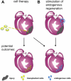
Approaches to stimulating heart regeneration. (A) One strategy for heart regeneration involves the injection of exogenous cells into the infarcted area of the heart. These transplanted cells could proliferate and repopulate the injured area with myocardium, or may signal to endogenous cardiac cells and promote their proliferation. (B) A second fundamental approach relies on the regenerative capacity of uninjured progenitor cells or cardiomyocytes adjacent to the infarcted area. These cells proliferate and/or differentiate in situ to build new cardiac muscle.
Figure 2
Figure 2
Injury and regeneration in zebrafish. (A) In the resection model, 20% of the ventricular apex is surgically removed. Within two months, the resected tissue is replaced through local cardiomyocyte proliferation. (B) In the cryoablation model, a cooled probe is applied directly to the heart to induce localized cell death affecting up to 25% of the ventricle. The lesions can remain detectable past 60 days post-injury, but the majority of collagen is eventually cleared coincident with local cardiomyocyte proliferation. (C) Using a conditional genetic approach, over 60% of the ventricular (and atrial) myocardium is destroyed throughout the heart. Regeneration is particularly rapid and robust, with full recovery within one month after ablation.
Figure 3
Figure 3
Cellular source of regenerating cardiomyocytes. (A) Genetic fate-mapping to determine the source of new cardiomyocytes during zebrafish heart regeneration. Treatment with tamoxifen leads to fluorescent EGFP marking of nearly all cardiomyocytes. Following resection, new cardiomyocytes expressed EGFP, indicating derivation from existing cardiomyocytes rather than a different unmarked cell source. (B) A similar fate-mapping experiment was performed in the neonatal mouse heart, in this case with an inducible LacZ reporter. Although the labeling of cardiomyocytes with this system was ~60%, the ratio of labeled cells was maintained between the regenerate and uninjured myocardium. This result indicates that the new cardiomyocytes are derived from existing cardiomyocytes rather than a progenitor cell.
Figure 4
Figure 4
Three major cardiac cell types activate developmental gene expression in a chamber-wide manner after resection injury. (A) Myocardial activation, represented by the activation of a gata4:EGFP reporter, is first detected throughout the compact layer of the myocardium by 7 days post-amputation (dpa) and becomes localized to the regenerating myocardium by 14–30 dpa (arrowheads). Dotted lines indicate amputation planes. (B) Ostensibly the entire epicardium activates developmental genes such as raldh2 (violet) within several days following injury. Enhanced gene expression localizes to the wound area by two weeks post-injury (arrowheads). (C) Endocardial cells undergo structural remodeling following injury and upregulate raldh2 (violet) within hours of amputation. Modified from Lepilina et al., 2006, Kikuchi et al., 2010, and Kikuchi et al., 2011b.
Figure 5
Figure 5
Transient regenerative capacity of the neonatal mouse heart after ventricular resection. (A) Rather than inducing fibrosis, resection injuries to the 1-day neonate stimulate replacement with new cardiomyocytes, as assessed 21 days later. Cardiomyocyte proliferation stimulated away from the injury is also likely to be a key component of this regenerative potential. (B) By contrast, fibrosis is the dominant response after resection of ventricular muscle in the 7-day neonate.

References

    1. Adler CP. Recent Adv Stud Cardiac Struct Metab. 1975;8:373–86. - VSports在线直播 - PubMed
    1. Adler CP, Costabel U. Recent Adv Stud Cardiac Struct Metab. 1975;6:343–55. - PubMed
    1. Bader D, Oberpriller J. J Exp Zool. 1979;208:177–93. - "V体育安卓版" PubMed
    1. Bader D, Oberpriller JO. J Morphol. 1978;155:349–57. - PubMed (V体育2025版)
    1. Baliga RR, Pimental DR, Zhao YY, Simmons WW, Marchionni MA, Sawyer DB, Kelly RA. Am J Physiol. 1999;277:H2026–37. - "V体育平台登录" PubMed

Publication types

"VSports最新版本" LinkOut - more resources