Skip to main page content
U.S. flag

An official website of the United States government

Dot gov

The . gov means it’s official. Federal government websites often end in VSports app下载. gov or . mil. Before sharing sensitive information, make sure you’re on a federal government site. .

Https

The site is secure. The https:// ensures that you are connecting to the official website and that any information you provide is encrypted and transmitted securely V体育官网. .

. 2012 May 18;287(21):17693-17705.
doi: 10.1074/jbc.M111.300012. Epub 2012 Mar 20.

"VSports" Oxidatively truncated phospholipids are required agents of tumor necrosis factor α (TNFα)-induced apoptosis

Affiliations

Oxidatively truncated phospholipids are required agents of tumor necrosis factor α (TNFα)-induced apoptosis

Calivarathan Latchoumycandane et al. J Biol Chem. .

Abstract

TNFα generates reactive oxygen species (ROS) at the cell surface that induce cell death, but how ROS communicate to mitochondria and their specific apoptotic action(s) are both undefined. ROS oxidize phospholipids to hydroperoxides that are friable and fragment adjacent to the (hydro)peroxide function, forming truncated phospholipids, such as azelaoyl phosphatidylcholine (Az-PC). Az-PC is relatively soluble, and exogenous Az-PC rapidly enters cells to damage mitochondrial integrity and initiate intrinsic apoptosis. We determined whether this toxic phospholipid is formed within cells during TNFα stimulation in sufficient quantities to induce apoptosis and if they are essential in TNFα-induced cytotoxicity. We found that TNFα induced ROS formation and phospholipid peroxidation in Jurkat cells, and either chemical interference with NADPH oxidase activity or siRNA suppression of the NADPH oxidase-4 subunit blocked ROS accumulation and phospholipid peroxidation. Mass spectrometry showed that phospholipid peroxides and then Az-PC increased after TNFα exposure, whereas ROS inhibition abolished Az-PC accumulation and TNFα-induced cell death. Glutathione peroxidase-4 (GPx4), which specifically metabolizes lipid hydroperoxides, fell in TNFα-stimulated cells prior to death VSports手机版. Ectopic GPx4 overcame this, reduced peroxidized phospholipid accumulation, blocked Az-PC accumulation, and prevented death. Conversely, GPx4 siRNA knockdown enhanced phospholipid peroxidation, increasing TNFα-stimulated Az-PC formation and apoptosis. Truncated phospholipids were essential elements of TNFα-induced apoptosis because overexpression of PAFAH2 (a phospholipase A(2) that selectively hydrolyzes truncated phospholipids) blocked TNFα-induced Az-PC accumulation without affecting phospholipid peroxidation. PAFAH2 also abolished apoptosis. Thus, phospholipid oxidation and truncation to apoptotic phospholipids comprise an essential element connecting TNFα receptor signaling to mitochondrial damage and apoptotic death. .


V体育安卓版 - Figures
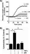
FIGURE 1.
FIGURE 1.
TNFα stimulates cytotoxic ROS formation in Jurkat cells. A, TNFα rapidly stimulates ROS formation in cultured Jurkat lymphoid cells. Amplex Red® fluorescence at 590 nm in the absence of or after the addition of 40 ng/ml TNFα with or without DPI addition. n = 3. B, DPI suppresses TNFα cytotoxicity. Jurkat cells were treated with TNFα for 24 h or not after a 30-min prior exposure to 20 μm DPI. Cell death was assessed as described under “Materials and Methods.” n = 3; *, p < 0.05. Error bars, S.E.
FIGURE 2.
FIGURE 2.
TNFα induces apoptotic cell death. A, TNFα induced a time-dependent increase in cell death. Jurkat cells were treated with TNFα (40 ng/ml) for the stated times before cytotoxicity was determined by the CytoTox One homogenous membrane integrity assay (Promega). n = 3; *, p < 0.05. B, DNA fragmentation is reduced by DPI. DNA was extracted after treatment or not with TNFα for 24 h with or without a 30-min preincubation with 20 μm DPI, which remained in the buffer, and resolved by electrophoresis before visualization after ethidium staining. C, phosphatidylserine appears on the outer aspect of TNFα-exposed cells. Jurkat cells were stained for Annexin V for 30 min after 24 h of TNFα treatment, and positive cells were analyzed using flow cytometry (FACScan). The data were analyzed using FlowJo software. n = 3; *, p < 0.05. D, TNFα induced cell death via activation of caspase-3. Jurkat cells were treated with TNFα for the indicated time, and then proteins were extracted and resolved in SDS-PAGE. Caspase 3 (Cell Signaling) fragments were detected by Western blot. E, caspase-3 inhibitor Z-DEVD-fmk abolished TNFα-induced cell death. Jurkat cells were preincubated with Z-DEVD-fmk for 30 min and then incubated with 40 ng/ml TNFα for 24 h, and cell death was assessed by LDH release assay as above. n = 3; *, p < 0.05. Error bars, S.E.
FIGURE 3.
FIGURE 3.
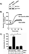
TNFα stimulated NADPH oxidase-4 generates cytotoxic ROS. A, NOx4 is reduced by targeted siRNA. Jurkat cells treated with nonspecific (NS) or NOx4 siRNA before NOx4 or β-actin were detected by Western blotting. n = 2. B, NOx4 produces half of TNFα-stimulated ROS. H2O2 was detected by Amplex Red® as in Fig. 1. n = 3. C, NOx4 knockdown reduces TNFα-stimulated cell death. Jurkat cells transfected with nonspecific or NOx4-directed siRNA were stimulated with TNFα before stimulation with TNFα, and the level of cytotoxicity was determined 24 h later. n = 3; *, p < 0.05. Error bars, S.E.
FIGURE 4.
FIGURE 4.
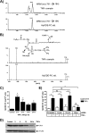
Phospholipid hydroperoxides and glutathione peroxidase-4 are inversely related after TNFα stimulation. A, phosphatidylcholine hydroperoxide is present in Jurkat cells after TNFα stimulation. Lipid extracted from Jurkat cells after treatment with TNFα (40 ng/ml) for 24 h or a synthetic HpODE-PC was analyzed by LC/MS/MS using the [M + H]+ m/z 791 → 184 transition as described under “Materials and Methods.” B, palmitoyl HpODE-PC is present after TNFα stimulation. Product ion scans in the positive mode were performed for both synthetic HpODE-PC and the resolved lipids from TNFα-treated cells that co-eluted with the standard. The fragmentation spectrum pattern establishes the presence of phosphocholine (m/z 184), whereas m/z 773 is produced from the loss of a hydroxyl moiety from palmitoyl HpODE-PC in both the standard and the co-eluting peak in TNFα-treated cells. C, TNFα induces a time-dependent increase in endogenous phosphatidylcholine hydroperoxide. HpODE-PC was determined as in B in relation to the standard as a function of the time of TNFα treatment of Jurkat cells. n = 6; *, p < 0.05 relative to time 0. D, TNFα stimulation depletes cellular GPx4. Western blot visualization of GPx4 and β-actin in control or TNFα-treated Jurkat cells. n = 2. E, NADPH oxidase, not lipoxygenase, promoted phospholipid peroxidation after TNFα activation. HpODE-PC was determined as in D using Jurkat cells previously transfected for 48 h with the stated siRNA or scrambled siRNA and then treated with TNFα for 3 h. n = 6; *, p < 0.05; NS, not significant. Error bars, S.E.
FIGURE 5.
FIGURE 5.
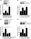
Glutathione peroxidase-4 blocks cell death by depleting cellular phospholipid hydroperoxides. A, GPx4 is reduced by targeted siRNA. GPx4 and β-actin were detected by immunoblotting Jurkat cells after transfection with nonspecific (NS) or GPx4-targeted siRNA. n = 2. Loss of endogenous GPx4 enhances TNFα-induced cytotoxicity determined by LDH release. n = 3; *, p < 0.05. B, ectopic GPx4 expression increased GPx4 protein. GPx4, GPx1, and β-actin were assessed by immunoblotting after transient transfection with pCMV-GPx4 or empty vector. n = 2. Overexpression of GPx4 blocked TNFα-induced cytotoxicity. Jurkat cells transiently expressing GPx4 were stimulated with 40 ng/ml TNFα or buffer, and 24 h later, the increase in released LDH was determined. n = 3; *, p < 0.05. C, loss of 12-lipoxygenase does not reduce TNFα cytotoxicity. siRNA directed to 12-lipoxygenase reduced cellular protein expression 48 h after transfection but did not suppress TNFα-induced cytotoxicity. n = 6; *, p < 0.05. D, loss of 15-lipoxygenase does not reduce TNFα cytotoxicity. siRNA directed to 15-lipoxygenase reduced cellular protein expression 48 h after transfection but did not suppress TNFα-induced cytotoxicity. n = 6; *, p < 0.05. Error bars, S.E.
FIGURE 6.
FIGURE 6.
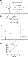
TNFα stimulation generates oxidatively truncated phospholipid. A, Az-PC is present in Jurkat cells after TNFα stimulation. Lipids extracted from Jurkat cells after treatment with TNFα (40 ng/ml) for 24 h or a synthetic Az-PC were analyzed by LC/MS/MS using the [M + H]+ m/z 661 → m/z 184 transition. B, palmitoyl Az-PC is present after TNFα stimulation. Product ion scans in the negative mode were performed for both synthetic Az-PC standard and the co-eluting peak from TNFα-treated cells. The fragmentation spectrum pattern establishes the presence of the azelaoyl residue from the intramolecularly methylated azelaoyl fragment (m/z 201) in both the standard and co-eluting cellular peak. C, Az-PC increases over time of TNFα stimulation. Jurkat cells were treated with TNFα for the stated times before the cells were lysed, and Az-PC content was determined by mass spectrometry in relation to the synthetic standard. n = 3; *, p < 0.05 from time 0. Error bars, S.E.
FIGURE 7.
FIGURE 7.
Phospholipid hydroperoxides are required precursors for TNFα-stimulated Az-PC accumulation. A, DPI abolishes TNFα-induced Az-PC accumulation. Jurkat cells were treated as stated with DPI and TNFα or not for 24 h before Az-PC was determined by mass spectrometry as in Fig. 6. n = 3; *, p < 0.05. B, NOx4 knockdown suppressed TNFα-initiated phospholipid fragmentation. Az-PC was quantified by mass spectrometry in cells with reduced NOx4 or cells expressing scrambled siRNA. n = 6; *, p < 0.05. C, siRNA knockdown of GPx4 enhances TNFα-initiated phospholipid truncation. GPx4 RNA and protein were reduced by siRNA before Az-PC was quantified. n = 6; *, p < 0.05. D, GPx4 overexpression abolishes TNFα-induced accumulation of Az-PC. Az-PC content after transient GPx4 or empty vector transfection with or without TNFα stimulation. n = 6; *, p < 0.05. Error bars, S.E.
FIGURE 8.
FIGURE 8.
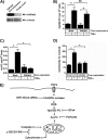
Oxidatively truncated phospholipids are required for TNFα-induced cytotoxicity. A, transient transfection increases PAFAH2. Jurkat cells were transfected by electroporation with PAFAH2 under control of a CMV promoter or an empty construct for 48 h before control lysates were resolved, and PAFAH2 and β-actin were analyzed by Western blotting. B, PAFAH2 overexpression does not reduce cellular phospholipid hydroperoxide content. HpODE-PC was determined as in Fig. 4. n = 3. C, overexpression of PAFAH2 abolishes TNFα-stimulated Az-PC accumulation. Jurkat cells transiently overexpressing PAFAH2 or not were stimulated with TNFα as above before Az-PC was quantitated by mass spectrometry. n = 3; *, p < 0.05. D, transient expression of PAFAH2 prevents apoptosis. Viability of Jurkat cells expressing ectopic PAFAH2 or not was determined by LDH release as in Fig. 1. n = 3; *, p < 0.05. E, schematic representation of TNFα stimulation of phospholipid oxidation and fragmentation to species that damage mitochondria, allowing caspase activation that promotes cell death. Error bars, S.E.

References

    1. Parameswaran N., Patial S. (2010) Tumor necrosis factor-α signaling in macrophages. Crit. Rev. Eukaryot. Gene Expr. 20, 87–103 - PMC - PubMed
    1. Gordon J. W., Shaw J. A., Kirshenbaum L. A. (2011) Multiple facets of NF-κB in the heart. To be or not to NF-κB. Circ. Res. 108, 1122–1132 - PubMed
    1. Shakibaei M., Sung B., Sethi G., Aggarwal B. B. (2010) TNF-α-induced mitochondrial alterations in human T cells requires FADD and caspase-8 activation but not RIP and caspase-3 activation. Antioxid. Redox Signal. 13, 821–831 - PMC - PubMed
    1. Falschlehner C., Emmerich C. H., Gerlach B., Walczak H. (2007) TRAIL signaling. Decisions between life and death. Int. J. Biochem. Cell Biol. 39, 1462–1475 - VSports注册入口 - PubMed
    1. Schafer Z. T., Kornbluth S. (2006) The apoptosome. Physiological, developmental, and pathological modes of regulation. Dev. Cell 10, 549–561 - PubMed

Publication types

MeSH terms

"V体育平台登录" LinkOut - more resources