Skip to main page content (V体育ios版)
U.S. flag

An official website of the United States government

Dot gov

The . gov means it’s official. Federal government websites often end in . gov or VSports app下载. mil. Before sharing sensitive information, make sure you’re on a federal government site. .

Https

The site is secure V体育官网. The https:// ensures that you are connecting to the official website and that any information you provide is encrypted and transmitted securely. .

. 2012 Apr 9;209(4):679-96.
doi: 10.1084/jem.20111512. Epub 2012 Mar 19.

MYC pathway activation in triple-negative breast cancer is synthetic lethal with CDK inhibition

Affiliations

MYC pathway activation in triple-negative breast cancer is synthetic lethal with CDK inhibition

Dai Horiuchi (VSports) et al. J Exp Med. .

Abstract

Estrogen, progesterone, and HER2 receptor-negative triple-negative breast cancers encompass the most clinically challenging subtype for which targeted therapeutics are lacking VSports手机版. We find that triple-negative tumors exhibit elevated MYC expression, as well as altered expression of MYC regulatory genes, resulting in increased activity of the MYC pathway. In primary breast tumors, MYC signaling did not predict response to neoadjuvant chemotherapy but was associated with poor prognosis. We exploit the increased MYC expression found in triple-negative breast cancers by using a synthetic-lethal approach dependent on cyclin-dependent kinase (CDK) inhibition. CDK inhibition effectively induced tumor regression in triple-negative tumor xenografts. The proapoptotic BCL-2 family member BIM is up-regulated after CDK inhibition and contributes to this synthetic-lethal mechanism. These results indicate that aggressive breast tumors with elevated MYC are uniquely sensitive to CDK inhibitors. .


Figures

Figure 1.
Figure 1.
Elevated MYC expression in human triple-negative cancers. (A) MYC mRNA expression in triple-negative versus receptor-positive primary breast tumors collected through the I-SPY TRIAL (P < 0.0001). (B) MYC and phospho-MYC (T58/S62) protein expression in triple-negative primary tumors. Shown is an independent cohort of 208 patients for which quantitative reverse-phase protein arrays were performed. (C) Relative expression of MYC mRNA in a panel of established human breast cell lines (Neve et al., 2006). The error bars represent means ± SEM. P-values were calculated by two-tailed Student’s t test.
Figure 2.
Figure 2.
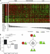
Elevated MYC signaling in human triple-negative breast cancers. (A) 149 primary tumors ordered by each tumor’s Pearson correlation to a 352 gene MYC signature centroid (P < 0.005; Fisher’s exact test). Triple-negative tumor samples are indicated with a red dot, whereas molecular subtypes are indicated with colored bars. (B) Correlation between MYC gene expression signature and breast cancer molecular subtypes. Relative MYC gene expression was based on each tumor’s Pearson correlation to the MYC gene signature. The error bars represent means ± SEM. P-values were calculated by two-tailed Student’s t test for each comparison. (C) Expression of multiple genes within the MYC signaling pathway is altered in triple-negative breast cancers. Schema shows various MAX-interacting genes that are deregulated and can positively or negatively modulate MYC transcriptional activity. Genes are shaded green if expression is suppressed in triple-negative versus receptor-positive tumors, and red if expression is increased in triple-negative tumors. MAX, P = 0.02; MYC, P < 0.0001; MYCN, P = 0.06; MXD4, P = 0.03 (two-tailed Student’s t test).
Figure 3.
Figure 3.
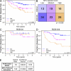
Elevated MYC signaling is associated with poor outcome. (A) Overall risk of breast cancer recurrence in patients with varying levels of MYC pathway activation. I-SPY TRIAL patients (n = 149) were divided into tertiles based on the relative expression of the genes in the MYC signature in their pretreatment biopsy samples. Differences in risk of disease recurrence between these groups were assessed using a Cox proportional hazards model and Wald’s test. (B) Correlation of MYC pathway activation with tumor burden after neoadjuvant chemotherapy. The MYC pathway activation tertile for the I-SPY TRIAL patients with gene expression data, and for which RCB (0-III; Symmans et al., 2007) was determined at the time of surgery (n = 133), was examined. The association between residual tumor burden and MYC pathway activation tertile was assessed using Fisher’s exact test. (C) Disease recurrence by MYC signature in RCB 0/I patients. (D) Disease recurrence by MYC signature in RCB II/III patients. (E) Multivariate analysis considering receptor status and MYC pathway activation as a continuous variable.
Figure 4.
Figure 4.
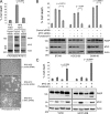
Elevated MYC expression sensitizes triple-negative cancers to CDK inhibition. (A) A panel of triple-negative as well as receptor-positive breast cells, together with a matched pair of nontumorigenic model epithelial cells (RPE cells) engineered to overexpress MYC, was treated with CDK inhibitors purvalanol A (10 µM) or dinaciclib (10 nM) for 72 h and subjected to viability assay. The dashed line indicates the relative starting cell number at the time of adding CDK inhibitors (time 0). Positive numbers indicate cell growth and negative numbers indicate cell death. The experiment was independently repeated five times. The error bars represent means ± SEM. P-values were calculated by two-tailed Student’s t test for comparisons of cell lines treated with each of the two inhibitors. Western blots showing MYC and actin protein expression from the indicated cell lines are shown. (B) Cell cycle profiles of three triple-negative cell lines and three receptor-positive cell lines after treatment with 10 µM purvalanol A or 10 nM dinaciclib for 72 h. The percentage of cells in G1 and G2-M phases of the cell cycle, as determined by DNA content based on propidium iodide staining, is indicated. (C) A panel of triple-negative, as well as receptor-positive, breast cancer cells was treated with siRNA against CDK1 or CDK2 for 72 h and assessed for cell viability. The experiment was independently repeated three times. The error bars represent means ± SEM. P-values were calculated by two-tailed Student’s t test. Western blots showing CDK1, CDK2, and actin protein expression are shown.
Figure 5.
Figure 5.
CDK inhibitor induced cell death is MYC dependent. (A) MYC dependency of cell death induced by CDK inhibition in RPE cells. RPE-MYC cells were first treated with either MYC siRNA or control nonspecific siRNA for 24 h, and then treated with purvalanol A for 72 h. The collected cells were analyzed for cell viability by Guava ViaCount assay and for PARP activation by Western blotting. The experiments were repeated three times in triplicate. The error bars represent means ± SEM. P-values were calculated by two-tailed Student’s t test. (B) siRNA-mediated MYC knockdown in triple-negative cells undergoing purvalanol A treatment. The experiment was independently repeated three times. The error bars represent means ± SEM. P-values were calculated by two-tailed Student’s t test. (C) Receptor-positive cell lines T47D and HCC1428, engineered to overexpress MYC and exposed for 72 h to purvalanol A treatment. The experiments were repeated three times in triplicate. The error bars represent means ± SEM. P-values were calculated by two-tailed Student’s t test.
Figure 6.
Figure 6.
CDK inhibition decreases colony size in triple-negative cancer cells in 3D Matrigel matrices. (A) Low- and high-magnification images of 3D Matrigel cultures of T47D, HCC1428, BT549, and SUM149 cell lines at day 0 of treatment and day 6 of DMSO-treated, purvalanol A–treated, and dinaciclib-treated cultures. Nonrefractile dark particles indicated the presence of cell death. Bars, 10 µm. (B) Quantification of colony size of 3D Matrigel cultures. Low-magnification images of Matrigel cultures were obtained, and the mean size of the colonies in each culture was determined both at the initial day of treatment (day 0) and at the final day of treatment (day 6). The mean colony size at day 0 was set to 100% for each cell line and the colony size at day 6 was compared with the day 0 size. The experiment was independently repeated three times. The error bars represent means ± SEM. P-values were calculated by two-tailed Student’s t test.
Figure 7.
Figure 7.
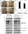
CDK inhibition is effective in treating xenografted triple-negative tumors in mice. (A) Tumor growth in mouse xenograft models of triple-negative breast cancer. Representative photos of tumors after 2 wk of treatment with either vehicle alone or with dinaciclib (50 mg/kg i.p. twice weekly) are shown. (B) Growth of triple-negative tumors in nude mice treated with the CDK inhibitor dinaciclib (50 mg/kg i.p. twice weekly) for 2 wk. Each treatment group per tumor cell type contained the indicated number of mice. The error bars represent means ± SEM. P-values were calculated by two-tailed Student’s t test. (C) PARP cleavage and serine phosphorylation of presumptive CDK substrates occurs in tumors within 24 h of dinaciclib administration. This antibody recognizes amino acid sequences that contain phosphorylated CDK consensus epitope, which is (K/R)(phosphorylated-S)(PX)(K/R) where X can be any amino acid. Two independent tumor samples for each treatment (vehicle or dinaciclib) are shown per cell line.
Figure 8.
Figure 8.
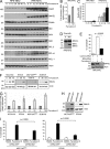
BIM contributes to CDK inhibition-induced cell death in cells with elevated MYC expression. (A) RPE cells, with or without constitutive MYC overexpression, were treated with 10 µM purvalanol A for 72 h and were tested for protein expression of multiple pro- and antiapoptotic BCL2 family members, as well as cleaved PARP and loading control β-actin. The results shown are representatives of at least five independent experiments. (B) Relative fold change in BIM protein expression in RPE-MYC cells treated with purvalanol A for 72 h. The purvalanol A time course experiments were repeated at least five times and BIM protein was quantified as described in Materials and methods. The error bars represent means ± SEM. (C) BIM mRNA expression after purvalanol A treatment. BIM mRNA levels were quantified by qPCR in RPE cells with or without constitutive MYC overexpression. Mean of three experiments ± SEM are shown. (D) BIM expression in RPE-MYC cells treated with dinaciclib for 36 h. (E) RPE-MYC cells were transfected with either a pool of specific BIM siRNAs or a pool of control nontargeting siRNAs and cell viability was determined after treatment with 10 µM purvalanol A for 72 h. The experiment was repeated three times. The error bars represent ± SEM. (F) BIM protein expression in a panel of triple-negative cells. Cell lines were tested for BIM protein expression after treatment with 10 µM purvalanol A for 72 h. The results shown are the representatives of at least three independent experiments. (G) Quantification of BIM protein expression. Shown are the means from at least three independent experiments that yielded the following SEMs, respectively: ±0.2 (HCC3153), ±0.88 (BT549), ±0.48 (MCF10AMYC), and ±0.57 (SUM149PT). (H) shRNA-mediated BIM knockdown in two triple-negative cell lines, MCF10AMYC and BT549. shRNA against GFP was used as negative control. (I) BIM knockdown effects on cell death of MCF10AMYC and BT549 cells after purvalanol A treatment. The experiments were repeated at least three times in triplicate. Error bars represent means ± SEM. P-values were calculated by two-tailed Student’s t test.

"VSports注册入口" References

    1. Alles M.C., Gardiner-Garden M., Nott D.J., Wang Y., Foekens J.A., Sutherland R.L., Musgrove E.A., Ormandy C.J. 2009. Meta-analysis and gene set enrichment relative to er status reveal elevated activity of MYC and E2F in the “basal” breast cancer subgroup. PLoS ONE. 4:e4710 10.1371/journal.pone.0004710 - DOI - PMC - PubMed
    1. Barker A.D., Sigman C.C., Kelloff G.J., Hylton N.M., Berry D.A., Esserman L.J. 2009. I-SPY 2: an adaptive breast cancer trial design in the setting of neoadjuvant chemotherapy. Clin. Pharmacol. Ther. 86:97–100. 10.1038/clpt.2009.68 - DOI - PubMed
    1. Bauer K.R., Brown M., Cress R.D., Parise C.A., Caggiano V. 2007. Descriptive analysis of estrogen receptor (ER)-negative, progesterone receptor (PR)-negative, and HER2-negative invasive breast cancer, the so-called triple-negative phenotype: a population-based study from the California cancer Registry. Cancer. 109:1721–1728. 10.1002/cncr.22618 - DOI - PubMed
    1. Ben-Porath I., Thomson M.W., Carey V.J., Ge R., Bell G.W., Regev A., Weinberg R.A. 2008. An embryonic stem cell-like gene expression signature in poorly differentiated aggressive human tumors. Nat. Genet. 40:499–507. 10.1038/ng.127 - DOI - PMC - PubMed
    1. Bertucci F., Finetti P., Cervera N., Esterni B., Hermitte F., Viens P., Birnbaum D. 2008. How basal are triple-negative breast cancers? Int. J. Cancer. 123:236–240. 10.1002/ijc.23518 - DOI - PubMed

Publication types

MeSH terms