Skip to main page content
U.S. flag

An official website of the United States government

Dot gov

The . gov means it’s official VSports app下载. Federal government websites often end in . gov or . mil. Before sharing sensitive information, make sure you’re on a federal government site. .

Https

The site is secure. The https:// ensures that you are connecting to the official website and that any information you provide is encrypted and transmitted securely. V体育官网.

. 2012 Mar 23;36(3):374-87.
doi: 10.1016/j.immuni.2012.01.015. Epub 2012 Mar 15.

Transcription factor Foxo1 represses T-bet-mediated effector functions and promotes memory CD8(+) T cell differentiation

Affiliations

Transcription factor Foxo1 represses T-bet-mediated effector functions and promotes memory CD8(+) T cell differentiation

"V体育官网" Rajesh R Rao et al. Immunity. .

Abstract

The evolutionary conserved Foxo transcription factors are important regulators of quiescence and longevity. Although, Foxo1 is known to be important in regulating CD8(+) T cell trafficking and homeostasis, its role in functional differentiation of antigen-stimulated CD8(+) T cells is unclear. Herein, we demonstrate that inactivation of Foxo1 was essential for instructing T-bet transcription factor-mediated effector differentiation of CD8(+) T cells. The Foxo1 inactivation was dependent on mTORC1 kinase, given that blockade of mTORC1 abrogated mTORC2-mediated Akt (Ser473) kinase phosphorylation, resulting in Foxo1-dependent switch from T-bet to Eomesodermin transcription factor activation and increase in memory precursors. Silencing Foxo1 ablated interleukin-12- and rapamycin-enhanced CD8(+) T cell memory responses and restored T-bet-mediated effector functions. These results demonstrate an essential role of Foxo1 in actively repressing effector or terminal differentiation processes to promote memory CD8(+) T cell development and identify the functionally diverse mechanisms utilized by Foxo1 to promote quiescence and longevity VSports手机版. .


VSports手机版 - Figures

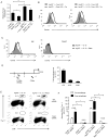
Figure 1
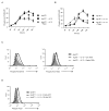
Figure 1. Instructions that program naïve CD8+ T cell for type I effector maturation inactivate transcription factor Foxo1
(A–E) Naïve OT-I cells stimulated with Ag (SIINFEKL, 10nM) plus B7.1 (Ag+B7.1) (+/−) IL-12 (2ng/ml) were evaluated for (A) phosphorylation of Foxo1 by intracytoplasmic staining (ICS) and flow cytometry at the indicated time points, (B) sub-cellular localization of Foxo1 by Image-stream based flow cytometry at 48h. Representative examples of bright-field (BF), total Foxo1-FITC, DAPI, and a merge image of the two stains is shown. The far right-top bar graph represents the similarity co-efficient between total Foxo1 and DAPI, obtained from pixel by pixel statistical analysis of each cell (n=10000) analyzed. The far right-bottom bar graph represents the intensity of Foxo1 protein in the cytoplasm; *p< 0.019 (C) total Foxo1 protein by ICS at 72h, (D) mRNA for Klf-2 at 48h by RT-PCR; *p< 0.029, and (E) cell cycle analysis by propidium iodide labeling at 48h; *p< 0.033. Experiments shown are representative of at least three (A–C) and two (D and E) independent experiments with similar outcomes. (Data are represented at mean +\− SEM)
Figure 2
Figure 2. mTORC1 activity is required for enhanced mTORC2 activity
(A–B) Naive OT-I cells stimulated with Ag+B7.1 (+/−) IL-12 and rapamycin (20ng/ml) were evaluated by ICS at the indicated time-points for (A) Akt phosphorylation at T308, and (B) Akt phosphorylation at S473. For mTOR inhibition, rapamycin was added 30 minutes prior to addition of Ag+B7.1+IL-12. (C) Naive OT-I cells stimulated with Ag+B7.1 (and/or) IL-12 were transduced with control or Raptor RNAi-GFP retroviral vector (RV), cultured for 4 days, re-stimulated and evaluated for Akt T308 and S473 phosphorylation. (D) Naive OT-I cells stimulated with Ag+B7.1 (and/or) IL-12 were transduced with control or DN-Akt GFP RV, cultured for 4 days, re-stimulated and evaluated for Foxo1 phosphorylation. Experiments shown are representative of at least three (A–B) and two (C–D) independent experiments with similar outcomes. (See also Figure S1)
Figure 3
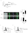
Figure 3. mTORC1 activity is required for inactivation of Foxo1
(A–C) Naive OT-I cells stimulated with Ag+B7.1 (+/−) IL-12 and rapamycin were evaluated for (A) Foxo1 phosphorylation by ICS at the indicated time-points, (B) sub-cellular localization of Foxo1 by Image-stream based flow Cytometry at 48h. Representative examples of bright-field (BF), total Foxo1-FITC, DAPI, and a merge image of the two stains is shown. The far right-top bar graph represents the similarity co-efficient between total Foxo1 and DAPI, obtained from pixel by pixel statistical analysis of each cell (n=10000) analyzed. The far right-bottom bar graph represents the intensity of Foxo1 protein in the cytoplasm; *p<0.011 and *p<0.0086 and (C) total Foxo1 protein expression by ICS at 72h. (D) Naive OT-I cells stimulated with Ag+B7.1 (+/−) IL-12 and rapamycin were transduced with control or Myr-Akt GFP RV, cultured for 4 days, re-stimulated and evaluated for Foxo1 phosphorylation by ICS. Experiments shown are representative of at least three (A–C) and two (D) independent experiments with similar outcomes. (See also Figure S2)
Figure 4
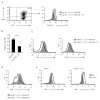
Figure 4. Foxo1 represses T-bet expression in CD8+ T cells
(A) Naive OT-I cells stimulated with Ag+B7.1+IL-12 were transduced with Foxo1-ER-Thy1.1 RV, cultured for 4 days (+/−) Tm (10nM), and evaluated for total Foxo1 protein expression on Thy1.1+ cells by ICS. (B–C) Transduced OT-I cells from (A) were re-stimulated (+/−) Tm, and evaluated for (B) T-bet mRNA expression by RT-PCR on sorted Thy1.1+ OT-I cells; **p<0.0023; and (C) T-bet protein and CD122 expression on Thy1.1+ cells (D) Naive OT-I cells stimulated with Ag+B7.1+IL-12 were transduced with control or DN-Akt GFP RV, cultured for 4 days, re-stimulated and evaluated for Foxo1 phosphorylation. (E) Purified CD8 SP thymocytes from WT and Foxo1−/− mice were stimulated with anti-CD3 and anti-CD28 (+/−) IL-12 and evaluated 72h post-stimulation for T-bet protein expression. Experiments shown are representative of three (A–C) and two (D,E) independent experiments with similar outcomes. (Data are represented at mean +\− SEM). (See also Figure S3)
Figure 5
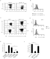
Figure 5. Foxo1 inhibits type I effector differentiation in a T-bet dependent manner
(A–B) Naive OT-I cells stimulated with Ag+B7.1/+IL-12 were transduced with Foxo1-ER-Thy1.1 RV, cultured for 4 days (+/−) Tm (10nM), re-stimulated and evaluated on gated Thy1.1+ cells for (A) IFN-γ production, and (B) Gzmb expression. (C–D) Purified CD8 SP thymocytes from WT and Foxo1−/− mice were stimulated with anti-CD3 and anti-CD28 (+/−) IL-12 and evaluated 72h post-stimulation for (C) IFN-γ production, and (D) Gzmb expression. (E) Naive WT or Tbx21−/− OT-I cells stimulated with Ag+B7.1 (+/−) IL-12 were transduced with control or Foxo1 shRNA-hCD2 RV, cultured for 4 days, re-stimulated and evaluated for IFN-γ production on gated hCD2+ cells; *p<0.0275, **p<0.0061 and n.s.-not significant. (F) Naïve OT-I cells stimulated with Ag+B7.1/+IL-12 (+/−) rapamycin were transduced with T-bet-ER RV, cultured for 4 days (+/−) 4-HT (10nM), re-stimulated and evaluated for IFN-γ production; ***p< 0.0005 and **p<0.0025. Experiments shown are representative of three (A, B and F) and two (C–E) independent experiments with similar outcomes. (Data are represented at mean +\− SEM) (See also Figure S4)
Figure 6
Figure 6. Rapamycin inhibits T-bet expression via Foxo1 dependent mechanisms
(A–C) Purified CD8 SP thymocytes from WT and Foxo1−/− mice were stimulated with anti-CD3 and anti-CD28 (+/−) IL-12 and rapamycin, and evaluated 72h post-stimulation for (A) T-bet protein expression, (B) IFN-γ production, and (C) CD62L protein expression. (D) Naive OT-I cells stimulated with Ag+B7.1 (+/−) IL-12 and rapamycin were transduced with control or Myr-Akt GFP RV, cultured for 4 days, re-stimulated and evaluated for T-bet protein expression. Experiments shown are representative of two independent experiments with similar outcomes.
Figure 7
Figure 7. Foxo1 regulates Eomes gene transcription and is essential for memory CD8+ T cell functions
(A–E) Naive OT-I cells stimulated with Ag+B7.1 (+/−) IL-12 were transduced with Foxo1-ER-Thy1.1 RV, cultured for 4 days (+/−) Tm (10nM), and evaluated on Thy1.1+ cells for (A) Eomes mRNA expression by RT-PCR; *p<0.021,**p<0.0091, and (B) Eomes protein expression. (C) Purified CD8+ T cells from WT and Foxo1−/− mice were stimulated with anti-CD3 and anti-CD28 (+/−) IL-12 and rapamycin, and evaluated 72h post-stimulation for Eomes protein expression. (D) Schematic structure of the Eomes promoter with putative Forkhead binding sites, and ChIP analysis of Foxo1 binding to the Eomes promoter in OT-I cells. Results analyzed by RT-PCR are presented as fold of template enrichment in immunoprecipitates of Foxo1 antibody relative to control immunoprecipitation (Ig). (E–F) Naive OT-I cells (Thy1.1+) stimulated with Ag+B7.1 (+/−) IL-12 and rapamycin were transduced with control or Foxo1 shRNA-hCD2 RV, sorted for hCD2-Thy1.1+ cells, adoptively transferred (2 × 10⁶ cells) into BL/6 recipients, and (E) evaluated for the frequency of adoptively transferred cells in the spleen at day 40 post adoptive transfer. The circles indicate OT-I/Thy1.1 population and the numbers indicate percent frequency. (F) The recipient mice were immunized with IFA-OVA on day 40 post transfer and secondary CD8+ T cell responses were measured 3 days later. The absolute numbers of adoptively transferred cells before (pre-rechallenge) and after (post-rechallenge) immunization in the spleen, is shown. The numbers in parenthesis indicate fold expansion of CD8α+Thy1.1+ from day 40 to day 43; *p< 0.0125 and **p< 0.0087. Experiments shown are representative of three (A,B) and two (C–F) independent experiments with similar outcomes. (See also Figure S5)

"V体育安卓版" References

    1. Araki K, Turner AP, Shaffer VO, Gangappa S, Keller SA, Bachmann MF, Larsen CP, Ahmed R. mTOR regulates memory CD8 T-cell differentiation. Nature. 2009;460:108–112. - V体育官网入口 - PMC - PubMed
    1. Burgering BM. A brief introduction to FOXOlogy. Oncogene. 2008;27:2258–2262. - PubMed
    1. Chen CC, Jeon SM, Bhaskar PT, Nogueira V, Sundararajan D, Tonic I, Park Y, Hay N. FoxOs inhibit mTORC1 and activate Akt by inducing the expression of Sestrin3 and Rictor. Dev Cell. 2010;18:592–604. - PMC - PubMed
    1. Curtsinger JM, Lins DC, Mescher MF. Signal 3 determines tolerance versus full activation of naive CD8 T cells: dissociating proliferation and development of effector function. J Exp Med. 2003;197:1141–1151. - PMC - PubMed
    1. Delgoffe GM, Pollizzi KN, Waickman AT, Heikamp E, Meyers DJ, Horton MR, Xiao B, Worley PF, Powell JD. The kinase mTOR regulates the differentiation of helper T cells through the selective activation of signaling by mTORC1 and mTORC2. Nat Immunol. 2011;12:295–303. - PMC - PubMed

Publication types (V体育安卓版)

MeSH terms

VSports在线直播 - Substances

V体育平台登录 - LinkOut - more resources