Skip to main page content
U.S. flag

An official website of the United States government

Dot gov

The . gov means it’s official VSports app下载. Federal government websites often end in . gov or . mil. Before sharing sensitive information, make sure you’re on a federal government site. .

Https

The site is secure. The https:// ensures that you are connecting to the official website and that any information you provide is encrypted and transmitted securely. V体育官网.

. 2012 Jul;5(4):492-502.
doi: 10.1242/dmm.008730. Epub 2012 Mar 15.

Liver hyperplasia after tamoxifen induction of Myc in a transgenic medaka model

Affiliations

Liver hyperplasia after tamoxifen induction of Myc in a transgenic medaka model

Luciana A Menescal et al. Dis Model Mech. 2012 Jul.

Abstract

Myc is a global transcriptional regulator and one of the most frequently overexpressed oncoproteins in human tumors. It is well established that activation of Myc leads to enhanced cell proliferation but can also lead to increased apoptosis VSports手机版. The use of animal models expressing deregulated levels of Myc has helped to both elucidate its function in normal cells and give insight into how Myc initiates and maintains tumorigenesis. Analyses of the medaka (Oryzias latipes) genome uncovered the unexpected presence of two Myc gene copies in this teleost species. Comparison of these Myc versions to other vertebrate species revealed that one gene, myc17, differs by the loss of some conserved regulatory protein motifs present in all other known Myc genes. To investigate how such differences might affect the basic biological functions of Myc, we generated a tamoxifen-inducible in vivo model utilizing a natural, fish-specific Myc gene. Using this model we show that, when activated, Myc17 leads to increased proliferation and to apoptosis in a dose-dependent manner, similar to human Myc. We have also shown that long-term Myc17 activation triggers liver hyperplasia in adult fish, allowing this newly established transgenic medaka model to be used to study the transition from hyperplasia to liver cancer and to identify Myc-induced tumorigenesis modifiers. .


"VSports手机版" Figures

Fig. 1.
Fig. 1.
Unrooted phylogenetic tree of Myc family genes. Amino acid sequences were aligned by the maximum likelihood method. Numbers on nodes are bootstrap values out of 100 iterations.
Fig. 2.
Fig. 2.
Expression of the myc17ER transgene in two medaka transgenic lines. (A) Real-time PCR analysis of the myc17ER transgene from liver, brain, eyes, gills and muscle from adult fish. Levels from the transgene were normalized against ef1a1 in each tissue. Histograms with T-bars indicate the mean standard deviation based on triplicate assays. (B) Western blot analysis of the Myc17ER fusion proteins. Protein samples extracted from pooled wild type (WT) and two transgenic fish lines were reacted with anti-ER antibody (30 hatched embryos per line). Protein extract containing human MycER (95 kDa), which has a higher molecular weight than medaka Myc17 (85 kDa), was used as control (crtl). Smaller and weaker bands in wild type control are due to unspecific antibody binding.
Fig. 3.
Fig. 3.
Functionality activity of the transgene product of myc17ER transgenic lines. (A) Immunofluorescence of myc17ER line 2 in medaka fin primary cell culture in the presence or absence of 4-OHT using anti-ER antibody and nuclear counterstaining with Hoechst. Scale bar: 10 μm. (B) Quantification of mean intensity differences (n=7 cells per treatment). Each cell was measured at two independent regions in the nucleus and the cytoplasm. Mean values were subtracted to calculate cytoplasmic to nuclear difference. Student’s t-tests show no significant differences between different −4-OHT controls (line 1 compared with WT: P=0.10; line 2 compared with WT: P=0.36). (C) Direct myc17 target gene expression (ODC1, CBX3, CCT5, EIF3S8 and MTLL1) in different organs via qPCR analysis after 4-OHT treatment. Relative fold change levels for each gene were normalized against ef1a1 in each tissue separately. Histograms with T-bars indicate the mean standard deviation based on duplicate assays.
Fig. 4.
Fig. 4.
Cell proliferation after myc17 activation in vivo. (A) BrdU staining on sections of liver (upper panels) and gills (lower panels) in the presence or absence of 4-OHT. Scale bar: 10 μm. (B) Quantification of BrdU-positive cells in liver (left graphic) and gills (right graphic) in both myc17ER lines. Histograms with T-bars indicate the mean percentage of BrdU-positive cells of two independent experiments with their corresponding standard deviation. C N indicates average cell number counted from five different sections.
Fig. 5.
Fig. 5.
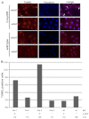
Effects of myc17 activation on cell death in vitro. (A) Detection of apoptosis in primary cell culture (arrows indicate TUNEL-positive cells). Cells were treated for 5 hours with 4-OHT before fixation. Nuclei were counterstained with Hoechst. Scale bar: 100 μm. (B) Quantification of the percentage of apoptotic cells in primary cultures from both myc17ER lines and wild-type fish in the presence or absence of 4-OHT in the media. C N indicates total cell number for each assay.
Fig. 6.
Fig. 6.
Induction of apoptosis after myc17 activation in vivo. (A) Detection of apoptosis by TUNEL assays in liver and gill sections from adult fish. Fish were treated for 24 hours with 4-OHT. DNA fragmentation is visible as red spots colocalizing with nuclei, which are stained with Hoechst. Scale bar: 10 μm. (B) Quantification of the percentage of apoptotic cells in liver (left panel) and gills (right panel) of both myc17ER lines in the presence or absence of 4-OHT. C N indicates total cell number for each assay.
Fig. 7.
Fig. 7.
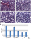
Constant myc17 activation triggers liver cell hyperplasia in vivo. (A-D) Images show hematoxylin- and eosin-stained sections of liver from adult transgenic fish from line 1 (A), line 2 (B) and wild type (C) treated for 4 weeks with 4-OHT or non-treated wild type (D). Scale bar: 20 μm. (E) Cell number per section (87,410 μm2) in both myc17ER lines and in wild-type fish treated or not treated with 4-OHT.

References

    1. Adhikary S., Eilers M. (2005). Transcriptional regulation and transformation by Myc proteins. Nat. Rev. Mol. Cell Biol. 6, 635–645 - "V体育官网" PubMed
    1. Amati B., Frank S. R., Donjerkovic D., Taubert S. (2001). Function of the c-Myc oncoprotein in chromatin remodeling and transcription. Biochim. Biophys. Acta 1471, 135–145 - PubMed
    1. Bhatia K., Huppi K., Spangler G., Siwarski D., Iyer R., Magrath I. (1993). Point mutations in the c-Myc transactivation domain are common in Burkitt’s lymphoma and mouse plasmacytomas. Nat. Genet. 5, 56–61 - PubMed (VSports)
    1. Blyth K., Terry A., O’Hara M., Baxter E. W., Campbell M., Stewart M., Donehower L. A., Onions D. E., Neil J. C., Cameron E. R. (1995). Synergy between a human c-myc transgene and p53 null genotype in murine thymic lymphomas: contrasting effects of homozygous and heterozygous p53 loss. Oncogene 10, 1717–1723 - PubMed
    1. Blyth K., Stewart M., Bell M., James C., Evan G., Neil J. C., Cameron E. R. (2000). Sensitivity to myc-induced apoptosis is retained in spontaneous and transplanted lymphomas of CD2-mycER mice. Oncogene 19, 773–782 - PubMed

Publication types

MeSH terms

"VSports在线直播" Substances