Skip to main page content
U.S. flag

An official website of the United States government

Dot gov

The . gov means it’s official. Federal government websites often end in . gov or VSports app下载. mil. Before sharing sensitive information, make sure you’re on a federal government site. .

Https

The site is secure V体育官网. The https:// ensures that you are connecting to the official website and that any information you provide is encrypted and transmitted securely. .

. 2012 Mar 1;26(5):433-8.
doi: 10.1101/gad.179416.111.

"V体育2025版" ATRX-mediated chromatin association of histone variant macroH2A1 regulates α-globin expression

Affiliations

ATRX-mediated chromatin association of histone variant macroH2A1 regulates α-globin expression (V体育ios版)

Kajan Ratnakumar (V体育安卓版) et al. Genes Dev. .

"V体育安卓版" Abstract

The histone variant macroH2A generally associates with transcriptionally inert chromatin; however, the factors that regulate its chromatin incorporation remain elusive. Here, we identify the SWI/SNF helicase ATRX (α-thalassemia/MR, X-linked) as a novel macroH2A-interacting protein. Unlike its role in assisting H3. 3 chromatin deposition, ATRX acts as a negative regulator of macroH2A's chromatin association. In human erythroleukemic cells deficient for ATRX, macroH2A accumulates at the HBA gene cluster on the subtelomere of chromosome 16, coinciding with the loss of α-globin expression VSports手机版. Collectively, our results implicate deregulation of macroH2A's distribution as a contributing factor to the α-thalassemia phenotype of ATRX syndrome. .


Figures
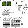
Figure 1.
Figure 1.
Identification of mH2A1.2 chromatin-free interacting factors. (A) Fluorescence microscopy of HEK293 cells stably expressing GFP-H2A and mH2A1.2-GFP. Arrowhead indicates Xi. (B) Procedure used to isolate chromatin-free H2A- and mH2A1.2-interacting factors. (C) αGFP immunoblot confirms expression and immunoprecipitation of histones in stable cell lines. (D) Immunoblots of Imp9, Npm, and NAP1 association with GFP-H2A or mH2A-GFP isoforms. Arrows on all αGFP blots indicate GFP-H2A (bottom) and mH2A-GFP (top), and asterisks indicate degradation products.
Figure 2.
Figure 2.
ATRX interacts with mH2A isoforms in chromatin-free extracts. (A,B) Immunoblots of GFP and ATRX from chromatin-free extracts. The absence of H3 in immunoprecipitations confirms chromatin-free interactions; see the long exposure. (B) Co-IP of GFP-H2A and all mH2A isoforms in chromatin-free extracts for ATRX. Immunoblots for Imp9 and Parp1 demonstrate chromatin-free interactions. (C) Whole-cell immunoprecipitations of GFP-tagged ATRX constructs (full-length, N-terminal [1–841], middle region [800–1670], and C-terminal [1670–2492]), followed by mH2A1, H3, and Daxx immunoblots. (D) Chromatin-free co-IP of N-ATRX-GFP with mH2A1.
Figure 3.
Figure 3.
mH2A and H3.3 are in distinct ATRX complexes. Co-IPs of chromatin-free association of H2A and mH2A isoforms (A) or H3 variants (B) with Daxx. Immunoblots detected the presence of Daxx specifically with H3.3. (C) Ethidium bromide-stained (left) and Coomassie-stained (right) mononucleosomes from H2A-Flag and mH2A1.2-Flag immunoprecipitations. (D) H3 composition of H2A-Flag and mH2A1.2-Flag mononucleosomes as analyzed by MS. The pie chart depicts the abundance of H3.1/H3.2 (gray) and H3.3 (black) in immunoprecipitated nucleosomes.
Figure 4.
Figure 4.
ATRX knockdown results in increased levels and stability of mH2A1 in chromatin. (A) shRNA-mediated knockdown of ATRX (sh90 and sh92) in HEK293 cells results in the loss of ATRX protein and mRNA, compared with shluc, without affecting mH2A1 mRNA. β-Actin was used for loading. (B) Loss of ATRX results in accumulation of mH2A1 in chromatin (top panel), and the whole-cell content remains unaffected (bottom panel). H2B was used for loading. (C) mH2A1 immunoblot of chromatin-extracted histones analyzed by qMS. (D, left) Quantitation of FRAP experiments indicates slower recovery of mH2A1.2-GFP in HeLa1.2.11 cells depleted of ATRX (sh92; green line) compared with control (shluc; blue line). (Right) Representative images of FRAP time series pre- and post-bleach are shown.
Figure 5.
Figure 5.
ATRX loss results in increased association of mH2A1 at telomeres and the α-globin cluster, concomitant with loss of α-globin expression. (A) shRNA-mediated knockdown of ATRX (sh90 and sh92) in K562 cells. β-Actin was used for loading. (B) ChIP reveals increased association of mH2A1 with telomeres in the absence of ATRX. (Right) Densitometry quantitation. One of two biological replicates is shown. (C) Loss of ATRX results in decreased α-globin protein and HBA mRNA. (D) Capture of the University of California at Santa Cruz Genome Browser showing an ∼50-kb region around the α-globin locus. ChIP-seq-enriched peaks are shown for mH2A1 (sh92 and shluc), ATRX (Law et al. 2010), and input (shluc). Significant peaks (MACS) are shown below each panel as black bars. (Bottom) RefSeq annotated genes. The threshold line was set at 35 to facilitate visualization.

References

    1. Angelov D, Molla A, Perche PY, Hans F, Cote J, Khochbin S, Bouvet P, Dimitrov S 2003. The histone variant macroH2A interferes with transcription factor binding and SWI/SNF nucleosome remodeling. Mol Cell 11: 1033–1041 - PubMed
    1. Banaszynski LA, Allis CD, Lewis PW 2010. Histone variants in metazoan development. Dev Cell 19: 662–674 - PMC (V体育安卓版) - PubMed
    1. Berube NG 2011. ATRX in chromatin assembly and genome architecture during development and disease. Biochem Cell Biol 89: 435–444 - PubMed
    1. Berube NG, Smeenk CA, Picketts DJ 2000. Cell cycle-dependent phosphorylation of the ATRX protein correlates with changes in nuclear matrix and chromatin association. Hum Mol Genet 9: 539–547 - PubMed
    1. Buschbeck M, Uribesalgo I, Wibowo I, Rue P, Martin D, Gutierrez A, Morey L, Guigo R, Lopez-Schier H, Di Croce L 2009. The histone variant macroH2A is an epigenetic regulator of key developmental genes. Nat Struct Mol Biol 16: 1074–1079 - "V体育官网" PubMed

Publication types

MeSH terms