<kbd dir="2RjNgq"></kbd> Skip to main page content
U.S. flag

An official website of the United States government

Dot gov

The . gov means it’s official VSports app下载. Federal government websites often end in . gov or . mil. Before sharing sensitive information, make sure you’re on a federal government site. .

Https

The site is secure. The https:// ensures that you are connecting to the official website and that any information you provide is encrypted and transmitted securely. V体育官网.

. 2012 Feb;10(2):e1001255.
doi: 10.1371/journal.pbio.1001255. Epub 2012 Feb 7.

Development and function of invariant natural killer T cells producing T(h)2- and T(h)17-cytokines

Affiliations

"VSports注册入口" Development and function of invariant natural killer T cells producing T(h)2- and T(h)17-cytokines

Hiroshi Watarai et al. PLoS Biol. 2012 Feb.

"VSports app下载" Abstract

There is heterogeneity in invariant natural killer T (iNKT) cells based on the expression of CD4 and the IL-17 receptor B (IL-17RB), a receptor for IL-25 which is a key factor in T(H)2 immunity. However, the development pathway and precise function of these iNKT cell subtypes remain unknown. IL-17RB⁺iNKT cells are present in the thymic CD44⁺/⁻ NK1 VSports手机版. 1⁻ population and develop normally even in the absence of IL-15, which is required for maturation and homeostasis of IL-17RB⁻iNKT cells producing IFN-γ. These results suggest that iNKT cells contain at least two subtypes, IL-17RB⁺ and IL-17RB⁻ subsets. The IL-17RB⁺iNKT subtypes can be further divided into two subtypes on the basis of CD4 expression both in the thymus and in the periphery. CD4⁺ IL-17RB⁺iNKT cells produce T(H)2 (IL-13), T(H)9 (IL-9 and IL-10), and T(H)17 (IL-17A and IL-22) cytokines in response to IL-25 in an E4BP4-dependent fashion, whereas CD4⁻ IL-17RB⁺iNKT cells are a retinoic acid receptor-related orphan receptor (ROR)γt⁺ subset producing T(H)17 cytokines upon stimulation with IL-23 in an E4BP4-independent fashion. These IL-17RB⁺iNKT cell subtypes are abundantly present in the lung in the steady state and mediate the pathogenesis in virus-induced airway hyperreactivity (AHR). In this study we demonstrated that the IL-17RB⁺iNKT cell subsets develop distinct from classical iNKT cell developmental stages in the thymus and play important roles in the pathogenesis of airway diseases. .


Conflict of interest statement

The authors have declared that no competing interests exist.

Figures
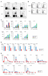
Figure 1
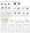
Figure 1. Function of iNKT cells in the spleen and liver from Il17rb −/− and Il15 L117P mice.
(A) FACS profile of spleen and liver mononuclear cells in WT, Il17rb −/− and Il15 L117P mice on a B6 background. Numbers are percentage of gated cells. α-GalCer/CD1d dimer+ iNKT cells and α-GalCer/CD1d dimer NK1.1+ NK cells were slightly decreased in Il17rb −/− mice and markedly reduced in Il15 L117P mice. (B) IL-17RB expression in spleen and liver iNKT cells of WT B6, Il17rb −/− and Il15 L117P mice. Shaded profiles in the histograms indicate the background staining with isotype matched control mAb. (C, D) In vitro cytokine production by spleen iNKT cells from Il17rb −/− and Il15 L117P mice (C) and by liver iNKT cells from Il17rb −/− mice (D). Sorted iNKT cells (5×10⁴/100 µL) from spleen and liver of WT B6 and Il17rb −/− mice were co-cultured with BM-DCs (5×10³/100 µL) for 48 h in the presence of the indicated doses of α-GalCer. The Il17rb −/− iNKT cells produced IFN-γ at levels equivalent to WT, while TH2 and TH17 cytokine production, except for IL-4, were severely impaired. (E) iNKT cell-dependent cytokine production in WT B6 and Il17rb −/− mice in vivo. α-GalCer (2 µg) was i.v. injected and the levels of cytokines in serum were analyzed at the indicated time points. The serum IFN-γ levels were similar in both mice, whereas production of TH2 and TH17 cytokines, except for IL-4, was significantly reduced in the Il17rb −/− mice. Cytokines were measured by ELISA or a cytometric bead array system at the indicated time points. Data are mean ± SDs from three mice and repeated three times with similar results.
Figure 2
Figure 2. Profile of iNKT cells in the thymus of Il17rb −/− and Il15 L117P mice.
(A, B) FACS profiles of thymus (A) and enriched thymic iNKT cells (B) in WT, Il17rb −/− and Il15 L117P mice on a B6 background. (A) α-GalCer/CD1d dimer+ iNKT cells were slightly decreased in Il17rb −/− mice and markedly reduced in Il15 L117P mice. (B) There was a loss of the NK1.1 population in Il17rb −/− thymic iNKT cells, while Il15 L117P thymic iNKT cells showed impairment of the NK1.1+ population. (C) IL-17RB and CD122 expression by thymic iNKT cell populations of B6 mice. IL-17RB and CD122 expression in CD4 and CD4+ of CD44lo NK1.1 (Stage 1), CD44lo NK1.1+ (Stage 2), and CD44hi NK1.1+ (Stage 3) populations were analyzed. IL-17RB expression was observed in Stages 1/2, while CD122 expression was in the Stage 3 cells. (D, E) Profiles of thymic iNKT cells in B6 and Il15 L117P mice showing expression of IL-17RB and CD4 (D) and further divided into CD44 and NK1.1 subpopulations (E). The percentage of IL-17RB+ iNKT cells was increased due to the loss of expansion of IL17RB iNKT cells in Il15 L117P mice. CD4 and CD4+, IL-17RB+ iNKT cells were almost all Stage 1 and Stage 2 in both WT B6 and Il15 L117P mice. On the other hand, the majority of CD4 and CD4+, IL-17RB iNKT cells were Stage 3 in both WT B6 and Il15 L117P mice. Loss of expansion of CD4 and CD4+, IL-17RB iNKT cells was also observed in Il15 L117P mice. (F, G) Percentage (F) and cell number (G) of the total iNKT cells and the four subtypes (i.e. IL-17RB+/− and CD4+/−) in B6, Il17rb −/− and Il15 L117P mice based on their CD44 and NK1.1 expression patterns. The number of CD4 and CD4+, IL-17RB iNKT cells was significantly decreased especially in Stage 3 in Il15 L117P mice compared to WT B6 mice. By contrast, CD4 and CD4+, IL-17RB+ iNKT cells in Il15 L117P mice were present in numbers comparable to WT. Results are representative of those from three independent experiments. (H, I) Development of iNKT subtypes in Stages 1 and 2. Stage 1 and 2 cells in the four iNKT subtypes (i.e. IL-17RB+/− and CD4+/−) from WT B6 mice were sorted and cocultured with dGuo treated 15 dpc FT lobes from Jα18 −/− mice (1,000 cells/well). 10 d after culture, cells were recovered and analyzed the surface expression pattern in CD44 versus NK1.1 (H) and CD4 versus IL-17RB (I). IL-17RB precursors gave rise through Stage 2 to Stage 3 cells with IL-17RB, while IL-17RB+ subtypes gave rise to Stage 2 cells with IL-17RB+. Results are representative of those from three independent experiments.
Figure 3
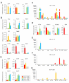
Figure 3. Differential gene expression and cytokine production among thymic iNKT cell subtypes from B6 mice.
(A, B, D, H) Quantitative PCR analysis of thymic iNKT subtypes. Thymic iNKT cells further divided into four subtypes based on the expression of CD4 and IL-17RB (red, CD4 IL-17RB+; orange, CD4+ IL-17RB+; blue, CD4 IL-17RB; green, CD4+ IL-17RB). One representative out of three experiments is shown (mean ± SEM). (A) The purity of the sorted cells was confirmed by the relative Il17rb and Cd4 mRNA expression levels in the respective subtypes. Il2rb ( = Cd122) expression was restricted to CD4 and CD4+, IL-17RB iNKT cells. (B) Expression of TH1/TH2/TH17 related genes. TH1 related: Ifng, Tbx21 and Stat4, TH2 related; Il4 and Gata3, and TH17 related: Il17a, Il22 and Rorc transcripts were analyzed. (D) Expression of cytokine receptor genes. Receptor for IL-12, IL-23, and IL-25 were analyzed. The component chains of the various receptors are IL-12 receptor: IL-12Rβ2/IL-12Rβ1; IL-23 receptor: IL-23R/IL-12Rβ1; IL-25 receptor: IL-17RB/IL-17RA. (H) Expression of chemokine receptor genes. Ccr4, Ccr6, Ccr7, Cxcr3, and Cxcr6. (C, E, F, G) In vitro cytokine production by thymic iNKT cell subtypes (red, CD4 IL-17RB+; orange, CD4+ IL-17RB+; blue, CD4 IL-17RB; green, CD4+ IL-17RB). Sorted thymic iNKT subtypes (5×10⁴ cells/100 µL) were co-cultured with BM-DCs (5×10³/100 µL) for 48 h in the presence of α-GalCer (100 ng/µL) (C), IL-12 (10 ng/µL) (E), IL-23 (10 ng/µL) (F), or IL-25 (10 ng/µL) (G). Levels of IFN-γ, IL-4, IL-9, IL-10, IL-13, IL-17A, and IL-22 were analyzed. The data are representative of three independent experiments (mean ± SEM).
Figure 4
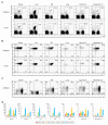
Figure 4. iNKT cell subtypes in the periphery.
(A–C) FACS profile of peripheral iNKT cells in B6 mice. α-GalCer/CD1d dimer+ TCRβ+ iNKT cells (A) and iNKT subtypes based on the expression of CD44 and NK1.1 (B) or CD4 and IL-17RB (C) in spleen, liver, BM, lung, inguinal LN, and mesenteric LN from B6 or Il17rb −/− mice. Numbers indicate percentage of total mononuclear cells (A) and iNKT cells (B, C). (D) Number of cells of each iNKT subtype based on the expression of IL-17RB and CD4 in thymus and periphery of B6 and BALB/c mice. Cell numbers were calculated based on the results from Figures 4A, 4C, S8A, and S8B. IL-17RB+ iNKT cells were mainly localized in spleen, lung, inguinal LN, and mesenteric LN, whereas hardly any were observed in liver and BM. One representative experiment of three is shown.
Figure 5
Figure 5. Function of iNKT cell subtypes in the spleen.
(A) Global gene expression profiles in iNKT subtypes in the thymus and spleen. Tree view representation of clustering analysis among the four iNKT subtypes in thymus and spleen from B6 and BALB/c. The values represent coefficients between the indicated panels. r 2>0.95 in red, 0.85<r 2<0.95 in orange, and r 2<0.85 in blue. One representative experiment of three is shown. (B) Plasticity and stability of iNKT subtypes. The four iNKT cell subtypes in the thymus were sorted and each subtype (5×10⁵) was i.v. transferred into independent Jα18 −/− mice (n = 3). 10 d after transfer, α-GalCer/CD1d dimer+ TCRβ+ cells in spleen were analyzed by FACS for the expression of IL-17RB and CD4. Representative data from three experiments are shown. (C–F) In vitro cytokine production by splenic iNKT cell subtypes (red, CD4 IL-17RB+; orange, CD4+ IL-17RB+; blue, CD4 IL-17RB; green, CD4+ IL-17RB). Sorted splenic iNKT subtypes (5×10⁴ cells/100 µL) were co-cultured with BM-DCs (5×10³/100 µL) for 48 h in the presence of α-GalCer (100 ng/µL) (C), IL-12 (10 ng/µL) (D), IL-23 (10 ng/µL) (E), and IL-25 (10 ng/µL) (F). Levels of IFN-γ, IL-4, IL-9, IL-10, IL-13, IL-17A, and IL-22 in the supernatants were analyzed by ELISA or CBA. Data are mean ± SD of triplicate wells. One representative experiment of three is shown.
Figure 6
Figure 6. Involvement of E4bp4 in cytokine production by CD4+ IL-17RB+ iNKT cells in response to IL-25.
(A, B) Quantitative analysis of Rorc (A) and E4bp4 (B) in iNKT cell subtypes after cytokine treatment. Sorted iNKT cell subtypes (5×10⁴/100 µL) from thymus (left) or spleen (right) were co-cultured with BM-DCs (5×10³/100 µL) in the presence or absence of IL-23 (10 ng/ml) or IL-25 (10 ng/ml) for 24 h. The iNKT cell subtypes were then sorted again and analyzed for expression of the indicated genes by quantitative real-time PCR. The data are representative of three independent experiments (mean ± SEM). (C, D) Cytokine production by CD4+ IL-17RB+ iNKT cells in response to IL-25. Sorted CD4+ IL-17RB+ iNKT cells (5×10⁴/100 µL) from thymus (C) or spleen (D) of B6 or E4bp4 −/− mice were co-cultured with BM-DCs (5×10³/100 µL) in the presence of IL-25 (10 ng/ml) for 48 h and then the levels of the indicated cytokines in the tissue culture media were analyzed. iNKT cells from B6 were compared to those from E4bp4 −/− mice. ** p<0.01 calculated by t test. The data are representative of three independent experiments (mean ± SD).
Figure 7
Figure 7. Involvement of IL-17RB+ iNKT cells in the development of RSV-induced AHR.
(A) Schematic showing the protocol for RSV-induced AHR. Mice were i.n. administered with RSV (106 pfu) or PBS alone as a control 4 times at 10-day intervals. Mice were i.p. immunized with rec Gs/alum (50 µg/2 mg) 4 d after first RSV infections. Three days after the last RSV administration, mice were exposed i.n. to rec Gs and were measured 1 d later. (B) Development of RSV-induced AHR in BALB/c, but not in Jα18 −/− or Il17rb −/− mice. Changes in RL are depicted. The RSV-infected, rec Gs immunized, BALB/c mice had a greatly increased AHR compared to the other three groups. Results are expressed as the mean ± SEM. * p<0.05 and ** p<0.01. (C, D) Total and differential cell counts (C) and cytokines (D) in BAL fluid. BAL fluid was collected 24 h after challenge of the mice depicted in (B) with intranasal rec Gs. Results are expressed as the mean ± SEM. * p<0.05 and ** p<0.01. (E) Histological examination of lung tissues by H&E and PAS staining. RSV infected, rec Gs immunized, BALB/c, Jα18 −/− or IL17rb −/−, mice were compared with control BALB/c mice (rec Gs alone). Bars indicate 100 µm. (F) AHR development after cell transfer of spleen IL-17RB+ iNKT cells into Jα18 −/− mice. Indicated cell numbers of sorted IL-17RB+, IL-17RB iNKT cells or total iNKT cells from spleen, or PBS control, were i.v. transferred into rec-Gs/alum-sensitized Jα18 −/− mice 24 h before RSV treatment (on the day 9, 19, and 29), and then challenged with rec Gs (24 h) and measurement of lung resistance (48 h). Each group of IL-17RB+ iNKT cell-transferred mice was compared to other three groups. * p<0.05, ** p<0.01 calculated by Kruskal Wallis test. The results represent one out of four experiments with five mice in each group.

References

    1. Taniguchi M, Harada M, Kojo S, Nakayama T, Wakao H. The regulatory role of Valpha14 NKT cells in innate and acquired immune response. Annu Rev Immunol. 2003;21:483–513. - PubMed
    1. Lantz O, Bendelac A. An invariant T cell receptor alpha chain is used by a unique subset of major histocompatibility complex class I-specific CD4+ and CD4−8− T cells in mice and humans. J Exp Med. 1994;180:1097–1106. - "V体育平台登录" PMC - PubMed
    1. Exley M, Garcia J, Balk S. P, Porcelli S. Requirements for CD1d recognition by human invariant Va24+CD4−CD8− T cells. J Exp Med. 1997;186:109–120. - PMC - PubMed
    1. Bendelac A. Positive selection of mouse NK1+ T cells by CD1-expressing cortical thymocytes. J Exp Med. 1995;182:2091–2096. - PMC - PubMed
    1. Bendelac A, Lantz O, Quimby M. E, Yewdell J. W, Bennink J. R, et al. CD1 recognition by mouse NK1+ T lymphocytes. Science. 1995;268:863–865. - PubMed

Publication types

MeSH terms

Substances

VSports最新版本 - LinkOut - more resources