Skip to main page content
U.S. flag

An official website of the United States government

Dot gov

The VSports app下载. gov means it’s official. Federal government websites often end in . gov or . mil. Before sharing sensitive information, make sure you’re on a federal government site. .

Https

The site is secure. The https:// ensures that you are connecting to the official website and that any information you provide is encrypted and transmitted securely. V体育官网.

. 2012 Apr 15;21(8):1782-93.
doi: 10.1093/hmg/ddr611. Epub 2012 Jan 6.

"VSports手机版" Balancing neural crest cell intrinsic processes with those of the microenvironment in Tcof1 haploinsufficient mice enables complete enteric nervous system formation

Affiliations

Balancing neural crest cell intrinsic processes with those of the microenvironment in Tcof1 haploinsufficient mice enables complete enteric nervous system formation

Amanda J Barlow et al. Hum Mol Genet. .

Abstract

The enteric nervous system (ENS) comprises a complex neuronal network that regulates peristalsis of the gut wall and secretions into the lumen. The ENS is formed from a multipotent progenitor cell population called the neural crest, which is derived from the neuroepithelium. Neural crest cells (NCCs) migrate over incredible distances to colonize the entire length of the gut and during their migration they must survive, proliferate and ultimately differentiate VSports手机版. The absence of an ENS from variable lengths of the colon results in Hirschsprung's disease (HSCR) or colonic aganglionosis. Mutations in about 12 different genes have been identified in HSCR patients but the complex pattern of inheritance and variable penetrance suggests that additional genes or modifiers must be involved in the etiology and pathogenesis of this disease. We discovered that Tcof1 haploinsufficiency in mice models many of the early features of HSCR. Neuroepithelial apoptosis diminished the size of the neural stem cell pool resulting in reduced NCC numbers and their delayed migration along the gut from E10. 5 to E14. 5. Surprisingly however, we observe continued and complete colonization of the entire colon throughout E14. 5-E18. 5, a period in which the gut is considered to be non- or less-permissive to NCC. Thus, we reveal for the first time that reduced NCC progenitor numbers and delayed migration do not unequivocally equate with a predisposition for the pathogenesis of HSCR. In fact, these deficiencies can be overcome by balancing NCC intrinsic processes of proliferation and differentiation with extrinsic influences of the gut microenvironment. .


Figures
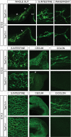
Figure 1.
Figure 1.
Delayed NCC migration in E10.5, E12.5 and E14.5 Tcof1+/− guts. TuJ1 immunostaining of E10.5 whole guts shows that NC-derived cells have colonized ≈50% of the SI in Tcof1+/− guts in comparison with wild-type guts where the migration wavefront is at the end of the SI (arrowhead). The cells appear to have migrated collectively in a thick chain in the SI in Tcof1+/− guts rather than extending evenly across the entire wall of the gut in Tcof1+/+. At E12.5, the migration wavefront is in the colon in Tcof1+/+ guts but has only reached the cecum in Tcof1+/− guts (arrowhead). There appears to be reduced or delayed NCC differentiation in Tcof1+/− guts at this stage. By E14.5, the characteristic network of NC-derived cells is present along the entire length of the gut in Tcof1+/+ mice, while <50% of Tcof1+/− guts contain aganglionic regions. A similar network is present in the SI in both genotypes. c, colon; D, distal; si, small intestine; st, stomach.
Figure 2.
Figure 2.
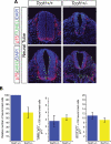
Neural tube apoptosis reduces the NCC numbers that migrate towards and into the foregut in Tcof1+/− mice. (A) Examination of apoptosis and proliferation was performed using immunostaining of cryosections of Tcof1+/+ and Tcof1+/− embryos with the NCC marker p75 and either TUNEL or pHH3, respectively. DAPI stained all nuclei. TUNEL staining was observed in the neural tube of Tcof1+/− embryos which are also smaller than their wild-type counterparts. No apparent proliferative differences were observed between genotypes. (B) Histograms showing that NCC numbers were reduced by 40% in Tcof1+/− embryos in comparison with Tcof1+/+. Similar percentages of apoptotic or proliferating NCC were found between genotypes.
Figure 3.
Figure 3.
A complete ENS network is seen in Tcof1+/− guts at E18.5. TuJ1 immunostaining of whole guts at E18.5 shows the ENS network within the distal colon of E18.5 Tcof1+/+ and Tcof1+/− untreated and H2O2-treated guts. D, distal.
Figure 4.
Figure 4.
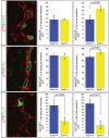
Increased proliferation at the NCC migration wavefront and reduced neuronal differentiation along the gut at E11.5 in Tcof1+/− embryos. Immunostaining of E11.5 whole guts with p75 (red) and either pHH3 (green) or BrdU (green) revealed no difference in proliferation between genotypes along the intestines. Dividing cells can be identified by the presence of green staining in the nucleus. However, a significant increase in NCC proliferation at the migration wavefront was counted in Tcof1+/− embryos compared with wild-type. Reduced neuronal differentiation was scored throughout the intestines in Tcof1+/− compared with Tcof1+/+ guts immunostained with p75 (red) and TuJ1 (green). *P < 0.05 (Student's t-test).
Figure 5.
Figure 5.
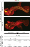
Oxidation treatment reduces the NCC migration in Tcof1+/− guts. Injection of pregnant females with 1% H2O2 prior to NCC delamination results in reduced NCC colonization of the gut in Tcof1+/− mice compared with wild-type and PBS-injected Tcof1+/− embryos. Immunostaining of E11.5 Tcof1+/+ and Tcof1+/− whole guts with p75 (red) and TuJ1 (green) showed a migration delay in 47% of Tcof1+/− injected with PBS which increased to ≈70% in H2O2-treated embryos. The percentage of SI and colon length colonized by NCC is represented by arrows. The number of guts with NCC at this position is above the arrow. (+/+), Tcof1+/+; (+/−), Tcof1+/−.
Figure 6.
Figure 6.
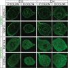
Increased laminin and reduced β1 integrin expression in Tcof1+/− guts compared with Tcof1+/+. Immunostaining of E13 and E14 colon cryosections with laminin and β1 integrin antibodies. Greater levels and more diffuse laminin staining in Tcof1+/− colon compared with wild-type at both stages. The expression of β1 integrin increased with developmental age in both genotypes; however, β1 integrin was initially reduced in the proximal colon at E13, but later at E14, was increased in this region in Tcof1+/− colon in comparison with Tcof1+/+. P, proximal; D, distal.

References

    1. Le Douarin N.M., Teillet M.A. The migration of neural crest cells to the wall of the digestive tract in avian embryo. J. Embryol. Exp. Morphol. 1973;30:31–48. - PubMed
    1. Yntema C.L., Hammond W.S. The origin of intrinsic ganglia of trunk viscera from vagal neural crest in the chick embryo. J. Comp. Neurol. 1954;101:515–541. doi:10.1002/cne.901010212. - DOI - PubMed
    1. Durbec P.L., Larsson-Blomberg L.B., Schuchardt A., Costantini F., Pachnis V. Common origin and developmental dependence on c-ret of subsets of enteric and sympathetic neuroblasts. Development. 1996;122:349–358. - PubMed
    1. Kapur R.P., Yost C., Palmiter R.D. A transgenic model for studying development of the enteric nervous system in normal and aganglionic mice. Development. 1992;116:167–175. - PubMed
    1. Natarajan D., Marcos-Gutierrez C., Pachnis V., de Graaff E. Requirement of signalling by receptor tyrosine kinase RET for the directed migration of enteric nervous system progenitor cells during mammalian embryogenesis. Development. 2002;129:5151–5160. - PubMed

"VSports" Publication types

MeSH terms

"VSports注册入口" Substances