Skip to main page content
U.S. flag

An official website of the United States government

Dot gov

The . gov means it’s official. Federal government websites often end in . gov or VSports app下载. mil. Before sharing sensitive information, make sure you’re on a federal government site. .

Https

The site is secure V体育官网. The https:// ensures that you are connecting to the official website and that any information you provide is encrypted and transmitted securely. .

. 2012 Apr;302(7):G645-54.
doi: 10.1152/ajpgi.00223.2011. Epub 2011 Dec 29.

Significance of para-esophageal lymph nodes in food or aeroallergen-induced iNKT cell-mediated experimental eosinophilic esophagitis

Affiliations

V体育平台登录 - Significance of para-esophageal lymph nodes in food or aeroallergen-induced iNKT cell-mediated experimental eosinophilic esophagitis

Priya Rajavelu et al. Am J Physiol Gastrointest Liver Physiol. 2012 Apr.

VSports - Abstract

Eosinophilic esophagitis (EoE) is a recently recognized inflammatory disorder driven by food hypersensitivity; however, the specific foods and mechanisms involved are unclear. In patients with EoE, we have found that hypersensitivities to corn and peanuts are the most common. Accordingly, we sensitized and exposed mice either intranasally or intragastrically with corn or peanut extract or saline. Esophageal eosinophilia, the genes of eosinophil-directed cytokines, and allergen-induced antibodies were examined in mice challenged with corn or peanut extract or saline. A high number of esophageal lamina propria eosinophils as well as eosinophilic microabscesses, intraepithelial eosinophils, extracellular eosinophilic granules, thickened and disrupted epithelial mucosa, and mast cell hyperplasia were observed in the esophagus of peanut or corn allergen-challenged mice. Mechanistic analysis indicated that para-esophageal lymph nodes might be critical in the trafficking of eosinophils to the esophagus and in EoE association to airway eosinophilia VSports手机版. Furthermore, experimentation with gene-targeted mice revealed that peanut allergen-induced EoE was dependent on eotaxin and invariant natural killer T (iNKT) cells, as CD1d and eotaxin-1/2 gene-deficient mice were protected from disease induction. Thus we provide evidence that para-esophageal lymph nodes are involved in food- or aeroallergen-induced eosinophilia and patchy EoE pathogenesis, likely a mechanism dependent on eotaxins and iNKT cells. .


"VSports app下载" Figures
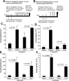
Fig. 1.
Fig. 1.
Esophageal eosinophil quantification following intranasal or intragastric corn or peanut allergen exposure. Mice sensitized intraperitoneally with 200 μg peanut or corn allergen with 1 mg alum and challenged orally or intragastrically or intranasally with 100 μg of corn or peanut allergen were analyzed for the number of eosinophils in the lung and esophagus following the protocol shown (A, B). EoE, Eosinophilic esophagitis. The number of eosinophils in the esophagus (C, E) and bronchoalveolar lavage fluid (D, F) were analyzed from mice euthanized 20–24 h after the last saline or allergen challenge. Data are expressed as means ± SD, n = 12 mice/group.
Fig. 2.
Fig. 2.
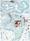
Eosinophil analysis in the esophageal lymph node and esophageal mucosa following saline or peanut allergen-induced experimental eosinophilic esophagitis (EoE). Immunohistochemical identification (by anti-major basic protein, MBP, staining) of eosinophils in the esophageal lymph nodes (ELN), along with muscularis mucosa (MM), lamina propria (LP), and epithelial mucosa (EM) of intranasally peanut allergen-challenged mice are shown (A, original magnification ×100). Eosinophils in the esophagus of intranasally saline-challenged (B, original magnification ×100) and peanut allergen-challenged Balb/c mice (C, original magnification ×100 and D, original magnification ×400). Eosinophils (arrows) are located in the LP of intranasally saline-challenged mice (B) and the LP and EM of intranasally peanut allergen-challenged mice (C and D). Higher-power magnification photomicrographs reveal eosinophil microabscesses in the basal epithelial layer and extracellular anti-MBP staining (D, inset of the area designated by the arrowhead, original magnification ×1,000). Intragastrically saline- (E, original magnification ×400) and peanut allergen- (F, original magnification ×400) challenged mice had eosinophils in the LP.
Fig. 3.
Fig. 3.
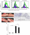
Antigen detection in the lung, mediastinal lymph node, and esophagus following intratracheal allergen challenge and the role of para-esophageal lymph nodes in EoE. A representative fluorescence-activated cell sorting (FACS) histogram demonstrates the presence of Alexafluor-conjugated Aspergillus allergen in the lung, mediastinal lymph node, and esophagus (AC). Antigen-positive cells from the populations isolated from each of these organs were analyzed on FACS; forward and side scatter are shown for the lung (A), mediastinal lymph node (B) and esophagus (C). The mean fluorescence shift 3 h after the last intratracheal saline or Alexafluor-conjugated allergen challenge is shown. Alexafluor-conjugated Aspergillus antigen-challenged mice showed higher mean florescence compared with saline-challenged mice. The filled histograms represent saline-challenged mice cells from the isolated cells of each respective organ. The dissecting microscopic photograph of para-esophageal lymph node in wild-type (WT) and Ltα mutant mice are shown and marked by arrows (D, af) and a microscopic photomicrograph of para-esophageal lymph node in WT and Ltα mutant mice (D, ef) are shown (original magnification ×200). A comparable esophageal eosinophilia in wild-type and Ltα gene-mutated mice are shown following allergen challenge (E). Data are shown as means ± SD, n = 10.
Fig. 4.
Fig. 4.
Mast cell accumulation in the esophagus following peanut exposure. Peanut/alum-sensitized mice exposed intranasally to saline or peanut allergen were analyzed for esophageal mast cells by chloroacetate esterase staining. The esophageal mast cells (arrows) of mice challenged intranasally with saline (A) or peanut allergen (B) are shown 18–20 h after the last saline or allergen exposure. Original magnifications of photomicrograph are ×125. The number of mast cells was quantified by morphometric analysis following digital microscopy of the esophageal sections of saline- or peanut allergen-challenged mice (C). The levels of total serum IgE following intranasally (D) or intragastrically (E) delivered saline or peanut allergen challenge are shown. Data are obtained 18–20 h after the last treatment. Data are expressed as means ± SD, n = 14 mice/group.
Fig. 5.
Fig. 5.
Peanut allergen-induced mRNA expression of eosinophil active cytokines in the esophagus. The quantitative real-time PCR analyses of the mRNA levels of the eosinophil active cytokines IL-5 (A), IL-13 (B), and IL-15 (C) following saline or peanut allergen exposure in mice are shown. The mRNA levels of the eosinophil active chemokines eotaxin-1 (D) and eotaxin-2 (E) following saline or peanut allergen exposure in mice are shown. Data are expressed as means ± SD, n = 10 mice/group.
Fig. 6.
Fig. 6.
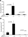
Peanut extract-induced EoE in eotaxin-1/-2 gene-deficient mice. The levels of eosinophils in the esophagus of wild-type and eotaxin-1/-2 double gene-deficient mice were analyzed following peanut extract exposure. Wild-type (+/+) and eotaxin-1/-2 double-deficient (−/−) mice were sensitized and challenged with intranasally (A) or intragastrically (B) delivered saline (−) or peanut extract (+), and the number of eosinophils in the esophagus was determined by anti-MBP staining. The data are expressed as mean ± SD, n = 12 mice/group. NS, not significant.
Fig. 7.
Fig. 7.
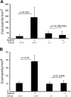
Peanut extract-induced EoE in CD1d gene-deficient mice. The levels of eosinophils in the esophagus of wild-type and CD1d gene-deficient mice were analyzed following peanut extract exposure. Wild-type (+/+) and CD1d-null (−/−) mice were sensitized and challenged with intranasally (A) or intragastrically (B) delivered saline (−) or peanut extract (+), and the number of eosinophils in the esophagus was determined by anti-MBP staining. The data are expressed as means ± SD, n = 12 mice/group. NS, not significant.

References

    1. Arora AS, Yamazaki K. Eosinophilic esophagitis: asthma of the esophagus? Clin Gastroenterol Hepatol 2: 523–530, 2004. - PubMed (V体育安卓版)
    1. Bachurski CJ, Pryhuber GS, Glasser SW, Kelly SE, Whitsett JA. Tumor necrosis factor-alpha inhibits surfactant protein C gene transcription. J Biol Chem 270: 19402–19407, 1995. - PubMed
    1. Becky Kelly EA, Busse WW, Jarjour NN. Inhaled budesonide decreases airway inflammatory response to allergen. Am J Respir Crit Care Med 162: 883–890, 2000. - PubMed
    1. Blanchard C, Wang N, Stringer KF, Mishra A, Fulkerson PC, Abonia JP, Jameson SC, Kirby C, Konikoff MR, Collins MH, Cohen MB, Akers R, Hogan SP, Assa'ad AH, Putnam PE, Aronow BJ, Rothenberg ME. Eotaxin-3 and a uniquely conserved gene-expression profile in eosinophilic esophagitis. J Clin Invest 116: 536–547, 2006. - PMC - PubMed
    1. Bock SA. Prospective appraisal of complaints of adverse reactions to foods in children during the first 3 years of life. Pediatrics 79: 683–688, 1987. - PubMed

Publication types

"V体育ios版" MeSH terms

LinkOut - more resources