Skip to main page content
U.S. flag

An official website of the United States government

Dot gov

The VSports app下载. gov means it’s official. Federal government websites often end in . gov or . mil. Before sharing sensitive information, make sure you’re on a federal government site. .

Https

The site is secure. The https:// ensures that you are connecting to the official website and that any information you provide is encrypted and transmitted securely. V体育官网.

. 2012 Feb 17;287(8):5819-32.
doi: 10.1074/jbc.M111.295964. Epub 2011 Dec 28.

Roles of STAT3 and ZEB1 proteins in E-cadherin down-regulation and human colorectal cancer epithelial-mesenchymal transition

Affiliations

Roles of STAT3 and ZEB1 proteins in E-cadherin down-regulation and human colorectal cancer epithelial-mesenchymal transition

Hua Xiong et al. J Biol Chem. .

Abstract

The progression of colorectal carcinoma (CRC) to invasive and metastatic disease may involve localized occurrences of epithelial-mesenchymal transition (EMT). However, mechanisms of the EMT process in CRC progression are not fully understood. We previously showed that knockdown of signal transducer and activator of transcription 3 (STAT3) up-regulated E-cadherin (a key component in EMT progression) in CRC. In this study, we examined the roles of STAT3 in CRC EMT and ZEB1, an EMT inducer, in STAT3-induced down-regulation of E-cadherin. Knockdown of STAT3 significantly increased E-cadherin and decreased N-cadherin and vimentin expressions in highly invasive LoVo CRC cells. Meanwhile, overexpression of STAT3 significantly reduced E-cadherin and enhanced N-cadherin and vimentin expressions in weakly invasive SW1116 CRC cells. Activation of STAT3 significantly increased CRC cell invasiveness and resistance to apoptosis. Knockdown of STAT3 dramatically enhanced chemosensitivity of CRC cells to fluorouracil. STAT3 regulated ZEB1 expression in CRC cells, and the STAT3-induced decrease in E-cadherin and cell invasion depended on activation of ZEB1 in CRC cells. Additionally, pSTAT3(Tyr-705) and ZEB1 expressions were significantly correlated with TNM (tumor, lymph node, and metastasis stages) (p < 0 VSports手机版. 01). In conclusion, STAT3 may directly mediate EMT progression and regulate ZEB1 expression in CRC. ZEB1 may participate in STAT3-induced cell invasion and E-cadherin down-regulation in CRC cells. The expressions of pSTAT3(Tyr-705) and ZEB1 may be positively associated with CRC metastasis. Our data may provide potential targets to prevent and/or treat CRC invasion and metastasis. .


Figures

FIGURE 1.
FIGURE 1.
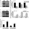
Effect of STAT3 on expression of epithelial and mesenchymal markers in LoVo and SW1116 cells. A representative Western blot (A) and the summarized data (B) show that knockdown of STAT3 expression significantly decreased the phosphorylation of STAT3 and the expressions of STAT3, N-cadherin, and vimentin in LoVo cells, whereas E-cadherin expression was dramatically increased, indicating that STAT3 may participate in the regulation of epithelial and mesenchymal markers in colon cancer. Another representative Western blot (C) and the summarized data (D) show that STAT3 overexpression significantly increased the phosphorylation of STAT3 and expressions of STAT3, N-cadherin, and vimentin in SW1116 cells, whereas E-cadherin expression was dramatically reduced. Seventy-five pmol of siRNA duplexes of STAT3, control siRNA, or plasmids complexed with liposomes were applied to each well. After 72 h of transfection, the cells were collected for analysis. n = 3, t test; *, p < 0.01; **, p < 0.05, compared with the pCDNA3.1 or control siRNA groups. Error bars, S.E.
FIGURE 2.
FIGURE 2.
Role of STAT3 in CRC cell EMT phenotypes. Transwell Matrigel invasion assays were performed in LoVo cells transfected with control siRNA or STAT3 siRNA (A) and in SW1116 cells transfected with pCDNA3.1 or pCDNA3.1-STAT3 (B). Twenty-four h after transfection, the cells were plated on transwell inserts, and invasion was assessed after incubation for 72 h. Cells were observed under a microscope and photographed. Cells were counted from five random microscopic fields (200×) per insert in triplicate. The migrated cell numbers were normalized to that of the control group. Data are shown as mean ± S.D. (error bars) from three separate experiments (*, p < 0.05 compared with control siRNA group; **, p < 0.01 compared with the pCDNA3.1 group). Representative colony-forming assays and relative quantitations show inhibition of growth in LoVo cells transfected with STAT3 siRNA compared with control siRNA (C) and increase of growth of SW1116 cells transfected with pCDNA3.1-STAT3 compared with pCDNA3.1 (D). Colony numbers following transfection with STAT3 siRNA or pCDNA3.1-STAT3 are expressed as the relative percentages of colonies compared with the corresponding control groups. Data are means ± S.E. (error bars) of five randomly selected microscopic fields from three independent wells in each group. *, p < 0.05 compared with the control siRNA group; **, p < 0.01 compared with the pCDNA3.1 group. E, dose-response curve of a representative experiment showing relative fluorouracil sensitivity determined by Cell Counting Kit 8 cell proliferation. LoVo cells were treated with fluorouracil after transfection with control or STAT3 siRNA. n = 3, t test; *, p < 0.05, compared with control siRNA group. F, apoptosis of SW116 cells were analyzed by flow cytometric analysis after transfection with pCDNA3.1-STAT3 or pCDNA3.1 following treatment with different doses of etopside. n = 3, t test; ▴, p < 0.01, compared with the pCDNA3.1 group.
FIGURE 3.
FIGURE 3.
Role of JAK/STAT3 pathway in ZEB1 expression in CRC cells. Real-time RT-PCR showed that the JAK2 inhibitor AG490 significantly decreased the expression of ZEB1 in SW1116 (A) and LoVo (B) cells, when compared with the control cells. AG490 treatment did not dramatically affect the expression of ZEB2, Snail1, Snail2, Twist1, Twist2, or E12/E47. C, knockdown of STAT3 significantly decreased the ZEB1 mRNA level in SW1116 cells. D, knockdown of STAT3 significantly decreased the ZEB1 mRNA level in LoVo cells. Cells were collected for analysis after treatment with 100 μm AG490 for 24 h. Seventy-five pmol of siRNA duplex of STAT3 or control siRNA complexed with liposomes were applied in each well. After 48 h of transfection, the cells were collected for analysis. n = 3, ANOVA; *, p < 0.05, compared with control; n = 3, t test; **, p < 0.05; ▴, p < 0.01, compared with control siRNA. Error bars, S.E.
FIGURE 4.
FIGURE 4.
Effect of STAT3 on ZEB1 expression and function in CRC cells. A representative Western blot (A) and the summarized data (B) show that transfection with STAT3 siRNA significantly decreased STAT3 phosphorylation and expressions of STAT3 and ZEB1 in LoVo cells. Another Western blot (C) and the summarized data (D) show that STAT3 phosphorylation and expressions of STAT3 and ZEB1 were boosted in STAT3-overexpressing SW1116 cells. E, stable knockdown of ZEB1 dramatically decreased cell invasion in basal condition and blocked STAT3-induced cell invasion in LoVo cells. n = 3, t test; *, p < 0.05, compared with control siRNA. n = 3, t test; **, p < 0.05, compared with pCDNA3.1. n = 3, ANOVA; ▴, p < 0.01, compared with pCDNA3.1+ control shRNA lentivirus; ▴▴, p < 0.05, compared with pCDNA3.1-STAT3+ control shRNA lentivirus. Error bars, S.E.
FIGURE 5.
FIGURE 5.
Role of STAT3 in the regulation of ZEB1 expression in CRC cells. A, bioinformatic analysis of STAT3 transcriptional factor binding site in part of the ZEB1 gene promoter region. The numbers on the left side indicate the locations upstream of the first base of the initial transcription site. STAT3-binding sites are highlighted, and the DNA sequence encompassed by two arrows was amplified in the ChIP assay. B, representative results from three experiments showed that ZEB1 DNA was detectable in the chromatin sample immunoprecipitated from SW1116 cells using an antibody against STAT3, suggesting that STAT3 binds to the ZEB1 promoter. Input DNA was used as a positive control; rabbit IgG and cell lysates without antibody were used as negative controls. C, overexpression of STAT3 caused a nearly 40% increase in ZEB1 promoter luciferase activity in SW1116 cells. D, knockdown of STAT3 significantly decreased ZEB1 promoter luciferase activity in SW1116 cells. E, two putative STAT3 binding sites were located between nt −500 and +100. White and black rhombuses indicate wild type and mutant sequences for STAT3 binding sites, respectively. WT, wild type; MT, mutant type. Mutation of STAT3 binding sites significantly decreased transcriptional activity of the ZEB1 promoter in a luciferase assay. n = 3, t test; **, p < 0.05, compared with pCDNA3.1+pGL3-basic-ZEB1PWT. n = 3, t test; *, p < 0.05, compared with control siRNA+pGL3-basic-ZEB1PWT; ▴, p < 0.01, compared with pGL3-basic-ZEB1PWT. Error bars, S.E.
FIGURE 6.
FIGURE 6.
Role of ZEB1 in STAT3-regulated E-cadherin expression in CRC cells. A, a representative Western blot analysis showed that transfection of ZEB1 siRNA significantly reduced the expression of ZEB1. A representative Western blot (B) and the summarized data (C) showed that STAT3 overexpression did not down-regulate E-cadherin expression after ZEB1 knockdown. D, knockdown of STAT3 or ZEB1 expression caused a nearly 2-fold increase in E-cadherin promoter luciferase activity in SW1116 cells. E, overexpression of STAT3 significantly decreased E-cadherin promoter luciferase activity in SW1116 cells. ZEB1 siRNA significantly blocked STAT3-induced down-regulation of E-cadherin promoter luciferase activity in SW1116 cells; n = 3, ANOVA; ▴, p < 0.05, compared with pCDNA3.1+control siRNA; ▴▴, p < 0.05, compared with pCDNA3.1-STAT3+control siRNA; n = 3, ANOVA; *, p < 0.05, compared with control siRNA+pGL3-basic-E-cadherinPWT; **, p < 0.05, compared with pCDNA3.1+control siRNA+pGL3-basic-E-cadherinPWT; ***, p < 0.05, compared with pCDNA3.1-STAT3+control siRNA+pGL3-basic-E-cadherinPWT. Error bars, S.E.
FIGURE 7.
FIGURE 7.
A, bioinformatic analysis of STAT3 and ZEB1 transcriptional factor binding site in part of the E-cadherin gene promoter region. The numbers on the left indicate the locations upstream of the first base of the initial transcription site. STAT3-binding sites and ZEB1-binding sites are highlighted, and the DNA sequence surrounded by two arrows was for ChIP. Representative results from three experiments show that E-cadherin DNA was detected in the chromatin sample immunoprecipitated from SW1116 (B) and LoVo (C) cells using an antibody against ZEB1, respectively, suggesting that ZEB1 binds to the E-cadherin promoter. Input DNA was used as a positive control; rabbit IgG and cell lysates without antibody were used as negative controls. Real-time PCR of the ChIP samples showed that overexpression of STAT3 dramatically increased the binding efficiency of ZEB1 to the E-cadherin promoter in SW1116 (D) and LoVo (E) cells. Knockdown of STAT3 significantly decreased the binding efficiency of ZEB1 to the E-cadherin promoter in SW1116 (F) and LoVo (G) cells. n = 3, t test; *, p < 0.01, compared with pCDNA3.1. n = 3, t test; **, p < 0.05, compared with control siRNA. Error bars, S.E.
FIGURE 8.
FIGURE 8.
Two putative STAT3 binding sites and four putative ZEB1 binding sites were located between nt −520 and +70 of the E-cadherin 5′-flanking region. White and black rhombuses indicate a wild or mutant sequence for STAT3 binding sites, respectively. White and black triangles indicate a wild type or mutant sequence for ZEB1 binding sites, respectively. WT, wild type; STAT3B MT, mutant type of each STAT3 mutation binding site; ZEB1B MT, mutant type of each ZEB1 mutation binding site; ZEB1B and STAT3B MT, mutant type of each ZEB1 and STAT3 mutation binding site. Mutation of ZEB1 binding sites or dual mutation of STAT3 and ZEB1 binding sites significantly increased the transcriptional activity of E-cadherin promoter in the luciferase assay. Mutation of STAT3 binding sites had no obvious effect on the transcriptional activity of the E-cadherin promoter. n = 3, ANOVA; *, p < 0.05, compared with pGL3-basic-E-cadherinPWT. Error bars, S.E.
FIGURE 9.
FIGURE 9.
Expressions of STAT3, ZEB1, and E-cadherin in CRC. Shown is immunohistochemical analysis of consecutive tissue sections for ZEB1, pSTAT3Tyr-705, STAT3, and E-cadherin in normal colorectal mucosa and high grade CRC. A and B, nuclear staining of ZEB1 and pSTAT3Tyr-705 were mostly presented in adenocarcinoma. Absent expression of ZEB1 (E) or pSTAT3Tyr-705 (F) was evident in the stroma or epithelium of the normal colorectal mucosa. C and G, cytoplasmic and nuclear staining of STAT3 was frequently detected in normal mucosa and adenocarcinoma. H, cytoplasmic staining of E-cadherin was predominantly detected in normal colorectal mucosa. D, staining of E-cadherin was dramatically decreased in adenocarcinoma, compared with normal colorectal epithelium. A–H (original magnification, ×200), representative areas from A1–H1 (original magnification, ×40), respectively.

"VSports" References

    1. Bates R. C., Mercurio A. M. (2005) The epithelial-mesenchymal transition (EMT) and colorectal cancer progression. Cancer Biol. Ther. 4, 365–370 - PubMed
    1. Markowitz S. D., Dawson D. M., Willis J., Willson J. K. (2002) Focus on colon cancer. Cancer Cell 1, 233–236 - PubMed
    1. Thiery J. P., Acloque H., Huang R. Y., Nieto M. A. (2009) Epithelial-mesenchymal transitions in development and disease. Cell 139, 871–890 - V体育2025版 - PubMed
    1. Hay E. D. (1995) An overview of epithelio-mesenchymal transformation. Acta Anat. 154, 8–20 - PubMed
    1. Savagner P. (2001) Leaving the neighborhood. Molecular mechanisms involved during epithelial-mesenchymal transition. BioEssays 23, 912–923 - PubMed

Publication types

"V体育ios版" MeSH terms