Skip to main page content
U.S. flag

An official website of the United States government

Dot gov

The . gov means it’s official. Federal government websites often end in VSports app下载. gov or . mil. Before sharing sensitive information, make sure you’re on a federal government site. .

Https

The site is secure. The https:// ensures that you are connecting to the official website and that any information you provide is encrypted and transmitted securely V体育官网. .

. 2012 Jan 10;109(2):E59-67.
doi: 10.1073/pnas.1113251108. Epub 2011 Dec 27.

Lupus-associated causal mutation in neutrophil cytosolic factor 2 (NCF2) brings unique insights to the structure and function of NADPH oxidase

Affiliations

Lupus-associated causal mutation in neutrophil cytosolic factor 2 (NCF2) brings unique insights to the structure and function of NADPH oxidase

Chaim O Jacob et al. Proc Natl Acad Sci U S A. .

Abstract

Systemic lupus erythematosus (SLE), the prototypic systemic autoimmune disease, is a debilitating multisystem autoimmune disorder characterized by chronic inflammation and extensive immune dysregulation in multiple organ systems, resulting in significant morbidity and mortality. Here, we present a multidisciplinary approach resulting in the identification of neutrophil cytosolic factor 2 (NCF2) as an important risk factor for SLE and the detailed characterization of its causal variant. We show that NCF2 is strongly associated with increased SLE risk in two independent populations: childhood-onset SLE and adult-onset SLE. The association between NCF2 and SLE can be attributed to a single nonsynonymous coding mutation in exon 12, the effect of which is the substitution of histidine-389 with glutamine (H389Q) in the PB1 domain of the NCF2 protein, with glutamine being the risk allele. Computational modeling suggests that the NCF2 H389Q mutation reduces the binding efficiency of NCF2 with the guanine nucleotide exchange factor Vav1. The model predicts that NCF2/H389 residue interacts with Vav1 residues E509, N510, E556, and G559 in the ZF domain of Vav1. Furthermore, replacing H389 with Q results in 1 VSports手机版. 5 kcal/mol weaker binding. To examine the effect of the NCF2 H389Q mutation on NADPH oxidase function, site-specific mutations at the 389 position in NCF2 were tested. Results show that an H389Q mutation causes a twofold decrease in reactive oxygen species production induced by the activation of the Vav-dependent Fcγ receptor-elicited NADPH oxidase activity. Our study completes the chain of evidence from genetic association to specific molecular function. .


Conflict of interest statement (V体育ios版)

The authors declare no conflict of interest.

Figures
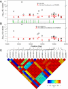
Fig. 1.
Fig. 1.
Association of NCF2 SNPs with SLE. Data are plotted with their P values (shown as −log10 values). Association of NCF2 SNPs with SLE in adult-onset (A) and childhood-onset (B) SLE cases in the EA subgroup with or without conditioning on rs17849502. The positions of exons (green rectangles) and introns (connecting lines) are indicated in the middle plot. The pale green rectangles at the two ends correspond to the UTRs. The dashed horizontal red lines correspond to P = 0.05. (C) LD heat map shows D′ between each of the typed SNPs in the region plotted, with D′ × 100 reported in each diamond. Chromosomal positions are according to National Center for Biotechnology Information build 37.1.
Fig. 2.
Fig. 2.
Relative contribution of different ethnicities to the overall OR for SNP rs17849502. Forest plot of additive OR ± 95% CI in indicated subgroups and overall OR (Mantel–Haenszel). AsA are not shown because the risk allele of rs17849502 was not found in this population. Size of rectangles is proportional to the weighting of the study in the meta-analysis, the top and bottom of diamond are the overall OR, the left and right whiskers (or left and right diamond edges) are the ±95% CI of the corresponding OR.
Fig. 3.
Fig. 3.
(A) Summary of interaction of truncated NCF2 derivatives with Vav1 as determined by Co-IP experiments and proposed binding region (vertical green bar) for Vav1 based on these experiments. (B and C) Immunoprecipitation (IP) of full-length or truncated NCF2 mutants with full-length Vav from lysates prepared from COS7 cells transiently transfected with corresponding expression constructs. Lysates were immunoprecipitated with a rabbit polyclonal antibody against Vav1 (Santa Cruz Biotechnology), and immunoprecipitates and lysates were analyzed by immunoblotting with the indicated antibodies. To detect NCF2, either a rabbit polyclonal antibody that reacts with the N terminus of NCF2 (Santa Cruz Biotechnology) or a mouse monoclonal antibody directed against the C terminus of NCF2 (BD Transduction Laboratories) was used in B and C, respectively. Samples shown in each panel were run on the same gel from the same experiment, but irrelevant lanes were excised for simplicity and clarity of presentation. Upon C-terminal truncation of NCF2, the removal of the PB1 domain led to a failure to coimmunoprecipitate with Vav1 (B, lanes ΔC299 and ΔC210), in contrast to an NCF2 derivative that included only the C-terminal PB1 and SH3 domains (C, lanes ΔN262 and ΔN350). WB, Western blot; Wt, wild type.
Fig. 4.
Fig. 4.
(A) Summary of interactions of truncated Vav1 derivatives with NCF2 as determined by Co-IP experiments and proposed binding region (vertical cyan bar) for NCF2. (BD) Immunoprecipitation of full-length or truncated Vav1 mutants with full-length NCF2 from lysates prepared from COS7 cells transiently transfected with corresponding expression constructs. Lysates were immunoprecipitated with antibodies directed against Vav1 or against epitope tags present on truncated Vav1 derivatives as indicated, and immunoprecipitates and lysates were analyzed by immunoblotting with the indicated antibodies. A Vav1 derivative lacking the ZF domain failed to coimmunoprecipitate with NCF2 (B, lane ΔN608), in contrast to full-length Vav1 or other truncated Vav1 proteins that included the ZF domain (BD, all lanes except ΔN608). IP, immunoprecipitation; WB, Western blot.
Fig. 5.
Fig. 5.
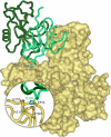
Model of the interaction of NCF2 with Vav1 via His389 in the presence of NCF4. Starting coordinates of Vav1 and NCF2 were taken from the PDB. The structure of the NCF2 PB1 domain was extracted from the complex with the PB1 domain of NCF4 [chain A in PDB entry 1oey (16)], and the structure of the DH-PH-ZF domains of Vav1 was taken from the complex with full-length Rac1 [chain B in PDB entry 2vrw (42)]. Coordinates of missing residues in the Vav1 domain DH were completed by superimposing the corresponding domain in the autoinhibited structure of Vav1 [PDB entry 3ky9 (43)]. The solvent-accessible surface of Vav1 is shown in yellow; NCF2 is shown as a ribbon diagram in green with H389 indicated. The nearby His anchoring spot is shown in magenta. NCF4 is shown in dark green. (Inset) Interaction site of H389 detected in the anchoring spots mapping is highlighted. The surface of Vav1 was made transparent to show the side chains of residues that are within 3 Å and interact with H389 in the docking model.
Fig. 6.
Fig. 6.
Conservation of H389 in NCF2 and its target amino acids in the binding pocket in the ZF domain of Vav. (A) H389 (boxed in yellow) is conserved from Gallus gallus (chicken) to human. (B) Sequence alignment of amino acid residues in the binding pocket in the ZF domain of the three Vav isoforms in the human and mouse are shown. Each human Vav isoform is identical to its relevant mouse homolog. The target amino acids (509, 510, 556, and 559) in Vav1 (yellow) are identical or have conservative substitution in Vav3 (yellow). Replacement of E by D in position 509 is considered a conserved charge; position 510 is less conserved, but the model suggests that NCF2 H389 binds Y510 similar to N510. The conservation of P508 and the general similarity of the character of the residues indicate that this fragment has a similar fold in the three Vav isoforms. The second fragment is even better conserved. E556 is identical in all three variants, and G559 is conserved in Vav1 and Vav3.
Fig. 7.
Fig. 7.
NCF2 H389Q mutation leads to decreased ROS release in K562 cells. K562 cells stably transfected with CYBB and NCF1 were cotransfected with plasmids for expression of NCF4 and NCF2 WT, NCF2 H389A mutant, or NCF2 H389Q mutant with the Amaxa Nucleofector kit V, under the T-16 program. Derivatives of NCF2 tagged at the C terminus with EYFP were also tested. According to our model, a Q in position 389 binds 1.5 kcal/mol weaker than H, whereas an A binds 2.0 kcal/mol weaker than H in this position. (A) ROS production in K562 cells in response to hIgG beads in the presence of luminol and HRP (n = 4; mean ± SEM; *P < 0.05). (B) ROS production in K562 cells in response to PMA in the presence of isoluminol and HRP (n = 4; mean ± SEM; In A and B, the total relative light unit (RLU) values over 60 min, measured at 1-min intervals, are expressed as the percentage of the RLU output of NCF2 WT-transfected cells in each experiment. Data were normalized to correct for interexperiment variability and log-transformed to stabilize variance; they were tested for significance using the Student t test. Representative kinetic curves are taken from one of four independent experiments for ROS production in K562 cells in response to hIgG beads (C) or PMA (D).
Fig. P1.
Fig. P1.
Schematic representation of NCF2 with domains and known binding partners (proteins) in their approximate binding positions. Human NCF2 harbors an N-terminal domain composed of several sections: four tricopeptide repeat (TPR) motifs, an AD (activation domain) and PR (proline-rich) region, an SH3 domain (SH3A), a PB1 domain, and a C-terminal SH3 domain (SH3B). Rac1/2 in the active (GTP-bound) state binds to the TPR domain, whereas other proteins (pictured) bind to other domains. Vav1 interacts with the PB1 domain of NCF2, which includes amino acid 389 based on data presented in our study.

References

    1. Tan W, et al. BIOLUPUS Network. GENLES Network Association of PPP2CA polymorphisms with systemic lupus erythematosus susceptibility in multiple ethnic groups. Arthritis Rheum. 2011;63:2755–2763. - "VSports" PMC - PubMed
    1. Deng Y, Tsao BP. Genetic susceptibility to systemic lupus erythematosus in the genomic era. Nat Rev Rheumatol. 2010;6:683–692. - VSports在线直播 - PMC - PubMed
    1. Harley JB, et al. International Consortium for Systemic Lupus Erythematosus Genetics (SLEGEN) Genome-wide association scan in women with systemic lupus erythematosus identifies susceptibility variants in ITGAM, PXK, KIAA1542 and other loci. Nat Genet. 2008;40:204–210. - PMC (V体育官网) - PubMed
    1. Armstrong DL, Jacob CO, Zidovetzki R. Function2Gene: A gene selection tool to increase the power of genetic association studies by utilizing public databases and expert knowledge. BMC Bioinformatics. 2008;9:311. - PMC - PubMed
    1. Jacob CO, et al. Identification of novel susceptibility genes in childhood-onset systemic lupus erythematosus using a uniquely designed candidate gene pathway platform. Arthritis Rheum. 2007;56:4164–4173. - PubMed

Publication types

"VSports在线直播" MeSH terms