Skip to main page content
U.S. flag

An official website of the United States government

Dot gov

The . gov means it’s official. Federal government websites often end in . gov or VSports app下载. mil. Before sharing sensitive information, make sure you’re on a federal government site. .

Https

The site is secure V体育官网. The https:// ensures that you are connecting to the official website and that any information you provide is encrypted and transmitted securely. .

. 2012 Jan 13;287(3):1861-73.
doi: 10.1074/jbc.M111.305789. Epub 2011 Dec 5.

Glucose and insulin induction of bile acid synthesis: mechanisms and implication in diabetes and obesity (VSports在线直播)

Affiliations

Glucose and insulin induction of bile acid synthesis: mechanisms and implication in diabetes and obesity

Tiangang Li et al. J Biol Chem. .

Abstract

Bile acids facilitate postprandial absorption of nutrients. Bile acids also activate the farnesoid X receptor (FXR) and the G protein-coupled receptor TGR5 and play a major role in regulating lipid, glucose, and energy metabolism. Transgenic expression of cholesterol 7α-hydroxylase (CYP7A1) prevented high fat diet-induced diabetes and obesity in mice. In this study, we investigated the nutrient effects on bile acid synthesis. Refeeding of a chow diet to fasted mice increased CYP7A1 expression, bile acid pool size, and serum bile acids in wild type and humanized CYP7A1-transgenic mice. Chromatin immunoprecipitation assays showed that glucose increased histone acetylation and decreased histone methylation on the CYP7A1 gene promoter. Refeeding also induced CYP7A1 in fxr-deficient mice, indicating that FXR signaling did not play a role in postprandial regulation of bile acid synthesis. In streptozocin-induced type I diabetic mice and genetically obese type II diabetic ob/ob mice, hyperglycemia increased histone acetylation status on the CYP7A1 gene promoter, leading to elevated basal Cyp7a1 expression and an enlarged bile acid pool with altered bile acid composition. However, refeeding did not further increase CYP7A1 expression in diabetic mice. In summary, this study demonstrates that glucose and insulin are major postprandial factors that induce CYP7A1 gene expression and bile acid synthesis. Glucose induces CYP7A1 gene expression mainly by epigenetic mechanisms. In diabetic mice, CYP7A1 chromatin is hyperacetylated, and fasting to refeeding response is impaired and may exacerbate metabolic disorders in diabetes VSports手机版. .


Figures

FIGURE 1.
FIGURE 1.
Feeding induces CYP7A1 in wild type C57BL6J mice. Male C57BL6J mice (12 weeks old) were fasted for 15 h from 9 a.m. to 12 a.m. and were either fasted or refed a regular chow diet for the time indicated. A and B, liver mRNA expression levels were measured by real-time PCR. C, CYP7A1 protein levels were determined by immunoblot of isolated liver microsomes from fasted and refed (3 h) mice. D, microsomal CYP7A1 enzyme activity was determined using an HPLC-based method as described under “Experimental Procedures.” Results are expressed as mean ± S.E. (error bars) (n = 4). *, statistical significance, p < 0.05 versus fasted mice. N.S., not significant.
FIGURE 2.
FIGURE 2.
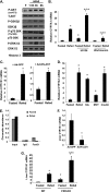
Insulin signaling mediates feeding induction of CYP7A1 in wild type C57BL6J mice. A, feeding effects on total and phosphorylated AKT, GSK3β p70 S6K, ERK1/2, and AMP-activated protein kinase in mouse liver were determined by immunoblot; samples were pooled from four mice. B, mice were fasted from 9 a.m. and given a single dose of the ERK inhibitor U0126 (5 mg/kg) or the PI3K inhibitor, wortmannin (0.8 mg/kg) by intravenous injection at 11 p.m. One h later, mice were either fasted or refed chow for an additional 3 h. Liver mRNA expression was determined by real-time PCR. C, mice were injected (intravenously) with adenovirus (2−9 pfu/mouse) expressing GFP (left) or a dominant negative form of AKT (Ad-DN-AKT) (right). Seven days later, mice were fasted for 15 h or refed chow for an additional 3 h, and liver CYP7A1 mRNA was measured. D, mice fasted for 15 h were refed chow or given a single dose of glucose (8 g/kg, oral gavage), medium chain triglycerides (MCT) (4 mg/kg, oral gavage), or insulin (0.8 unit/kg, intraperitoneal injection). Liver CYP7A1 mRNA levels were determined 3 h later. E, ChIP assay of FoxO1 binding to the cyp7a1 gene promoter in mouse livers. Liver nuclei were isolated from fasted and refed (3 h) mice. Pooled samples from four mice were used. ChIP primers detecting the proximal cyp7a1 promoter region (−1k) were used. Real-time PCR was done in triplicate, and mean values were plotted. F, effect of adenovirus-mediated transduction of constitutively active nuclear form of FoxO1 on CYP7A1 mRNA expression levels in mouse liver. Mice were injected (intravenously) with adenovirus (2−9 pfu/mouse) expressing either GFP as control or expressing a constitutively active nuclear form of FoxO1 (Ad-FoxO1). Seven days later, liver CYP7A1 mRNA was measured in overnight fasted mice. G, liver CYP7A1 mRNA was determined in fasted and refed (3 h) wild type and FXR knock-out (KO) mice. Results are expressed as mean ± S.E. (error bars) (n = 4). *, statistical significance versus fasted mice; **, statistical significance versus fasted + U0126 or fasted FXR knock-out mice (p < 0.05).
FIGURE 3.
FIGURE 3.
Feeding caused histone hyperacetylation and histone hypomethylation in the cyp7a1 gene chromatin in wild type C57BL6J mice. Mice were fasted or refed chow for 3 h. ChIP assays were used to determine the histone 3 (H3) acetylation (A), histone 4 (H4) acetylation (B), H3K9 dimethylation (C), and H3K9 trimethylation (D). Pooled samples from four mice were used for real-time PCR to detect acetylated histones 3 and 4, dimethylated H3K9, and trimethylated H3K9 in CYP7A1 chromatin. Assays were done in triplicate, and mean values were plotted.
FIGURE 4.
FIGURE 4.
Nutrients control the circadian variations of cyp7a1 gene expression in wild type C57BL6J mice. Mice were fasted at 9 a.m. (ZT3). For restricted feeding, regular chow was given at 12 a.m. (ZT18) for 3 h, and mice were moved to a clean cage without food. Liver CYP7A1 mRNA (A), liver Rev-erbα mRNA (B), and ileum FGF16 mRNA (C) expression levels were measured at the times indicated. A filled bar below each panel indicates the dark period. An arrow and open bar indicate the start of the restricted feeding period (3 h). Results are expressed as mean ± S.E. (error bars) (n = 4). *, statistical significance versus fasted mice at the same time point (p < 0.05). D, ChIP assays were used to determine histone 4-acetylation (AcH4) in the cyp7a1 gene promoter in mouse liver. Pooled samples from four mice were used. Real-time PCR was used to detect acetylated H4 in CYP7A1 chromatin. Assays were done in triplicate, and mean values were plotted.
FIGURE 5.
FIGURE 5.
Effects of restricted feeding on tissue and serum bile acid homeostasis. Serum bile acids (A), gallbladder bile acids (B), and intestine bile acids (C) were measured in fasted and food-restricted mice at different time points, as indicated. D, tissue bile acids were quantified in fasted and refed (3 h) mice. E, ileum bile acid-binding protein mRNA was determined by real-time PCR. A filled bar below each panel indicates the dark period. An arrow and open bar indicate the start of restricted feeding and period (3 h). Results are expressed as mean ± S.E. (error bars) (n = 4). *, statistical significance versus fasted mice at the same time point (p < 0.05).
FIGURE 6.
FIGURE 6.
Diabetes is associated with elevated CYP7A1 mRNA expression and histone hyperacetylation in cyp7a1 gene chromatin. STZ-treated mice, ob/ob mice, and their respective controls were fasted for 15 h and refed for 3 h. A, serum glucose in STZ mice. B, serum insulin in STZ mice. C, liver CYP7A1 mRNA in STZ mice. D, liver CYP7A1 mRNA expression levels in ob/ob mice. E, ChIP assays of histone 3 acetylation and H3K9 methylation in CYP7A1 gene promoter of ob/ob and STZ mice. F, liver SHP mRNA in ob/ob mice. G, liver SHP mRNA in STZ mice. H, ileum FGF15 mRNA in ob/ob mice. I, intestine FGF15 mRNA in STZ mice. Liver and ileum mRNA expression was determined by real-time PCR. Results are expressed as mean ± S.E. (error bars) (n = 4). *, statistical significance, p < 0.05 versus fasted control mice. **, statistical significance, p < 0.05 versus fasted STZ mice. N.S., not significant. ChIP assays were used to determine H3 acetylation and H3K9 dimethylation in fasted mouse livers using pooled samples (n = 4), and real-time PCR was used to detect acetylated histone 3 (AcH3) and dimethylated H3K9 (DiMeth-H3K9) in CYP7A1 chromatin. Assays were done in triplicate, and mean values were plotted.
FIGURE 7.
FIGURE 7.
Diabetic mice had an enlarged bile acid pool, altered bile acid composition, and increased intestine cholesterol absorption. A, serum bile acid concentration in STZ mice; B, serum bile acid concentration in ob/ob mice; C, total bile acid contents in liver, gallbladder (GB), intestine, and total bile acid pool in STZ mice. D, total bile acid contents in liver, gallbladder (GB), intestine, and total bile acid pool (liver + GB + intestine) in ob/ob mice. E, gallbladder bile acid composition in STZ mice. F, gallbladder bile acid composition in ob/ob mice. G, fecal bile acid composition in STZ and ob/ob mice. Pooled fecal bile acid extracts from 3–4 mice were used. H, intestinal fractional cholesterol absorption in STZ and ob/ob mice. Bile acid composition and intestine fractional cholesterol absorption were determined as described under “Experimental Procedures.” Results are expressed as mean ± S.E. (error bars) (n = 3–4). *, statistical significance versus control mice (p < 0.05). Fecal samples from four mice were pooled, and bile acids were extracted for composition analysis. T, tauro-conjugated bile acids; CA, cholic acid; MCA, α- and β-muricholic acids; CDCA, chenodeoxycholic acid; DCA, deoxycholic acid; UDCA, ursodeoxycholic acid; HDCA, hyodeoxycholic acid; LCA, lithocholic acid.
FIGURE 7.
FIGURE 7.
Diabetic mice had an enlarged bile acid pool, altered bile acid composition, and increased intestine cholesterol absorption. A, serum bile acid concentration in STZ mice; B, serum bile acid concentration in ob/ob mice; C, total bile acid contents in liver, gallbladder (GB), intestine, and total bile acid pool in STZ mice. D, total bile acid contents in liver, gallbladder (GB), intestine, and total bile acid pool (liver + GB + intestine) in ob/ob mice. E, gallbladder bile acid composition in STZ mice. F, gallbladder bile acid composition in ob/ob mice. G, fecal bile acid composition in STZ and ob/ob mice. Pooled fecal bile acid extracts from 3–4 mice were used. H, intestinal fractional cholesterol absorption in STZ and ob/ob mice. Bile acid composition and intestine fractional cholesterol absorption were determined as described under “Experimental Procedures.” Results are expressed as mean ± S.E. (error bars) (n = 3–4). *, statistical significance versus control mice (p < 0.05). Fecal samples from four mice were pooled, and bile acids were extracted for composition analysis. T, tauro-conjugated bile acids; CA, cholic acid; MCA, α- and β-muricholic acids; CDCA, chenodeoxycholic acid; DCA, deoxycholic acid; UDCA, ursodeoxycholic acid; HDCA, hyodeoxycholic acid; LCA, lithocholic acid.
FIGURE 8.
FIGURE 8.
Feeding induces CYP7A1 in humanized CYP7A1-tg mice. Total bile acid pool (A) and gallbladder bile acid composition (B) were determined in humanized CYP7A1-tg mice and wild type controls. C and D, human CYP7A1 mRNA levels in humanized CYP7A1-tg mice. Mice were fed chow or chow containing 5% cholestyramine or 2% cholesterol for 1 week. Human CYP7A1 mRNA was determined by real-time PCR. E, refeeding effect on human CYP7A1 mRNA expression in humanized CYP7A1-tg mice. Mice were fasted or refed for 3 h. Liver CYP7A1 mRNA was determined by real-time PCR. Results are expressed as mean ± S.E. (error bars), n = 3–4. *, statistical significance (p < 0.05). F, ChIP assay of the abundance of acetylated histone 3 (AcH3), trimethylated H3K9, and FoxO1 in the human CYP7A1 gene promoter of humanized CYP7A1-tg mice. Pooled liver samples from 3–4 mice were used in ChIP assays. Real-time PCR was done in triplicate, and mean values were plotted. Abbreviations for bile acids are as in Fig. 7.
FIGURE 9.
FIGURE 9.
Hyperglycemia in STZ-treated humanized CYP7A1-tg mice caused increased human CYP7A1 gene expression and enlarged bile acid pool. STZ-treated humanized CYP7A1-tg mice and their respective controls were used. A, human CYP7A1 mRNA level in STZ-treated diabetic humanized CYP7A1-tg mice. B, ChIP assay of histone 4-acetylation (AcH4) in the CYP7A1 gene promoter. Pooled liver samples from three mice were used in ChIP assays. Real-time PCR was done in triplicate, and mean values were plotted. C, tissue bile acid contents in intestine, gallbladder, liver, and total bile acid pool of STZ-treated and control humanized CYP7A1-tg mice. D, serum bile acid concentrations in STZ-treated and control humanized CYP7A1-tg mice. Results are expressed as mean ± S.E. (error bars) (n = 3–4). *, statistical significance versus control (p < 0.05).

References

    1. Chiang J. Y. (2009) Bile acids. Regulation of synthesis. J. Lipid Res. 50, 1955–1966 - PMC - PubMed
    1. Lefebvre P., Cariou B., Lien F., Kuipers F., Staels B. (2009) Role of bile acids and bile acid receptors in metabolic regulation. Physiol. Rev. 89, 147–191 - PubMed (VSports注册入口)
    1. Li T., Owsley E., Matozel M., Hsu P., Novak C. M., Chiang J. Y. (2010) Transgenic expression of cholesterol 7α-hydroxylase in the liver prevents high-fat diet-induced obesity and insulin resistance in mice. Hepatology 52, 678–690 - PMC - PubMed
    1. Goodwin B., Jones S. A., Price R. R., Watson M. A., McKee D. D., Moore L. B., Galardi C., Wilson J. G., Lewis M. C., Roth M. E., Maloney P. R., Willson T. M., Kliewer S. A. (2000) A regulatory cascade of the nuclear receptors FXR, SHP-1, and LRH-1 represses bile acid biosynthesis. Mol. Cell 6, 517–526 - PubMed
    1. Inagaki T., Choi M., Moschetta A., Peng L., Cummins C. L., McDonald J. G., Luo G., Jones S. A., Goodwin B., Richardson J. A., Gerard R. D., Repa J. J., Mangelsdorf D. J., Kliewer S. A. (2005) Fibroblast growth factor 15 functions as an enterohepatic signal to regulate bile acid homeostasis. Cell Metab. 2, 217–225 - PubMed

Publication types

VSports最新版本 - MeSH terms