Skip to main page content
U.S. flag

An official website of the United States government

Dot gov

The . gov means it’s official. Federal government websites often end in . gov or VSports app下载. mil. Before sharing sensitive information, make sure you’re on a federal government site. .

Https

The site is secure. The https:// ensures that you are connecting to the official website and that any information you provide is encrypted and transmitted securely V体育官网. .

. 2011 Dec 15;187(12):6335-45.
doi: 10.4049/jimmunol.1003965. Epub 2011 Nov 14.

V体育平台登录 - IL-15 regulates homeostasis and terminal maturation of NKT cells

Affiliations

VSports app下载 - IL-15 regulates homeostasis and terminal maturation of NKT cells

Laura E Gordy et al. J Immunol. .

Abstract

Semi-invariant NKT cells are thymus-derived innate-like lymphocytes that modulate microbial and tumor immunity as well as autoimmune diseases. These immunoregulatory properties of NKT cells are acquired during their development. Much has been learned regarding the molecular and cellular cues that promote NKT cell development, yet how these cells are maintained in the thymus and the periphery and how they acquire functional competence are incompletely understood. We found that IL-15 induced several Bcl-2 family survival factors in thymic and splenic NKT cells in vitro. Yet, IL-15-mediated thymic and peripheral NKT cell survival critically depended on Bcl-x(L) expression. Additionally, IL-15 regulated thymic developmental stage 2 to stage 3 lineage progression and terminal NKT cell differentiation VSports手机版. Global gene expression analyses and validation revealed that IL-15 regulated Tbx21 (T-bet) expression in thymic NKT cells. The loss of IL-15 also resulted in poor expression of key effector molecules such as IFN-γ, granzyme A and C, as well as several NK cell receptors, which are also regulated by T-bet in NKT cells. Taken together, our findings reveal a critical role for IL-15 in NKT cell survival, which is mediated by Bcl-x(L), and effector differentiation, which is consistent with a role of T-bet in regulating terminal maturation. .


Figures (VSports最新版本)

Figure 1
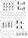
Figure 1. Defective NKT cell development and maintenance in IL-150 mice
(A) Thymic, splenic and hepatic NKT cells from B6 (n=13) and B6-IL-150 (n=9) mice were identified as CD3ε+tetramer+ cells within electronically gated CD8LO thymic, B220LO splenic or liver mononuclear cells (MNCs). Numbers are % NKT cells among total leukocytes within each organ. Data are representative of 9 independent experiments. (B) B6 and B6-IL-150 mice were injected i.p. with 2 mg BrdU daily for three consecutive days and sacrificed one day later. BrdU incorporation was determined by flow cytometry after extracellular lineage specific staining and intracellular labeling with anti-BrdU Alexa-647 mAb. Overlaid histograms are of NKT cells identified as in A from vehicle (shaded) or BrdU injected (open) mice. The histograms show the expression levels of BrdU within NK1.1NEG (left) and NK1.1+ (right) NKT cells within the thymus and spleen. Data are representative of 3 independent experiments; n=6. (C&D) Thymic and splenic MNCs from B6 and B6-IL-150 mice were assessed for intracellular expression of Bcl-2, Bcl-xL (C) and Mcl-1 (D), detected with specific mAb within electronically gated B220LOCD3ε+tetramer+ cells. Numbers refer to the difference in mean fluorescence intensity (ΔMFI) between isotype control (shaded) and specific mAb (open) staining. Data are representative of 3 independent experiments; n=5. The flow data were acquired on two different instruments (FACSCalibur and LSR-II) in different experiments. Even though the actual mean fluorescence intensity varied, the represented trend remained the same, nonetheless. Therefore, we have chosen to represent the ratio of expression between wt and IL-150 NKT cells. The ratio of Bcl-2, Bcl-xL and Mcl-1 expression by IL-150 and wt NKT cells is shown. A ratio of ~1 indicates no difference in expression; a ratio <1 indicates lower expression in IL-150 NKT cells.
Figure 2
Figure 2. IL-15 up-regulates expression of the survival factors Bcl-2, Bcl-xL, and Mcl-1 within thymic and splenic NKT cells
Thymic and splenic MNCs from B6 mice were cultivated in vitro in the absence or presence of 100 ng/mL of rhIL-15. After 5 days, intracellular expression of (A) Bcl-2, Bcl-xL and (B) Mcl-1 was detected with specific mAb within electronically gated B220LOCD3ε+tetramer+ cells. Numbers refer to the difference in mean fluorescence intensity (ΔMFI) between isotype control (shaded) and specific mAb (open). Data are representative of 3 independent experiments, n=5. As for Figure 1, the flow data were acquired on two different instruments in different experiments. Hence, the ratio of IL-15-induced Bcl-2, Bcl-xL and Mcl-1 expression NKT cells in comparison to un-stimulated, fresly isolated NKT cells is shown. A ratio of ≤1 indicates no induction; a ratio >1 indicates IL-15-induced expression.
Figure 3
Figure 3. Bcl-xL over expression restores NKT cell development in IL-150 mice
(A) Thymic, splenic and hepatic NKT cells from B6 (n=8), B6-IL-150 (n=10), B6-IL-150;Bcl-2tg (n=6), and B6-IL-150;Bcl-xLtg (n=16) mice were identified as CD3ε+tetramer+ cells within electronically gated CD8LO thymic, B220LO splenic orliver MNCs. Numbers are % NKT cells among total leukocytes. (B) Absolute numbers of NKT cells in the thymus and spleen of B6, B6-IL-150, B6-IL-150;Bcl-2tg, and B6-IL-150;Bcl-xLtg mice were calculated from % NKT cells in A and total cell count. Data are representative of 6 independent experiments showing mean±sem; n, as in A. p value was calculated by one-way ANOVA with Tukey’s post-test. (C) Intracellular expression of the hBcl-xL and hBcl-2 transgene within DN, DP, CD4+CD8NEG and CD8+CD4+ thymocytes of B6.C-Bcl-xLtg (right panels) and B6-Bcl-2tg (left panels) mice. Over-laid histograms represent isotype control (shaded) and specific mAb (open). Data are representative of 3 independent experiments; n=5. (D) Thymic and splenic NKT cells from IL-7Rα+/− (n=3), IL-7Rα0 (n=3), IL-7Rα0;Bcl-2tg (n=3) and IL-7Rα0;Bcl-xLtg (n=2) mice were identified as CD3ε+tetramer+ cells within electronically gated CD8LO thymocytes or B220LO splenocytes and liver MNCs. Numbers are % NKT cells among total leukocytes. Absolute NKT cell number (mean±sem) was calculated from % NKT cells and total MNC number. Data are representative of 2 independent experiments. Note that because IL-7Rα0 thymic size is small due to low cellularity, % NKT cells appears artificially high despite extremely low NKT cell numbers. P value was calculated by one-way ANOVA with Tukey’s post-test. (E) Intracellular expression of the hBcl-xL and hBcl-2 transgenes within NKT and NK cells of B6.C-Bcl-xLtg and B6-Bcl-2tg mice. Overlaid histograms represent isotype control (shaded) and specific mAb (open) staining. Data are representative of 2 independent experiments; n as in C. (F) Splenic and hepatic NK and NKT cells from B6 (n=6), B6-IL-150 (n=7), B6-IL-150;Bcl-2tg (n=4), and B6-IL-150;Bcl-xLtg (n=10) mice were identified as NK1.1+tetramerNEG and NK1.1+tetramer+ cells, respectively, within electronically gated B220LO splenic or hepatic MNCs. Data are representative of 4 independent experiments.
Figure 4
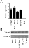
Figure 4. Normal TCRα rearrangement in IL-150 mice
(A) Vα14-to-Jα18 rearrangement within DP thymocytes flow sorted from the indicated strains was assessed by real-time qPCR represented as mean±sd of 2 independent experiments. β-actin was used as the internal control for normalisation. p value calculated by two-tailed, unpaired T-test indicated that Vα14-to-Jα18 rearrangement in B6, B6-IL-150 and B6-IL-150;Bcl-xLtg DP thymocytes was significant only when compared to that in B6-J180 DP cells. The ~1.5-fold increased Vα14-to-Jα18 rearrangement seen in B6-IL-150;Bcl-xLtg DP thymocytes compared to the others was statistically insignificant (p ≤0.0997. (B) RT-PCR assessment of Vα8-to-Jα5 and Vα14-to-Jα18 rearrangements. Data are representative of 2 experiments.
Figure 5
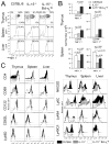
Figure 5. IL-15 regulates terminal maturation of NKT cells
(A) NKT cell developmental stages in B6 (n=9), B6-IL-150 (n=6), and B6-IL-150;Bcl-xLtg (n=13) mice were identified as CD44NEGNK1.1NEG ST1, CD44+NK1.1NEG ST2, or CD44+NK1.1+ ST3 in the thymus or as NK1.1NEGtetramer+ or NK1.1+tetramer+ within the splenic and hepatic MNCs. Numbers are % of cells among total NKT cells. Data are representative of 6 independent experiments. (B) Absolute numbers of thymic NKT cells within ST1, ST2 and ST3 were calculated as in Fig. 3D. Data are representative of 6 independent experiments showing the mean±sem; n, same as in A. p value was calculated by one-way ANOVA with Tukey’s post-test. (C) Expression of lineage-specific markers by thymic, splenic, and hepatic NKT cells from B6 (n=6), B6-IL-150 (n=6), and B6-IL-150;Bcl-xLtg (n=6) mice as determined by flow cytometry after staining with specific mAb. Data are representative of 3 independent experiments.
Figure 6
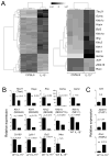
Figure 6. IL-15-induces Tbx21 and T-bet-regulated genes in developing NKT cells
A. Cluster analysis of all (left) or select genes (right) differentially expressed in B6 (n=3) and IL-150 (n=2) NKT cells a microarray experiment. Differential expression was defined as those genes that showed log2 fold change ≥1.5/≤−1.5 with a nominal p value <0.001. The microarray data can be accessed at http://www.ncbi.nlm.nih.gov/geo/query/acc.cgi?token=dtgxpkoosiokmpm&acc= using the accession number GSE32568. B&C. Select differentially expressed genes identified in A were validated by qPCR using RNA isolated from flow sorted B6 and B6-IL-150 mice. Genes up-regulated in B6-derived thymic NKT cells (B) and genes up-regulated in B6-IL-150-derived NKT cells (C) are shown. β-actin was used to normalise expression levels. Data represent mean±sd of 2 independent experiments; each qPCR was performed in duplicate for an n value of 4 to calculate the p value by unpaired T test.
Figure 7
Figure 7. IL-15 regulates functional maturation of NKT cells
A&B. B6 (n=6), B6-IL-150 (n=6), and B6-IL-150;Bcl-xLtg (n=12) mice were injected i.p. with 5 μg αGalCer (open) or vehicle (shaded). After 4 hrs, splenic (A) and hepatic (B) B220LOCD3ε+tetramer+ cells were electronically gated and intracellular IL-4 and IFN-γ expression was monitored. Numbers refer to ΔMFI. Data are representative of 4 independent experiments. C&D. Using ELISA, serum IFN-γ (C) and IL-4 (D) responses to in vivo NKT cell stimulation in the above experiment were measured. p value was calculated by one-way ANOVA with Tukey’s post-test. E&F. Thymic and splenic MNCs from B6 and B6-IL-150 mice were analyzed for intracellular T-bet (E) and Gata-3 (F) expression within electronically gated CD8LOCD3ε+tetramer0 thymocytes or B220LOCD3ε+tetramer+ splenocytes. Numbers refer to the ΔMFI between isotype control (shaded) and specific mAb (open). Data are representative of 2 independent experiments; n=5. (G) The ratio of T-bet and Gata-3 expression by IL-150 and wt NKT cells is shown. A ratio of ~1 (e.g., Gata-3) indicates no difference in expression; a ratio <1 (e.g., T-bet) indicates lower expression in IL-150 NKT cells.

References

    1. Bendelac A, Savage PB, Teyton L. The biology of NKT cells. Annu Rev Immunol. 2007;25:297–336. - PubMed
    1. Van Kaer L. α-Galactosylceramide therapy for autoimmune diseases: prospects and obstacles. Nat Rev Immunol. 2005;5:31–42. - PubMed
    1. Godfrey DI, Stankovic S, Baxter AG. Raising the NKT cell family. Nat Immunol. 2010;11:197–206. - PubMed
    1. Bezbradica JS, Hill T, Stanic AK, Van Kaer L, Joyce S. Commitment toward the natural T (iNKT) cell lineage occurs at the CD4+8+ stage of thymic ontogeny. Proc Natl Acad Sci U S A. 2005;102:5114–5119. - "VSports手机版" PMC - PubMed
    1. D’Cruz LM, Knell J, Fujimoto JK, Goldrath AW. An essential role for the transcription factor HEB in thymocyte survival, Tcra rearrangement and the development of natural killer T cells. Nat Immunol. 2010;11:240–249. - PMC - PubMed

"V体育安卓版" Publication types

MeSH terms

VSports最新版本 - Associated data