Skip to main page content
U.S. flag

An official website of the United States government

Dot gov

The . gov means it’s official. Federal government websites often end in . gov or . mil. Before sharing sensitive information, make sure you’re on a federal government site VSports app下载. .

Https

The site is secure V体育官网. The https:// ensures that you are connecting to the official website and that any information you provide is encrypted and transmitted securely. .

. 2011 Nov 29;108(48):19246-51.
doi: 10.1073/pnas.1114799108. Epub 2011 Nov 14.

Constitutive exposure of phosphatidylserine on viable cells

Affiliations

"V体育安卓版" Constitutive exposure of phosphatidylserine on viable cells

VSports - Katsumori Segawa et al. Proc Natl Acad Sci U S A. .

VSports最新版本 - Erratum in

  • Proc Natl Acad Sci U S A. 2012 Jan 17;109(3):995

V体育官网 - Abstract

Apoptotic cells are quickly recognized and engulfed by phagocytes to prevent the release of noxious materials from dying cells. Phosphatidylserine (PS) exposed on the surface of apoptotic cells is a proposed "eat-me" signal for the phagocytes. Transmembrane protein 16F (TMEM16F), a membrane protein with eight transmembrane segments, has the Ca-dependent phospholipid scramblase activity. Here we show that when lymphoma cells were transformed with a constitutively active form of TMEM16F, they exposed a high level of PS that was comparable to that observed on apoptotic cells VSports手机版. The PS-exposing cells were morphologically normal and grew normally. They efficiently responded to interleukin 3 and underwent apoptosis upon treatment with Fas ligand. The viable PS-exposing cells bound to peritoneal macrophages at 4 °C, but not at 25 °C. Accordingly, these cells were not engulfed by macrophages. When apoptotic cells were injected i. v. into mice, they were phagocytosed by CD11c(+)CD8(+) dendritic cells (DCs) in the spleen, but the PS-exposing living cells were not phagocytosed by these DCs. Furthermore, when PS-exposing lymphoma cells were transplanted s. c. into nude mice, they generated tumors as efficiently as parental lymphoma cells that did not expose PS. These results indicated that PS exposure alone is not sufficient to be recognized by macrophages as an eat-me signal. .

"V体育2025版" PubMed Disclaimer

Conflict of interest statement

The authors declare no conflict of interest.

Figures

Fig. 1.
Fig. 1.
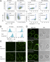
Constitutive exposure of phosphatidylserine on TMEM16F mutants. (A) Schematic diagrams of mouse TMEM16F (wild type) and its mutants D409G and D430G-L. The wild-type mouse TMEM16F is composed of 911 amino acids. The D409 mutant carries a glycine residue in place of an aspartic acid at codon 409. In the D430G-L mutant, a 21-aa peptide is inserted at amino acid position 24. (B) Splice variant produces TMEM16F carrying an extra peptide. A population of TMEM16F mRNA uses a sequence in intron 1 as an extra exon that codes for 21 amino acids. (C) Schematic representation of TMEM16F mutants. The Asp-to-Gly point mutation is in the first cytoplasmic loop, whereas the 21-aa insertion is in the N-terminal cytoplasmic domain. (D) (Left four panels) Ba/F3 cells were transformed with the empty vector, wild-type TMEM16F, its D409G mutant, or its D430G-L mutant. The amount of PS exposed on the cells was analyzed by Cy5-labeled annexin V, followed by FACS analysis. (Right) W3-Ildm cells were transformed with D430G-L, and the PS exposure was analyzed as above.
Fig. 2.
Fig. 2.
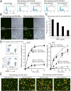
No effect of phosphatidylserine exposure on cell growth or cytokine responses. (A) Mouse Ba/F3 and Ba/F3–D430G-L cells were seeded at 104 cells/mL in 1 mL of RPMI1640 medium containing 10% FCS and 100 units/mL mouse IL-3, and cultured at 37 °C for 6 d. The cells were split 1:100 every 3 d, and their growth was followed. (B) Ba/F3 and Ba/F3–D430G-L cells were washed with RPMI1640 containing 10% FCS and cultured at 37 °C for 48 h in medium containing the indicated concentration of mouse IL-3. The cell viability was then determined by the WST-1 assay and is expressed as the percentage of that obtained with 64 units/mL IL-3. (C) Mouse W3-Ildm and W3-D430G-L cells were seeded at 3 × 10⁴ cells/mL in 1 mL of DMEM containing 10% FCS and cultured at 37 °C for 9 d. The cells were split 1:80 every 3 d, and the cell growth was followed. (D) W3-Ildm and W3-D430G-L cells were incubated at 37 °C for 4 h with the indicated concentration of human FasL. The cell viability was then determined by the WST-1 assay and is expressed as the percentage of that without FasL.
Fig. 3.
Fig. 3.
Exposure of phosphatidylserine on living W3-D430G-L cells. (A) W3-Ildm and W3-D430G-L cells with or without FasL treatment were stained with Cy5-labeled annexin V and Sytox blue and analyzed by flow cytometry. (Left four panels) FSC–SSC profile. (Right four panels) Profile for annexin V and Sytox blue staining. (B) W3-Ildm and W3-D430G-L cells with or without FasL treatment were stained with the FITC-labeled MFG-E8 and analyzed by FACS. The MFG-E8 staining profile in the Sytox blue population is shown. (C and D) W3-Ildm and W3-D430G-L cells with or without FasL treatment were stained with FITC-labeled MFG-E8 and observed by confocal fluorescence microscopy. Images for MFG-E8 (green) and differential interference contrast (DIC) are shown. Arrowheads indicate microvilli. (Scale bar, 10 μm.)
Fig. 4.
Fig. 4.
No engulfment of the PS-exposing living cells by macrophages. (A) TUNEL-staining profile of Mac-1+ thioglycollate-elicited peritoneal macrophages. W3-Ildm and W3-D430G-L cells that had been untreated or treated with FasL were coincubated with the thioglycollate-elicited peritoneal macrophages for 2 h, subjected to TUNEL staining, and analyzed by FACS. The TUNEL-staining profile of the Mac-1+ population is shown. The number indicates the percentage of TUNEL+ cells. (B) W3-Ildm and W3-D430G-L cells that had been untreated or treated with FasL were coincubated for 2 h with thioglycollate-elicited peritoneal macrophages, stained for TUNEL and DAPI, and observed by confocal microscopy. Images for TUNEL (green) and merged images for TUNEL, DAPI, and DIC are shown. (Scale bar, 10 μm.) (C) W3-Ildm cells were treated with FasL, incubated at 25 °C for 5 min with the indicated concentrations of the D89E mutant of mouse MFG-E8, and coincubated for 2 h with thioglycollate-elicited peritoneal macrophages. The macrophages were then subjected to TUNEL staining, followed by flow cytometry. The phagocytosis index indicates the percentage of TUNEL+ macrophages. (D) Binding of the PS-exposing cells to macrophages. Untreated or FasL-treated CMRA-labeled W3-Ildm and W3-D430G-L cells (5 × 10⁵ cells for 4 °C and 2.5 × 10⁵ cells for 25 °C incubation) were incubated at 4 °C or 25 °C for the indicated period with resident peritoneal cells (1 × 10⁵ cells). The cells were stained with APC-labeled anti–Mac-1 and analyzed by flow cytometry. The percentage of CMRA+ macrophages in the Mac-1+ population is plotted. (Left) Representative FACS profiles in which the FasL-treated W3-Ildm cells were incubated at 25 °C for 0 or 60 min are shown. (E) The CMRA-labeled W3-ldm and W3-D430G-L (red) cells, with or without FasL-treatment, were incubated at 4 °C for 60 min with resident peritoneal cells. The cells were thoroughly washed with cold PBS, stained with Alexa Fluor 488 anti–Mac-1 (green) and observed by fluorescence microscopy. Arrows indicate macrophages surrounded by CMRA-labeled cells. (Scale bar, 20 μm.)
Fig. 5.
Fig. 5.
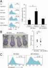
No clearance of the viable PS-exposing cells in vivo. (A) CMRA-labeled W3-Ildm and W3-D430G-L and FasL-treated W3-Ildm cells were injected into syngeneic female BALB/c mice. Thirty minutes after the injection, the splenic DCs were enriched by MACS sorting and analyzed by flow cytometry. The CMRA profile of the CD11c+CD8α+ population is shown. The percentages are of CMRA+ cells in the CD11c+CD8α+ population. Average values of three to four mice per group are shown with SD *P < 0.01. (B) Tumor development. W3-Ildm and W3-D430G-L cells (1.0 × 10⁶ cells) were transplanted s.c. into five BALB/c nude mice, respectively. Four weeks later, the tumors were dissected and their weight was determined. (Left) Picture of mice with tumors of the indicated cell line. (Right) Weights of tumors. The average values of the tumors from five mice are indicated by red horizontal bars. (C) PS exposure in the tumors developed in nude mice. Annexin V-staining profile of the Thy1.2+ tumor cells of the indicated cell line is shown. The number indicates the percentage of annexin V+ cells.

V体育官网 - References

    1. Jacobson MD, Weil M, Raff MC. Programmed cell death in animal development. Cell. 1997;88:347–354. - PubMed
    1. Nagata S. Apoptosis by death factor. Cell. 1997;88:355–365. - PubMed
    1. Ravichandran KS. Find-me and eat-me signals in apoptotic cell clearance: Progress and conundrums. J Exp Med. 2010;207:1807–1817. - PMC - PubMed
    1. Nagata S, Hanayama R, Kawane K. Autoimmunity and the clearance of dead cells. Cell. 2010;140:619–630. - PubMed (V体育官网)
    1. Fadok VA, de Cathelineau A, Daleke DL, Henson PM, Bratton DL. Loss of phospholipid asymmetry and surface exposure of phosphatidylserine is required for phagocytosis of apoptotic cells by macrophages and fibroblasts. J Biol Chem. 2001;276:1071–1077. - PubMed (VSports app下载)

Publication types

MeSH terms (V体育官网)