V体育平台登录 - Skip to main page content
U.S. flag

An official website of the United States government

Dot gov

The . gov means it’s official. Federal government websites often end in . gov or . mil. Before sharing sensitive information, make sure you’re on a federal government site. VSports app下载.

Https

The site is secure V体育官网. The https:// ensures that you are connecting to the official website and that any information you provide is encrypted and transmitted securely. .

. 2012 Jan;26(1):110-27.
doi: 10.1210/me.2011-1027. Epub 2011 Nov 10.

Mouse resistin modulates adipogenesis and glucose uptake in 3T3-L1 preadipocytes through the ROR1 receptor

Affiliations

VSports app下载 - Mouse resistin modulates adipogenesis and glucose uptake in 3T3-L1 preadipocytes through the ROR1 receptor

Beatriz Sánchez-Solana (V体育2025版) et al. Mol Endocrinol. 2012 Jan.

Abstract

Mouse resistin, a cysteine-rich protein primarily secreted from mature adipocytes, is involved in insulin resistance and type 2 diabetes. Human resistin, however, is mainly secreted by immune mononuclear cells, and it competes with lipopolysaccharide for the binding to Toll-like receptor 4, which could mediate some of the well-known proinflammatory effects of resistin in humans. In addition, resistin has been involved in the regulation of many cell differentiation and proliferation processes, suggesting that different receptors could be involved in mediating its numerous effects. Thus, a recent work identifies an isoform of Decorin (Δ Decorin) as a functional resistin receptor in adipocyte progenitors that may regulate white adipose tissue expansion. Our work shows that the mouse receptor tyrosine kinase-like orphan receptor (ROR)1 could mediate some of the described functions of resistin in 3T3-L1 adipogenesis and glucose uptake VSports手机版. We have demonstrated an interaction of mouse resistin with specific domains of the extracellular region of the ROR1 receptor. This interaction results in the inhibition of ROR1 phosphorylation, modulates ERK1/2 phosphorylation, and regulates suppressor of cytokine signaling 3, glucose transporter 4, and glucose transporter 1 expression. Moreover, mouse resistin modulates glucose uptake and promotes adipogenesis of 3T3-L1 cells through ROR1. In summary, our results identify mouse resistin as a potential inhibitory ligand for the receptor ROR1 and demonstrate, for the first time, that ROR1 plays an important role in adipogenesis and glucose homeostasis in 3T3-L1 cells. These data open a new line of research that could explain important questions about the resistin mechanism of action in adipogenesis and in the development of insulin resistance. .


Figures (V体育官网入口)

Fig. 1.
Fig. 1.
Interaction of mouse resistin with the ROR1 receptor. 3-AT assay (A) and quantitative β-galactosidase activity assay, by using ortho-nitrophenyl-β-galactoside as a substrate (B), of the cotransformant colonies coexpressing mouse resistin, lacking its signal peptide (RETN*), and each one of the indicated mouse ROR1 extracellular domains (IG, KR, or FZZ). I) +, Positive control, pVA3-1/pTD1-1; −, Negative control), pLAM5′-1/pTD1-1; V1/V2, pAS2–1/pACT2 empty vectors; V1/IG, pAS2-1/pAC-ROR1-IG; V1/FZZ, pAS2-1/pAC-ROR1-FZZ; V1/KR, pAS2-1/pAC-ROR1-KR; and RETN*/V2, pAS-RETN*/pACT2. II) Two colonies of each type of cotransformant cells coexpressing RETN* and one of the extracellular domains of ROR1 are shown: RETN*/IG, RETN*/FZZ, and RETN*/KR. CG1945 NT, Nontransformed yeast cells. Western blot analysis of the overexpression of entire RETN-6XHIS-HA (C) and 3XFLAG-ROR1e* (D) fusion proteins in soluble extracts from HEK 293T/17 cells was performed with α-HA and α-FLAG antibodies, respectively. V 6XHIS-HA and V 3XFLAG: empty vectors. Both C and D Western blot analyses show the signal corresponding to a 10% of the total protein concentration used for the coimmunoprecipitation assay in E. E, Western blot analysis (WB) of the coimmunoprecipitation of ROR1 and RETN proteins. A mixture of soluble cell extracts from HEK 293T/17 cells, overexpressing, or not, 3XFLAG-ROR1e* or RETN-6XHIS-HA proteins, were immunoprecipitated (IP) with an α-FLAG antibody. The α-resistin antibody revealed the coimmunoprecipitation of the ROR1 receptor and RETN-6XHIS-HA protein pulled down with the α-FLAG antibody. MM-LTH, Minimal medium without leucine, tryptophan, and histidine; MM-LT, minimal medium without leucine and tryptophan.
Fig. 1.
Fig. 1.
Interaction of mouse resistin with the ROR1 receptor. 3-AT assay (A) and quantitative β-galactosidase activity assay, by using ortho-nitrophenyl-β-galactoside as a substrate (B), of the cotransformant colonies coexpressing mouse resistin, lacking its signal peptide (RETN*), and each one of the indicated mouse ROR1 extracellular domains (IG, KR, or FZZ). I) +, Positive control, pVA3-1/pTD1-1; −, Negative control), pLAM5′-1/pTD1-1; V1/V2, pAS2–1/pACT2 empty vectors; V1/IG, pAS2-1/pAC-ROR1-IG; V1/FZZ, pAS2-1/pAC-ROR1-FZZ; V1/KR, pAS2-1/pAC-ROR1-KR; and RETN*/V2, pAS-RETN*/pACT2. II) Two colonies of each type of cotransformant cells coexpressing RETN* and one of the extracellular domains of ROR1 are shown: RETN*/IG, RETN*/FZZ, and RETN*/KR. CG1945 NT, Nontransformed yeast cells. Western blot analysis of the overexpression of entire RETN-6XHIS-HA (C) and 3XFLAG-ROR1e* (D) fusion proteins in soluble extracts from HEK 293T/17 cells was performed with α-HA and α-FLAG antibodies, respectively. V 6XHIS-HA and V 3XFLAG: empty vectors. Both C and D Western blot analyses show the signal corresponding to a 10% of the total protein concentration used for the coimmunoprecipitation assay in E. E, Western blot analysis (WB) of the coimmunoprecipitation of ROR1 and RETN proteins. A mixture of soluble cell extracts from HEK 293T/17 cells, overexpressing, or not, 3XFLAG-ROR1e* or RETN-6XHIS-HA proteins, were immunoprecipitated (IP) with an α-FLAG antibody. The α-resistin antibody revealed the coimmunoprecipitation of the ROR1 receptor and RETN-6XHIS-HA protein pulled down with the α-FLAG antibody. MM-LTH, Minimal medium without leucine, tryptophan, and histidine; MM-LT, minimal medium without leucine and tryptophan.
Fig. 2.
Fig. 2.
Mouse resistin binds to intact HEK 293T/17 cells overexpressing the ROR1 receptor and inhibits ROR1 tyrosine phosphorylation. A, Western blot analysis for the binding of purified rRETN to HEK 293T/17 cells overexpressing, or not, ROR1-FLAG fusion protein. Resistin bound to membrane fractions was detected with an α-resistin antibody. ROR1-FLAG was detected with the α-FLAG antibody. Sample load was estimated by Ponceau staining. B, In vitro binding assay of resistin and ROR1. HEK 293T/17 cells overexpressing, or not, the entire ROR1-FLAG protein were incubated with different concentrations of purified HA-tagged mouse resistin (RETN-6XHIS-HA), or purified mouse leptin, used as a negative control (concentrations ranging from 0 to 200 ng/ml in both cases). Relative binding was calculated by normalizing the amount of purified RETN-6XHIS-HA bound to the membrane fractions with the amount of ROR1-FLAG in each sample. Relative amounts of these proteins were obtained by densitometric analysis of the Western blot signals of at least three different assays. C, Western blot analysis of the competition between purified RETN-6XHIS-HA and rRETN proteins for ROR1 receptor in the binding assay in B. HEK 293T/17 cells, overexpressing the entire ROR1-FLAG protein, were incubated with 200 ng/ml purified RETN-6XHIS-HA and with, or without, 100-fold excess of purified rRETN. A representative Western blot analysis is shown. RETN-6XHIS-HA and ROR1-FLAG were detected with α-HA and α-FLAG antibodies, respectively. The α-tubulin signal was used as a loading control. D, Tyrosine phosphorylation of the ROR1 receptor in ROR1-FLAG transiently transfected HEK 293T/17 cells, in the presence or the absence of 100 ng/ml purified rRETN. To detect ROR1-FLAG, α-ROR1 and α-FLAG antibodies were used. To detect P-Tyr residues, an α-P-Tyr antibody was used. Representative Western blottings (WB) are shown. V FLAG: HEK 293T/17 cells transfected with empty vector. ROR1-FLAG, HEK 293T/17 cells overexpressing the complete ROR1 protein fused to the FLAG epitope at the C-terminal end. WB, Western blot; IP, immunoprecipitation.
Fig. 3.
Fig. 3.
Mouse ROR1 receptor modulates the expression of GLUT1 and GLUT4 in 3T3-L1 preadipocytes. Analysis of the expression of Ror1 (A) and Glut1/Glut4 (B) mRNAs in 3T3-L1 cells overexpressing, or not, Ror1. Data were normalized to P0 mRNA expression levels. The thin bars represent the sd of RT-PCR data. C, Western blot analysis of ROR1, GLUT1, and GLUT4 proteins in stably transfected 3T3-L1cells, overexpressing, or not, Ror1, in the presence or in the absence of purified rRETN 100 ng/ml. Analysis of ROR1 (D) and GLUT1 and GLUT4 (E) proteins in ROR1 knockdown 3T3-L1 cells by Western blotting. To detect the expression of the proteins, rabbit α-ROR1, mouse α-GLUT4, and rabbit α-GLUT1 antibodies were used. Representative Western blottings are shown. The expression of α-tubulin was used as a loading control and to normalize data. The bars correspond to the sd of data from three different assays. NT, Not treated cells.
Fig. 4.
Fig. 4.
Mouse resistin modulates glucose uptake stimulated by insulin in 3T3-L1 preadipocytes through ROR1 receptor. A, Culture medium glucose (mmol/liter) in control 3T3-L1 cells and cells overexpressing Ror1 at different times (hours) under cell confluence. B, Culture medium glucose (mmol/liter) at different times (hours) under cell confluence in control 3T3-L1 cells or 3T3-L1 cells with diminished ROR1 expression levels, obtained by shRNA methodology. Culture medium glucose (mmol/liter) in 3T3-L1 cells overexpressing, or not, Ror1 (C and D), or in control shRNA and Ror1 shRNA stable transfectants of these cells with diminished levels of ROR1 expression (E and F). These cells were incubated in the presence of 1 μm insulin or 1 μm insulin plus 100 ng/ml purified rRETN, at different times (hours) under cell confluence and compared with not treated cells. Curves represent the remaining glucose in culture medium at the indicated times. The data correspond to the average of three different assays.
Fig. 5.
Fig. 5.
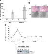
Mouse resistin enhances adipogenesis of 3T3-L1 preadipocytes through the ROR1 receptor. A, Quantitative RT-PCR analysis of Fabp4/aP2 gene expression levels in differentiated (DIF) and not differentiated (ND) 3T3-L1 preadipocytes, in the presence or in the absence of purified rRETN. B, Red Oil O staining analysis of differentiated and not differentiated 3T3-L1 preadipocytes, in the presence or in the absence of purified rRETN. Representative images of the adipocyte differentiation level of these cells are shown. Analysis by quantitative RT-PCR of Ror1 expression of 3T3-L1 preadipocytes along adipogenesis (C), and at different times under cell confluence, by Western blotting (D). BC, Before cell confluence; C, cell confluence; 1DC, 1-d confluent cells; D, days of cell confluence or treatment; DEX, dexamethasone; IBMX, 3-isobuthyl-1-methylxanthine. The expression of α-tubulin was used as a loading control. To detect the expression of ROR1, a rabbit α-ROR1 antibody was used. A representative Western blotting is shown. E, Quantitative RT-PCR analysis of Fabp4/aP2 expression in differentiated or not differentiated 3T3-L1 preadipocytes, overexpressing Ror1, in the presence or in the absence of purified rRETN. Quantitative RT-PCR analysis of Fabp4ABP4/aP2 expression (F) and analysis of the level of adipogenesis visualized by direct microscope observation, without adipocyte staining (G), in differentiated or not differentiated 3T3-L1 preadipocytes incubated with α-ROR1 or α-HA antibodies, and in the presence or in the absence of purified rRETN. The expression of P0 mRNA was used to normalize data from RT-PCR assays. The expression values obtained in not differentiated cells were used as a reference. The thin bars represent the sd of data from three different assays. NT, Not treated cells.
Fig. 5.
Fig. 5.
Mouse resistin enhances adipogenesis of 3T3-L1 preadipocytes through the ROR1 receptor. A, Quantitative RT-PCR analysis of Fabp4/aP2 gene expression levels in differentiated (DIF) and not differentiated (ND) 3T3-L1 preadipocytes, in the presence or in the absence of purified rRETN. B, Red Oil O staining analysis of differentiated and not differentiated 3T3-L1 preadipocytes, in the presence or in the absence of purified rRETN. Representative images of the adipocyte differentiation level of these cells are shown. Analysis by quantitative RT-PCR of Ror1 expression of 3T3-L1 preadipocytes along adipogenesis (C), and at different times under cell confluence, by Western blotting (D). BC, Before cell confluence; C, cell confluence; 1DC, 1-d confluent cells; D, days of cell confluence or treatment; DEX, dexamethasone; IBMX, 3-isobuthyl-1-methylxanthine. The expression of α-tubulin was used as a loading control. To detect the expression of ROR1, a rabbit α-ROR1 antibody was used. A representative Western blotting is shown. E, Quantitative RT-PCR analysis of Fabp4/aP2 expression in differentiated or not differentiated 3T3-L1 preadipocytes, overexpressing Ror1, in the presence or in the absence of purified rRETN. Quantitative RT-PCR analysis of Fabp4ABP4/aP2 expression (F) and analysis of the level of adipogenesis visualized by direct microscope observation, without adipocyte staining (G), in differentiated or not differentiated 3T3-L1 preadipocytes incubated with α-ROR1 or α-HA antibodies, and in the presence or in the absence of purified rRETN. The expression of P0 mRNA was used to normalize data from RT-PCR assays. The expression values obtained in not differentiated cells were used as a reference. The thin bars represent the sd of data from three different assays. NT, Not treated cells.
Fig. 6.
Fig. 6.
Mouse ROR1 receptor modulates SOCS3 expression and the phosphorylation of different kinases involved in glucose metabolism and adipogenesis in 3T3-L1 preadipocytes. A, Analysis of ERK1/2 MAPK, AKT, GSK3β, p38MAPK, and AMPK phosphorylation in stably 3T3-L1 transfectants overexpressing, or not, Ror1. Analysis of SOCS3 mRNA and protein expression levels in stable 3T3-L1 transfectants overexpressing, or not, Ror1, in the presence or the absence of purified rRETN, by quantitative RT-PCR (B), and by Western blotting (C). The right panel in C shows the densitometric analysis of the Western blot signals corresponding to three different assays. D, Analysis of SOCS3 expression and ERK1/2 MAPK, AKT, GSK3β, p38MAPK, and AMPK phosphorylation in a stable 3T3-L1 transfectant with diminished levels of ROR1 receptor (Ror1 shRNA) (2). Analysis of the expression and phosphorylation of ERK1/2 MAPK, by Western blotting, in stable 3T3-L1 transfectants overexpressing Ror1, in response to purified rRETN (E), or insulin treatments (F), at the indicated times and concentrations. Cells in F were previously treated overnight with 100 ng/ml purified rRETN. To detect the expression and phosphorylation of the different kinases analyzed, we used the antibodies described in Materials and Methods. The expression of α-tubulin and the expression of nonphosphorylated kinases were used as a loading control and to normalize data in densitometric analysis. The expression of P0 mRNA was used to normalize the data from the RT-PCR assays. The bars correspond to the sd of data. Ror1, Stably transfected 3T3-L1cells overexpressing Ror1; vector, 3T3-L1 cells transfected with empty vector; NT, not treated cells.
Fig. 6.
Fig. 6.
Mouse ROR1 receptor modulates SOCS3 expression and the phosphorylation of different kinases involved in glucose metabolism and adipogenesis in 3T3-L1 preadipocytes. A, Analysis of ERK1/2 MAPK, AKT, GSK3β, p38MAPK, and AMPK phosphorylation in stably 3T3-L1 transfectants overexpressing, or not, Ror1. Analysis of SOCS3 mRNA and protein expression levels in stable 3T3-L1 transfectants overexpressing, or not, Ror1, in the presence or the absence of purified rRETN, by quantitative RT-PCR (B), and by Western blotting (C). The right panel in C shows the densitometric analysis of the Western blot signals corresponding to three different assays. D, Analysis of SOCS3 expression and ERK1/2 MAPK, AKT, GSK3β, p38MAPK, and AMPK phosphorylation in a stable 3T3-L1 transfectant with diminished levels of ROR1 receptor (Ror1 shRNA) (2). Analysis of the expression and phosphorylation of ERK1/2 MAPK, by Western blotting, in stable 3T3-L1 transfectants overexpressing Ror1, in response to purified rRETN (E), or insulin treatments (F), at the indicated times and concentrations. Cells in F were previously treated overnight with 100 ng/ml purified rRETN. To detect the expression and phosphorylation of the different kinases analyzed, we used the antibodies described in Materials and Methods. The expression of α-tubulin and the expression of nonphosphorylated kinases were used as a loading control and to normalize data in densitometric analysis. The expression of P0 mRNA was used to normalize the data from the RT-PCR assays. The bars correspond to the sd of data. Ror1, Stably transfected 3T3-L1cells overexpressing Ror1; vector, 3T3-L1 cells transfected with empty vector; NT, not treated cells.

References

    1. Galic S , Oakhill JS , Steinberg GR. 2010. Adipose tissue as an endocrine organ. Mol Cell Endocrinol 316:129–139. - PubMed
    1. Steppan CM , Lazar MA. 2002. Resistin and obesity-associated insulin resistance. Trends Endocrinol Metab 13:18–23 - PubMed
    1. Steppan CM , Lazar MA. 2004. The current biology of resistin. J Intern Med 255:439–447 - PubMed
    1. Filková M , Haluzík M , Gay S , Senolt L. 2009. The role of resistin as a regulator of inflammation: implications for various human pathologies. Clin Immunol 133:157–170 - PubMed (V体育平台登录)
    1. Steppan CM , Brown EJ , Wright CM , Bhat S , Banerjee RR , Dai CY , Enders GH , Silberg DG , Wen X , Wu GD , Lazar MA. 2001. A family of tissue-specific resistin-like molecules. Proc Natl Acad Sci USA 98:502–506 - PMC - PubMed

Publication types

"V体育2025版" MeSH terms

Substances