Skip to main page content (V体育官网入口)
U.S. flag

An official website of the United States government

Dot gov

The . gov means it’s official. Federal government websites often end in . gov or VSports app下载. mil. Before sharing sensitive information, make sure you’re on a federal government site. .

Https

The site is secure V体育官网. The https:// ensures that you are connecting to the official website and that any information you provide is encrypted and transmitted securely. .

. 2012 Jul;1822(7):1169-79.
doi: 10.1016/j.bbadis.2011.10.007. Epub 2011 Oct 13.

Increased cone sensitivity to ABCA4 deficiency provides insight into macular vision loss in Stargardt's dystrophy

Affiliations

Increased cone sensitivity to ABCA4 deficiency provides insight into macular vision loss in Stargardt's dystrophy

Shannon M Conley (VSports最新版本) et al. Biochim Biophys Acta. 2012 Jul.

Abstract

Autosomal recessive Stargardt macular dystrophy is caused by mutations in the photoreceptor disc rim protein ABCA4/ABCR. Key clinical features of Stargardt disease include relatively mild rod defects such as delayed dark adaptation, coupled with severe cone defects reflected in macular atrophy and central vision loss. In spite of this clinical divergence, there has been no biochemical study of the effects of ABCA4 deficiency on cones vs. rods. Here we utilize the cone-dominant Abca4(-/-)/Nrl(-/-) double knockout mouse to study this issue. We show that as early as post-natal day (P) 30, Abca4(-/-)/Nrl(-/-) retinas have significantly fewer rosettes than Abca4(+/+)/Nrl(-/-) retinas, a phenotype often associated with accelerated degeneration. Abca4-deficient mice in both the wild-type and cone-dominant background accumulate more of the toxic bisretinoid A2E than their ABCA4-competent counterparts, but Abca4(-/-)/Nrl(-/-) eyes generate significantly more A2E per mole of 11-cis-retinal (11-cisRAL) than Abca4(-/-) eyes. At P120, Abca4(-/-)/Nrl(-/-) produced 340 ± 121 pmoles A2E/nmol 11-cisRAL while Abca4(-/-) produced 50. 4 ± 8 VSports手机版. 05 pmoles A2E/nmol 11-cisRAL. Nevertheless, the retinal pigment epithelium (RPE) of Abca4(-/-)/Nrl(-/-) eyes exhibits fewer lipofuscin granules than the RPE of Abca4(-/-) eyes; at P120: Abca4(-/-)/Nrl(-/-) exhibit 0. 045 ± 0. 013 lipofuscingranules/μm² of RPE vs. Abca4(-/-) 0. 17 ± 0. 030 lipofuscingranules/μm² of RPE. These data indicate that ABCA4-deficient cones simultaneously generate more A2E than rods and are less able to effectively clear it, and suggest that primary cone toxicity may contribute to Stargardt's-associated macular vision loss in addition to cone death secondary to RPE atrophy. .


Figures

Fig. 1
Fig. 1
Development of retinal flecking and atrophic areas in the Abca4−/− mouse. Fundus images were collected from anesthetized mice at the age of 12 months. On the left is shown a normal aged fundus (Abca4+/+), on the middle and right are shown two representative images collected from age-matched Abca4−/− mice. Note the appearance of yellowish flecks and atrophic areas (arrows).
Fig. 2
Fig. 2
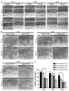
Structural effects of ABCA4 deficiency. Representative sections from Abca4+/+ and Abca4−/−(A), Abca4+/+/Nrl−/− and Abca4−/−/Nrl−/−(B) reared at 30 (top rows) or 300 lx (bottom rows) were collected at P30, P60 and P120. (A) No overt changes in retinal structure are observed in the absence of ABCA4 in the WT background. (B) In the Nrl−/− background, the primary structural defect arising from ABCA4 deficiency is a decrease in the number rosettes detected in the outer nuclear layer (ONL). Light rearing had no discernible effect on any genotype. (C) Rosettes were quantified in central retinal sections in 3–5 mice per group. Values are presented as mean total rosettes counted±SEM. * p<0.05. OS: outer segment, ONL: outer nuclear layer, INL: inner nuclear layer. Scale bar 25 μm.
Fig. 3
Fig. 3
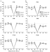
ABCA4 deficiency does not increase retinal degeneration up to P120. Measuring from the optic nerve head, sections (as in Fig. 2) were imaged at 100 μm intervals. Total nuclei in the ONL were counted in each image from 3 to 5 mice per group. Values are presented as mean±SEM. Horizontal gray lines represent average levels at P30. (A) WT mice and their ABCA4 deficient counterparts do not exhibit any retinal thinning up to P120 at either light level. (B) In the Nrl−/− background, a decrease in ONL thickness is observed by P120 but neither lack of ABCA4 nor light exposure has any significant effect on the decrease. N: nasal, T: temporal.
Fig. 4
Fig. 4
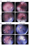
Development of retinal flecking in the absence of ABCA4. Shown are representative fundus images captured from animals raised at 30 lx at 2–3 months of age (top row of each panel) or 4–5 months of age (bottom row of each panel) in the WT (A) or Nrl−/−(B) background. (A) Normal fundus (left) and Abca4−/− fundus (right). At both ages, some Abca4−/− animals exhibited signs of retinal flecking (black arrows) while others exhibited a normal fundus. (B) Abca4+/+/Nrl−/− fundus (left) and Abca4−/−/Nrl−/− fundus (right). The Nrl−/− fundus at 2–3 months of age exhibits evenly spaced white-gray spots across the fundus (white arrows). By 4–5 months of age these spots are detected primarily in the periphery with very few in the central portion. In the Abca4−/−/Nrl−/− eye, the white-gray spots are detected at 2–3 months, although not to the same extent as in the Abca4+/+/Nrl−/−. Very few white-gray spots are detected in Abca4−/−/Nrl−/− eyes at 4–5 months. No signs of retinal flecking are observed in the Nrl−/− background at either age.
Fig. 5
Fig. 5
Retinal stress is exacerbated in the absence of ABCA4. Frozen retinal sections collected at P30, 60, and 120 were stained with the retinal stress marker, GFAP. At P30 (top) and P60 (middle) normal GFAP localization is observed in Abca4+/+ and Abca4+/+/Nrl−/− retinas, expression is limited to the inner limiting membrane. Very mild GFAP induction is observed at P30 age in Abca4−/− and Abca4−/−/Nrl−/− animals (arrowheads). At P60 patchy induction of GFAP is seen in Abca4−/− animals (arrows) and induction is seen throughout Abca4−/−/Nrl−/− retinas. By P120, all genotypes except Abca4+/+ show signs of GFAP induction, with the most severe being detected in the Abca4−/−/Nrl−/−. Scale bar 25 μm. ONL: outer nuclear layer, INL: inner nuclear layer, IPL: inner plexiform layer, GCL: ganglion cell layer.
Fig. 6
Fig. 6
Cone function is not severely affected by ABCA4 deficiency. Full-field scotopic ERGs were performed on 6–8 animals per group at P30, 60, and 120. Results shown are mean maximum amplitudes±SEM. (A) In the WT background, ABCA4 deficiency has no effect on cone function. (B) In the Nrl−/− background, a mild decrease in cone function is observed in Abca4−/−/Nrl−/− animals at P120 compared to P30. * p<0.05.
Fig. 7
Fig. 7
A2E levels are significantly increased in the absence of ABCA4. Total A2E levels were measured from eyecups harvested at P30, 60 or 120. 3–6 independent samples were measured for each group and values are presented as mean pmoles of A2E/eye±SEM. Rearing under 300 lx did not have a significant effect on A2E accumulation but at every timepoint, in both the WT and Nrl−/− background, animals lacking ABCA4 accumulated significantly more A2E than control animals. *p<0.05, **p<0.01, ***p<0.001.
Fig. 8
Fig. 8
11-cisRAL levels are decreased in the absence of ABCA4. 11-cisRAL levels were measured from eyecups harvested at P120 (30 lx), 4–5 measurements per group. (a) 11-cisRAL levels are significantly decreased in the Nrl−/− background. (b) For clarity’s sake, A2E values at P120 (30 lx) from Fig. 8 are shown here. (c) A2E values are shown normalized to 11-cisRAL levels. Per pmole of 11-cisRAL, animals in the Nrl−/− background, particularly when lacking ABCA4, accumulate significantly more A2E than animals in the WT background. *p<0.05, **p<0.01, ***p<0.001.
Fig. 9
Fig. 9
Fewer lipofuscin granules are detected in the RPE cells of ABCA4 deficient eyes in the Nrl−/− background when compared to the WT background. (A) Representative electron micrographs of the RPE cell layer at P120 showing lipofuscin granules (black arrows) and phagocytosed outer segments (white arrows). Scale bar 2 μm. For quantification, 3–5 animals per group were analyzed and at least 5 images per eye were counted, totals were summed for each eye and values shown are means±SEM. (B) Both the number of lipofuscin granules per μm of RPE area (left) and the fraction of RPE occupied by lipofuscin granules (right) are increased with significance in the Abca4−/− compared to all other genotypes, and these parameters are also non-significantly increased in Abca4−/−/Nrl−/− compared to Abca4+/+/Nrl−/−. (C) In both the WT and Nrl−/− backgrounds, the average size of each lipofuscin granule is larger in the absence of ABCA4. (D) In the Nrl−/− background the number of OS pieces detected inside the RPE appeared to be less than in the WT background, but the difference was not statistically significant. *p<0.05, **p<0.01, ***p<0.001.

References (VSports app下载)

    1. Sun H, Nathans J. Stargardt’s ABCR is localized to the disc membrane of retinal rod outer segments. Nat Genet. 1997;17:15–16. - PubMed
    1. Weng J, Mata NL, Azarian SM, Tzekov RT, Birch DG, Travis GH. Insights into the function of Rim protein in photoreceptors and etiology of Stargardt’s disease from the phenotype in abcr knockout mice. Cell. 1999;98:13–23. - PubMed
    1. Illing M, Molday LL, Molday RS. The 220-kDa rim protein of retinal rod outer segments is a member of the ABC transporter superfamily. J Biol Chem. 1997;272:10303–10310. - PubMed
    1. Molday RS. ATP-binding cassette transporter ABCA4: molecular properties and role in vision and macular degeneration. J Bioenerg Biomembr. 2007;39:507–517. - PubMed
    1. Molday LL, Rabin AR, Molday RS. ABCR expression in foveal cone photoreceptors and its role in Stargardt macular dystrophy. Nat Genet. 2000;25:257–258. - PubMed

"VSports手机版" Publication types

MeSH terms