"VSports" Skip to main page content
U.S. flag

An official website of the United States government

Dot gov

The . gov means it’s official. Federal government websites often end in VSports app下载. gov or . mil. Before sharing sensitive information, make sure you’re on a federal government site. .

Https

The site is secure. The https:// ensures that you are connecting to the official website and that any information you provide is encrypted and transmitted securely V体育官网. .

. 2011;6(10):e26332.
doi: 10.1371/journal.pone.0026332. Epub 2011 Oct 17.

From SOMAmer-based biomarker discovery to diagnostic and clinical applications: a SOMAmer-based, streamlined multiplex proteomic assay

Affiliations

From SOMAmer-based biomarker discovery to diagnostic and clinical applications: a SOMAmer-based, streamlined multiplex proteomic assay (V体育安卓版)

Stephan Kraemer et al. PLoS One. 2011.

VSports手机版 - Abstract

Recently, we reported a SOMAmer-based, highly multiplexed assay for the purpose of biomarker identification. To enable seamless transition from highly multiplexed biomarker discovery assays to a format suitable and convenient for diagnostic and life-science applications, we developed a streamlined, plate-based version of the assay. The plate-based version of the assay is robust, sensitive (sub-picomolar), rapid, can be highly multiplexed (upwards of 60 analytes), and fully automated. We demonstrate that quantification by microarray-based hybridization, Luminex bead-based methods, and qPCR are each compatible with our platform, further expanding the breadth of proteomic applications for a wide user community. VSports手机版.


Conflict of interest statement

Competing Interests: The authors have read the journal's policy and have the following conflict: the authors are all employees of SomaLogic, Inc. This fact does not alter the authors′ adherence to PLoS One policies on sharing of data and materials V体育安卓版.

Figures
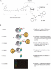
Figure 1
Figure 1. SOMAmer-based assay reagent and assay principles.
The primary analyte capture reagent and quantified component consists of an analyte-specific SOMAmer coupled to a Cy3 moiety and a biotin group joined to the fluorophore-substituted SOMAmer through a photocleavable linker (Panel A). The principal features of the assay consist of equilibration of capture reagent and analyte mixture in solution, followed by immobilization of the entire capture reagent population on immobilized streptavidin through the biotin moieties of the capture reagent population. The immobilized capture reagent population, including analyte/capture reagent complexes, is washed to remove non-complexed proteins. Immobilized protein analytes are then biotinylated using a conventional amine-reactive biotinylation reagent. The entire capture reagent population, including biotinylated analyte/capture reagent complexes, is then released into solution via photocleavage. Biotinylated analyte/capture reagent complexes are exclusively captured on immobilized streptavidin via the biotin moieties appended to the analyte population. Washes remove the capture reagent population at large, leaving only analyte/capture reagent complexes. The remaining capture reagent population is a surrogate for the analyte/capture reagent population. This material is eluted from immobilized analytes and quantified via conventional DNA quantification methods.
Figure 2
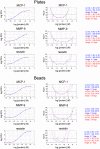
Figure 2. Diagram of manual plate-based assay steps and dose-response curves generated in manual plate-based assay format.
The five steps of the manual plate-based assay - equilibration, biotinylation, photocleavage, and elution, are punctuated by three wash-and-dump cycles and one liquid transfer. The total processing time is about 70 minutes (Panel A). A set of dose response curve generated in a nine-plex manual assay format at various capture reagent concentrations (Panel B). Dose-response curves were generated by spiking analytes into plasma at the indicated concentrations. Shown are resistin and MCP-1. Dose-response curves of MIP-4, RANTES, MMP-9, MMP-7, Lipocalin 2, tPA, and IL-8 can be found in Supporting Materials.
Figure 3
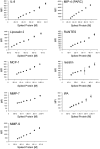
Figure 3. Precision profiles and limits of quantification of plate- and bead-based assays.
Eight individual measurements of fluorescent signal as a function of analyte concentration in buffer were made for each of nine analytes in multiplexed format. For the dose-response curves (left of each panel), the average RFU at each concentration is denoted by the blue markers and the eight individual measurements used to compute each average are denoted by the red markers plotted on the four parameter curve fit (solid blue line). Precision profiles (right of each panel) were computed with two different methods: (1) by calculating the variance in computed concentrations (blue, bottom left of each panel) and (2) by calculating the variance in log RFU (assay response, top right of each panel) combined with the slope of the standard curve (red). Limits of quantification were defined as points on the curves where the coefficient of variation (CV) exceeded 20%. Panels A, C, and E were generated in plate-based format. Panels B, D, and F were generated in bead-based format. Analytes measured were MCP-1 (Panels A and B), MMP9 (Panel C and D), resistin (Panels E and F) tPA (Figure S1B), MMP-7 (Figure S2B), IL-8 (Figure S2A), Lipocalin 2 (Figure S2A), MIP-4 (Figure S2A), Protein S (Figure S2B) and RANTES (Figure S2B).
Figure 4
Figure 4. High-resolution titration of analytes.
Analytes were titrated in 20% concentration increments in the region of signal generated by serum without spikes. Assay eluates were quantified by Luminex bead hybridization.
Figure 5
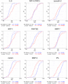
Figure 5. Differential expression between case and control populations.
Twenty-four case samples and twenty-four control samples were measured using the model 9-plex plate-based assay. Empirical CDFs were constructed for the control (blue) and case (red) populations separately for each analyte and are displayed in panels a–i. Spikes into the control samples (tPA, MMP-9, and Lipocalin 2) result in clear “down-regulation”, spikes into case samples (IL-8, MCP-1, resistin and RANTES) result in clear “up-regulation” and the two analytes not spiked (MIP-4 and MMP-7) show no differential expression.

References

    1. O'Farrell PH. The pre-omics era: the early days of two-dimensional gels. Proteomics. 2008;8:4842–4852. - PMC (V体育平台登录) - PubMed
    1. Zichi D, Eaton B, Singer B, Gold L. Proteomics and diagnostics: Let's Get Specific, again. Current Opinion in Chemical Biology. 2008;12:78–85. - PubMed (V体育官网)
    1. Gold L, Ayers D, Bertino J, Bock A, Bock C, et al. Aptamer-based multiplexed proteomic technology for biomarker discovery. PLoS One. 2010;Dec 7;5(12):e15004. - PMC - PubMed
    1. Ellington D, Szostak JW. In vitro selection of RNA molecules that bind specific ligands. Nature. 1990;346:818. - PubMed
    1. Tuerk C, Gold L. Systematic evolution of ligands by exponential enrichment: RNA ligands to bacteriophage T4 DNA polymerase. Science. 1990;249:505. - PubMed

"V体育官网入口" Publication types

MeSH terms