VSports在线直播 - Skip to main page content
U.S. flag

An official website of the United States government

Dot gov

The . gov means it’s official. Federal government websites often end in VSports app下载. gov or . mil. Before sharing sensitive information, make sure you’re on a federal government site. .

Https

The site is secure. The https:// ensures that you are connecting to the official website and that any information you provide is encrypted and transmitted securely V体育官网. .

. 2011 Nov 23;35(5):780-91.
doi: 10.1016/j.immuni.2011.08.013. Epub 2011 Oct 20.

Notch2 receptor signaling controls functional differentiation of dendritic cells in the spleen and intestine

Affiliations

Notch2 receptor signaling controls functional differentiation of dendritic cells in the spleen and intestine

Kanako L Lewis et al. Immunity. .

Abstract

Dendritic cells (DCs) in tissues and lymphoid organs comprise distinct functional subsets that differentiate in situ from circulating progenitors VSports手机版. Tissue-specific signals that regulate DC subset differentiation are poorly understood. We report that DC-specific deletion of the Notch2 receptor caused a reduction of DC populations in the spleen. Within the splenic CD11b(+) DC subset, Notch signaling blockade ablated a distinct population marked by high expression of the adhesion molecule Esam. The Notch-dependent Esam(hi) DC subset required lymphotoxin beta receptor signaling, proliferated in situ, and facilitated CD4(+) T cell priming. The Notch-independent Esam(lo) DCs expressed monocyte-related genes and showed superior cytokine responses. In addition, Notch2 deletion led to the loss of CD11b(+)CD103(+) DCs in the intestinal lamina propria and to a corresponding decrease of IL-17-producing CD4(+) T cells in the intestine. Thus, Notch2 is a common differentiation signal for T cell-priming CD11b(+) DC subsets in the spleen and intestine. .


"V体育2025版" Figures

Figure 1
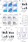
Figure 1. Notch2 signaling regulates splenic DC development
Mice with Itgax-cre -mediated deletion of Notch1 or Notch2 or DC-specific overexpression of DNMAML1 were analyzed along with the respective cre-negative littermate controls. Statistically significant differences are indicated as follows: ***, P<0.001; **, P<0.01; *, P<0.05. (A) Representative staining profiles of total splenocytes with the fraction of CD11chi MHC II+ DCs indicated. (B) The fraction (top) and absolute number (bottom) of splenic DCs (mean ± S.D. of 3–6 animals per group). (C) Staining profiles of gated CD11chi MHC II+ splenic DCs, with CD11b+ (blue), CD8+ (green), and double-negative (purple) subsets highlighted. The percentages represent mean ± S.D., n=3–6 for DC-Notch1Δ and DC-DNMAML1 and 11–12 for DC-Notch2Δ. (D) The percentage among total splenocytes (top) and absolute number (bottom) of CD11b+ and CD8+ DC subsets (mean ± S.D. of 8–10 animals per group). (E) The expression of SIRPα and CD24 in gated CD11chi MHC II+ CD11b CD8 double-negative DCs from conditional Notch2 (DC-Notch2Δ) and littermate control (Ctrl) spleens. The SIRPα+ and CD24+ populations indicative of the differentiation towards CD11b+ and CD8+ DC subsets, respectively, are indicated (representative of 2 animals). (F) Expression of surface markers in CD11b+ DCs from DC-Notch2Δ and control spleens.
Figure 2
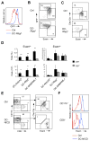
Figure 2. Notch2-RBPJ signaling specifies a distinct population of CD11b+ cDC
(A) The expression of Cx3cr1-GFP reporter in CD11b+ splenic DCs from conditional RBPJ-deficient (DC-RbpjΔ) mice. Shown is the histogram of GFP expression in gated CD11chi MHC II+ CD11b+ DCs from Cx3cr1-GFP+ control (Ctrl) or DC-RbpjΔ mice (representative of 4 animals per group). (B) The expression of Esam vs GFP in gated CD11b+ splenic DCs from Cx3cr1-GFP+ control or DC-RbpjΔ mice. (C) The expression of Esam vs Clec12a in gated CD11b+ splenic DCs from DC-RbpjΔ or control mice. (D) The percentage among total splenocytes (top) and absolute number (bottom) of the Esamhi and Esamlo CD11b+ DC subsets in mice with DC-specific targeting of the indicated genes (mean ± S.D. of 3–5 animals). Significance is indicated as in Fig. 1. (E) Splenic DC populations in mice with DC-specific expression of Notch1 intracellular domain (DC-NICD). Shown are staining profiles of gated splenic CD11chi MHC II+ DCs and of their CD11b+ subset from DC-NICD mice or Cre-negative littermate controls (Ctrl). Representative of 8 animals per genotype. (F) The expression of Esam in splenic CD11b+ and CD8+ DCs from DC-NICD and control mice.
Figure 3
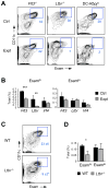
Figure 3. The development of Esamhi CD11b+ cDCs requires LTβR signaling
(A) Splenic CD11b+ Esamhi DC subset in experimental (Expt) Flt3−/− or Ltbr−/− animals or respective wild-type controls (Ctrl). The DC-RbpjΔ splenocytes that lack Esamhi DCs (Fig. 2D) are included for comparison. Shown are staining profiles of gated B220 CD11b+ CD8 splenocytes, with the CD11chi Esamhi DC population highlighted. (B) The fraction of Esamhi and Esamlo DC subsets among total splenocytes from Flt3−/−, Ltbr−/− or DC-Irf4Δ experimental (Expt) animals or controls (Ctrl). Significance is indicated as in Fig. 1. (C) Splenic CD11b+ Esamhi DC population among gated wild-type competitor (WT) or Ltbr−/− donor populations in competitively reconstituted BM chimeras, shown as in panel A (mean ± S.D. of 3 animals). (D) The fraction of WT competitor- or Ltbr−/− donor-derived CD11b+ DC subsets among total splenocytes in the BM chimeras (mean ± S.D. of 3 animals).
Figure 4
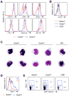
Figure 4. Phenotypic analysis of the splenic CD11b+ DC subsets
Unless indicated otherwise, Cx3cr1-GFP reporter mice were used to separate the Esamhi GFPlo and Esamlo GFPhi subsets of CD11chi CD11b+ CD8 splenic DCs. (A) Expression of the indicated surface markers in the gated Esamhi GFPlo and Esamlo GFPhi subsets. Dashed lines indicate positive staining threshold. (B) Expression of LTβR on the indicated DC subsets from wild-type mice. (C) Microphotographs of the sorted DC subsets stained by Giemsa stain on cytospin preparations. The CD11c CD11b+ F4/80+ Cx3cr1-GFP+ macrophages (MΦ) are shown for comparison (magnification, 400x). (D) Histograms of BrdU staining in the gated DC subsets after 2 days of in vivo BrdU pulse. The percentages of BrdU+ cells are indicated; representative of 4 animals. (E) Cell cycle profiles of DC subsets after 2 hr of in vivo BrdU pulse. Shown are gated DC subsets stained for DNA content (DAPI) and BrdU incorporation; the fractions of cells in S and G2/M phases are indicated (representative of 2 animals).
Figure 5
Figure 5. Gene expression profiles of splenic CD11b+ DC subsets
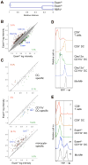
(A) The comparison of control and RBPJ-deficient CD11b+ DCs. Splenic CD11b+ DCs from Cx3cr1-GFP+ DC-RbpjΔ mice were sorted along with the Esamhi GFPlo and Esamlo GFPhi subsets of CD11b+ splenic DCs from control Cx3cr1-GFP+ mice. Shown is unsupervised clustering analysis of the resulting microarray expression profiles. (B) Pairwise comparison of the Esamhi and Esamlo subsets. The probes overexpressed >2 fold in the respective populations are shown in green and red, and their percentage among the total probes is indicated. Probes for select relevant genes are highlighted in blue. (C) The expression of signature probe sets in the Esamhi and Esamlo subsets. The signature sets of total DCs, CD11b+ DCs and monocytes were derived from Immgen database and their distribution among the Esamhi and Esamlo subsets is depicted as in panel B. (D) The expression of Ccr2-RFP reporter in DC subsets. Shown are histograms of RFP expression in the indicated splenic populations including CD11c CD11b+ monocytes and macrophages (Mo-MΦ), and Clec12a and Clec12a+ CD11b+ DCs corresponding to Esamhi and Esamlo subsets, respectively. (E) Efficiency of Lyz2-cre recombination in DC subsets. Shown are histograms of YFP expression in the indicated splenic populations of Lyz2-cre+ mice with cre-inducible YFP reporter allele.
Figure 6
Figure 6. Functional properties of splenic CD11b+ DC subsets
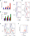
(A) The expression of indicated genes in GFPlo Esamhi and GFPhi Esamlo subsets of splenic CD11b+ DCs sorted from Cx3cr1-GFP mice. Data represent normalized expression values relative to the Esamhi sample as determined by qPCR (mean ± S.D. of triplicate reactions). (B) The expression of indicated genes in the same DC subsets as well as in CD11c CD11b+ F4/80+ GFP+ macrophages (MΦ), determined by qPCR as in (A) and shown on a log scale. (C) Cytokine production by CD11b+ DCs in response to TLR9 ligand CpG. Lymphocyte-depleted splenocytes from Cx3cr1-GFP mice were incubated for 6 hr with CpG and stained for surface markers and intracellular TNF-α or IL-12. Shown are cytokine expression profiles in GFPlo Esamhi or GFPhi Esamlo subsets of CD11chi CD11b+ DCs, or in CD11c CD11b+ GFPhi monocytes and macrophages (Mo-MΦ). Data are representative of three independent experiments. (D) TNF-α production by CD11b+ DCs in response to TLR ligands. Splenocytes from Cx3cr1-GFP mice were activated with CpG or TLR2 ligand heat-killed L.monocytogenes (HKLM) and stained as above. (E) Phenotypic maturation of CD11b+ DCs in vitro. Splenocytes from Cx3cr1-GFP mice were activated with CpG or HKLM and stained for DC markers as above. Shown are staining profiles of CD11b+ DC subsets for CD40, with mean fluorescence intensities indicated. (F) In vivo CD4+ T cell priming in the absence of Esamhi DCs. DC-RbpjΔ or littermate control (Ctrl) mice were administered CFSE-labeled OVA-specific OT-II T cells and immunized with OVA. Shown are CFSE staining profiles of gated OT-II T cells from the recipient spleens 3 days after immunization. The fractions of T cells that did not divide or divided 1–3 and >3 times are indicated (mean of two recipients).
Figure 7
Figure 7. Notch2 controls the development of intestinal CD11b+ CD103+ DCs
(A) DC populations in the intestinal lamina propria from DC-Notch2Δ mice and littermate controls. Shown are staining profiles of CD45+ CD11chi MHC II+ DCs and of the gated CD11b+ DCs, highlighting the CD11b+ CD103+ DC subset. Fractions represent average ± S.D. of 4 animals per group. Statistical significance is indicated as in Fig. 1. (B) Absolute numbers of the indicated DC subsets (mean ± S.D. of 4 animals). (C) Staining profiles of CD11chi MHC II+ DCs from the mesenteric lymph nodes. Fractions represent average ± S.D. of 7 animals per group. (D) Effector T cell populations in the intestine. Lymphocytes were isolated from the LP of small intestine and stained ex vivo for intracellular FoxP3 or activated in vitro with PMA plus ionomycin and stained for intracellular IL-17. Shown are profiles of CD11chi MHC II+ LP DCs and of gated TCRβ+ CD4+ T cells isolated ex vivo or activated in vitro (two pairs of littermates shown; representative of 4 DC-Notch2Δ animals).

References

    1. Bar-On L, Birnberg T, Lewis KL, Edelson BT, Bruder D, Hildner K, Buer J, Murphy KM, Reizis B, Jung S. CX3CR1+ CD8alpha+ dendritic cells are a steady-state population related to plasmacytoid dendritic cells. Proc Natl Acad Sci U S A. 2010;107:14745–14750. - PMC (V体育官网) - PubMed
    1. Barnden MJ, Allison J, Heath WR, Carbone FR. Defective TCR expression in transgenic mice constructed using cDNA-based alpha- and beta-chain genes under the control of heterologous regulatory elements. Immunol Cell Biol. 1998;76:34–40. - VSports注册入口 - PubMed
    1. Bogunovic M, Ginhoux F, Helft J, Shang L, Hashimoto D, Greter M, Liu K, Jakubzick C, Ingersoll MA, Leboeuf M, et al. Origin of the lamina propria dendritic cell network. Immunity. 2009;31:513–525. - PMC - PubMed
    1. Bray SJ. Notch signalling: a simple pathway becomes complex. Nat Rev Mol Cell Biol. 2006;7:678–689. - VSports app下载 - PubMed
    1. Buonamici S, Trimarchi T, Ruocco MG, Reavie L, Cathelin S, Mar BG, Klinakis A, Lukyanov Y, Tseng JC, Sen F, et al. CCR7 signalling as an essential regulator of CNS infiltration in T-cell leukaemia. Nature. 2009;459:1000–1004. - PMC - PubMed

Publication types (V体育平台登录)

MeSH terms

"V体育平台登录" Substances

Associated data (V体育官网)