VSports在线直播 - Skip to main page content
U.S. flag

An official website of the United States government

Dot gov

The . gov means it’s official. Federal government websites often end in . gov or . mil. Before sharing sensitive information, make sure you’re on a federal government site VSports app下载. .

Https

The site is secure. The https:// ensures that you are connecting to the official website and that any information you provide is encrypted and transmitted securely. V体育官网.

. 2011 Oct 21;44(2):290-303.
doi: 10.1016/j.molcel.2011.08.030.

mTOR drives its own activation via SCF(βTrCP)-dependent degradation of the mTOR inhibitor DEPTOR

Affiliations

mTOR drives its own activation via SCF(βTrCP)-dependent degradation of the mTOR inhibitor DEPTOR

V体育官网入口 - Daming Gao et al. Mol Cell. .

"VSports在线直播" Abstract

The activities of both mTORC1 and mTORC2 are negatively regulated by their endogenous inhibitor, DEPTOR VSports手机版. As such, the abundance of DEPTOR is a critical determinant in the activity status of the mTOR network. DEPTOR stability is governed by the 26S-proteasome through a largely unknown mechanism. Here we describe an mTOR-dependent phosphorylation-driven pathway for DEPTOR destruction via SCF(βTrCP). DEPTOR phosphorylation by mTOR in response to growth signals, and in collaboration with casein kinase I (CKI), generates a phosphodegron that binds βTrCP. Failure to degrade DEPTOR through either degron mutation or βTrCP depletion leads to reduced mTOR activity, reduced S6 kinase activity, and activation of autophagy to reduce cell growth. This work expands the current understanding of mTOR regulation by revealing a positive feedback loop involving mTOR and CKI-dependent turnover of its inhibitor, DEPTOR, suggesting that misregulation of the DEPTOR destruction pathway might contribute to aberrant activation of mTOR in disease. .


VSports在线直播 - Conflict of interest statement

Conflict of Interest Statement: J. W. H V体育ios版. is a consultant for Millennium Pharmaceuticals.

Figures
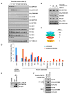
Figure 1
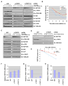
Figure 1. Identification of β-TRCP as a DEPTOR-interacting protein
A–B. Immunoblot (IB) analysis of whole cell lysates derived from T98G cells that were serum-starved for 72 hours and then collected at the indicated time periods following serum re-addition. Where indicated, MG132 was added to block the activity of 26S proteasome. C. Schematic illustration of the recovered DEPTOR-associated proteins. TSCs, total spectral counts. D. Normalized total spectral counts for β-TRCP-associated proteins identified in the presence or absence of bortezomib using CompPASS. In addition to DEPTOR, several known β-TRCP substrates were identified, as well as the USP47 binding protein. E. Immunoblot analysis of WCL and anti-DEPTOR immunoprecipitates (IP) derived from HeLa cells. Mouse IgG was used as a negative control for the IP process. Cells were pretreated with 15 μM MG132 for 10 hours to block the proteasome pathway before harvest. F. Immunoblot analysis of whole cell lysates and immunoprecipitates derived from HeLa cells transfected with HA-DEPTOR and the indicated FLAG-β-TRCP1 constructs. Thirty hours post-transfection, cells were pretreated with 15 μM MG132 for 10 hours to block the proteasome pathway before harvest. (See also Figure S1)
Figure 2
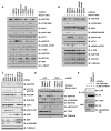
Figure 2. SCFβ-TRCP controls DEPTOR abundance
A. Immunoblot (IB) analysis of whole cell lysates derived from HeLa cells transfected with the indicated siRNA oligos. E2F1 scramble siRNA (SCRAMBLE) was used as a negative control. B–D. Immunoblot analysis of whole cell lysates derived from T98G cells infected with the indicated lentiviral shRNA vectors. The infected cells were selected by 2 μg/ml puromycin for 48 hours before harvesting for immunoblot analysis. E. Immunoblot analysis of whole cell lysates derived from HeLa cells infected with the indicated lentiviral shRNA vectors. The infected cells were selected by 1 μg/ml puromycin and 200 μg/ml hygromycin for 48 hours before harvesting for immunoblot analysis. F. Immunoblot analysis of whole cell lysates derived from HeLa cells infected with the indicated lentiviral shRNA vectors. The infected cells were selected by 2 μg/ml puromycin for 48 hours before harvesting for immunoblot analysis. (See also Figure S2)
Figure 3
Figure 3. SCFβ-TRCP governs mTOR signaling by controlling DEPTOR degradation
A. HeLa cells were infected with the indicated lentiviral shRNA constructs for 24 hours. Uninfected cells were eliminated by selection with 2 μg/ml puromycin for 48 hours. The resulting cell lines were then serum-starved for 24 hours and 10% FBS was added to the serum-starved cells for the indicated time period before harvesting. Equal amounts of whole cell lysates were immunoblotted with the indicated antibodies. B. Quantification of the band intensities in (A). DEPTOR bands were normalized to TUBULIN, then normalized to the t=0 time point. C. T98G cells were infected with the indicated lentiviral shRNA constructs for 24 hours. Uninfected cells were eliminated by selection with 2 μg/ml puromycin for 48 hours. The resulting cell lines were serum-starved for 72 hours and 10% FBS was added to the serum-starved cells for the indicated time period before harvesting. Equal amounts of whole cell lysates were immunoblotted with the indicated antibodies. D. HeLa cells were infected with the indicated lentiviral shRNA constructs for 24 hours. Non-infected cells were eliminated by selection with 2 μg/ml puromycin for 48 hours. The resulting cell lines were serum-starved for 24 hours and 10% FBS together with 20 μg/ml cycloheximide (CHX) was added to the serum-starved cells for the indicated time period before harvesting. Equal amounts of whole cell lysates were immunoblotted with the indicated antibodies. E. Quantification of the band intensities in (D). DEPTOR bands were normalized to VINCULIN, then normalized to the t=0 time point. F–H. Real-time RT-PCR analysis to examine the relative DEPTOR (F), β-TRCP1 (G) and β-TRCP2 (H) mRNA levels in HeLa cells infected with the indicated shRNA lentiviral vectors. Three independent sets of experiments were performed to generate the error bars. The error bars represent means ± SD.
Figure 4
Figure 4. DEPTOR stability is negatively regulated by the mTOR signaling pathway
A–B. Immunoblot (IB) analysis of whole cell lysates derived from T98G cells that were serum-starved for 72 hours and then collected at 16 hours following serum re-addition. Where indicated, proteasome inhibitor MG132 and the indicated kinase inhibitors were added together with serum. DMSO was used as a negative control. C. Immunoblot analysis of whole cell lysates derived from HeLa cells infected with the indicated lentiviral shRNA vectors. The infected cells were selected by 2 μg/ml puromycin for 48 hours before harvesting for immunoblot analysis. D. HeLa cells were infected with the lentiviral shTSC2 construct (with shGFP as a negative control) for 24 hours. Non-infected cells were eliminated by selection with 2 μg/ml puromycin for 48 hours. The resulting cell lines were serum-starved for 24 hours and 10% FBS, together with the indicated kinase inhibitors, were added to the serum-starved cells for 16 hours before harvesting. DMSO was used as a negative control. Equal amounts of whole cell lysates were immunoblotted with the indicated antibodies. E. Immunoblot analysis of whole cell lysates (WCL) and immunoprecipitates (IP) derived from HeLa cells transfected with FLAG-β-TRCP1 and HA-DEPTOR constructs. Thirty hours post-transfection, cells were pretreated with 15 μM MG132 for 10 hours to block the proteasome pathway before harvest. Where indicated, cells were treated with the indicated kinase inhibitors as well for 10 hours before harvest. (See also Figure S3)
Figure 5
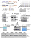
Figure 5. Casein Kinase I controls DEPTOR stability
A. Immunoblot (IB) analysis of whole cell lysates derived from HeLa cells treated with the indicated CKI inhibitor (50 μM D4476 and 50 μM IC261 for 12 hours). B. Immunoblot analysis of whole cell lysates derived from HeLa cells infected with the indicated lentiviral shRNA vectors. The infected cells were selected by 2 μg/ml puromycin for 48 hours before harvesting for immunoblot analysis. C. Immunoblot analysis of whole cell lysates (WCL) and immunoprecipitates (IP) derived from HeLa cells transfected with FLAG-β-TRCP1, GST-CKI and the indicated HA-DEPTOR constructs. Thirty hours post-transfection, cells were pretreated with 15 μM MG132 for 10 hours to block the proteasome pathway before harvest. Where indicated, PP242 or rapamycin was also added for 10 hours to inactivate mTOR kinase before harvest.
Figure 6
Figure 6. mTOR-dependent phosphorylation of DEPTOR triggers its recognition by β-TRCP and promotes DEPTOR turnover
A–B. Schematic representation of the candidate phospho-degron in DEPTOR and the previously reported 13A DEPTOR mutant (Peterson et al., 2009). C. Immunoblot (IB) analysis of whole cell lysates (WCL) and immunoprecipitates (IP) derived from HeLa cells transfected with FLAG-β-TRCP1 and the indicated HA-DEPTOR constructs. Thirty hours post-transfection, cells were pretreated with 15 μM MG132 for 10 hours to block the proteasome pathway before harvest. D. Immunoblot analysis of whole cell lysates derived from HeLa cells that were transfected with the indicated HA-DEPTOR constructs, and serum-starved for 24 hours before being collected at the indicated time period following serum re-addition. E. Immunoblot analysis of whole cell lysates and immunoprecipitates derived from HeLa cells transfected with FLAG-β-TRCP1, GST-CKI and the indicated HA-DEPTOR constructs. Thirty hours post-transfection, cells were pretreated with 15 μM MG132 for 10 hours to block the proteasome pathway before harvest. F. Immunoblot analysis of whole cell lysates and immunoprecipitates derived from HeLa cells transfected with FLAG-β-TRCP1 and the indicated HA-DEPTOR constructs. Thirty hours post-transfection, cells were pretreated with 15 μM MG132 for 10 hours to block the proteasome pathway before harvest. G. Autoradiograms showing the recovery of various 35S-labelled DEPTOR mutant proteins bound to GST-β-TRCP1 (with GST as a negative control) after incubation with mTOR catalytic domain. H. Immunoblot analysis of whole cell lysates derived from HeLa cells that were transfected with the indicated HA-DEPTOR constructs, and serum-starved for 24 hours before being collected at the indicated time period following serum re-addition. I. Immunoblot analysis of whole cell lysates and immunoprecipitates derived from HeLa cells transfected with FLAG-β-TRCP1, GST-CKI and the indicated HA-DEPTOR constructs. Thirty hours post-transfection, cells were pretreated with 15 μM MG132 for 10 hours to block the proteasome pathway before harvest. J. In the first step of kinase assay (labeled as 1st), purified GST-DEPTOR was incubated with mTOR (or kinase reaction buffer as a negative control) in the presence of cold ATP for 30 minutes. In the second step of kinase reaction (labeled as 2nd), the reaction product from the first step was further incubated with CKI kinase and γ-32P-ATP for 30 minutes. To eliminate mTOR activity in the second step kinase reaction, where indicated, mTOR inhibitor PP242 (0.2 μM) was added to the reaction. The kinase reaction products were separated by SDS-PAGE, and phosphorylation was detected by autoradiography. (See also Figure S4)
Figure 7
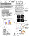
Figure 7. SCFβ-TRCP -mediated DEPTOR destruction participates in the regulation of cell growth and autophagy through modulating the activity of the mTOR signaling pathway
A. HeLa cells were transfected with wild-type DEPTOR and 3A DEPTOR mutant, along with empty vector (EV) as a control, cells were synchronized by serum deprivation (24h), and re-stimulated with serum to enter the cell cycle. Cells were harvested at various times and blotted for the indicated proteins or modifications. B. HeLa cells were infected with the indicated lentiviral shRNA constructs for 24 hours. Non-infected cells were eliminated by selection with 2 μg/ml puromycin for 48 hours. The resulting cell lines were grown in the indicated experimental conditions for 24 hours before harvesting. Equal amounts of whole cell lysates were immunoblotted with the indicated antibodies. C. Immunoblot analysis of whole cell lysates (WCL) derived from the engineered Tet-inducible 3A DEPTOR-expressing HeLa cells (with Tet-EV as a negative control) that were incubated in serum-free medium for 12 hours to induce autophagy. Where indicated, various dose of doxycycline was used to induce the expression of 3A DEPTOR. D. HeLa cells were infected with Hygro-pLKO-shDEPTOR (or hygro-pLKO-EV as a negative control) and then selected with 200 μg/ml Hygromycin for 72 hours to eliminate the non-infected cells. The resulting cell lines were then infected with pBabe-Puro-mCherry-GFP-LC3B retroviral construct and selected with 1 μg/ml puromycin for 48 hours to eliminate the non-infected cells before plating the resulting cells in either regular medium or glucose deprivation medium for 24 hours for confocal microscopy analysis. Scale bars, 10 μm. E. Quantification of the average percentage of the red-only fluorescent puncta, which represents the matured antophagosome shown in (D). For each experimental condition, more than 25 cells with over 800 puncta in total were counted to generate each data point. The error bars represent means ± SD. F. Immunoblot analysis of whole cell lysates derived from T98G cells infected with the indicated lentiviral shRNA vectors. The infected cells were selected by 200 μg/ml huygromycin for 72 hours before harvesting for immunoblot analysis. G. Proposed model for the ubiquitination and destruction of DEPTOR by SCFβ-TRCP in an mTOR and CKI-dependent manner. (See also Figure S5)

Comment in

References

    1. Alessi DR, Pearce LR, Garcia-Martinez JM. New insights into mTOR signaling: mTORC2 and beyond. Sci Signal. 2009;2:pe27. - V体育官网入口 - PubMed
    1. Behrends C, Sowa ME, Gygi SP, Harper JW. Network organization of the human autophagy system. Nature. 2010;466:68–76. - PMC - PubMed
    1. Boehm JS, Hession MT, Bulmer SE, Hahn WC. Transformation of human and murine fibroblasts without viral oncoproteins. Mol Cell Biol. 2005;25:6464–6474. - "VSports在线直播" PMC - PubMed
    1. Cardozo T, Pagano M. The SCF ubiquitin ligase: insights into a molecular machine. Nat Rev Mol Cell Biol. 2004;5:739–751. - PubMed
    1. Choo AY, Kim SG, Vander Heiden MG, Mahoney SJ, Vu H, Yoon SO, Cantley LC, Blenis J. Glucose addiction of TSC null cells is caused by failed mTORC1-dependent balancing of metabolic demand with supply. Mol Cell. 2010;38:487–499. - "V体育平台登录" PMC - PubMed

Publication types

MeSH terms

Substances