"V体育官网" Skip to main page content
U.S. flag

An official website of the United States government

Dot gov

The VSports app下载. gov means it’s official. Federal government websites often end in . gov or . mil. Before sharing sensitive information, make sure you’re on a federal government site. .

Https

The site is secure. The https:// ensures that you are connecting to the official website and that any information you provide is encrypted and transmitted securely. V体育官网.

. 2011 Dec;60(12):3246-55.
doi: 10.2337/db11-0375. Epub 2011 Oct 7.

Early treatment of NOD mice with B7-H4 reduces the incidence of autoimmune diabetes

Affiliations

Early treatment of NOD mice with B7-H4 reduces the incidence of autoimmune diabetes

VSports - Xiaojie Wang et al. Diabetes. 2011 Dec.

V体育平台登录 - Abstract

Objective: Autoimmune diabetes is a T cell-mediated disease in which insulin-producing β-cells are destroyed. Autoreactive T cells play a central role in mediating β-cell destruction. B7-H4 is a negative cosignaling molecule that downregulates T-cell responses. In this study, we aim to determine the role of B7-H4 on regulation of β-cell-specific autoimmune responses VSports手机版. .

Research design and methods: Prediabetic (aged 3 weeks) female NOD mice (group 1, n = 21) were treated with intraperitoneal injections of B7-H4. Ig at 7. 5 mg/kg, with the same amount of mouse IgG (group 2, n = 24), or with no protein injections (group 3, n = 24), every 3 days for 12 weeks. V体育安卓版.

Results: B7-H4. Ig reduced the incidence of autoimmune diabetes, compared with the control groups (diabetic mice 28. 6% of group 1, 66. 7% of group 2 [P = 0. 0081], and 70. 8% of group 3 [group 1 vs. 3, P = 0. 0035]). Histological analysis revealed that B7-H4 treatment did not block islet infiltration but rather suppressed further infiltrates after 9 weeks of treatment (group 1 vs. 2, P = 0. 0003). B7-H4 treatment also reduced T-cell proliferation in response to GAD65 stimulation ex vivo. The reduction of diabetes is not due to inhibition of activated T cells in the periphery but rather to a transient increase of Foxp3(+) CD4(+) T-cell population at one week posttreatment (12. 88 ± 1 V体育ios版. 29 vs. 11. 58 ± 1. 46%; n = 8; P = 0. 03). .

Conclusions: Our data demonstrate the protective role of B7-H4 in the development of autoimmune diabetes, suggesting a potential means of preventing type 1 diabetes by targeting the B7-H4 pathway. VSports最新版本.


Figures

FIG. 1.
FIG. 1.
B7-H4.Ig inhibits NOD T-cell proliferation and cytokine production. Purified CD3+ T cells were stimulated with various concentrations of plate-bound anti-CD3 for 18 h (for early T cell marker CD69 detection) (DG), 48 h (for cytokine detection) (C), or 72 h (for T-cell proliferation) (A and B). B7-H4.Ig or mouse Ig was added at indicated concentrations. For the T-cell proliferation assay, cultures were pulsed with 1 μCi of [3H]-thymidine in the last 18 h. B7-H4.Ig inhibited T-cell proliferation in a dose-dependent manner (P = 0.61, 0.04, 0.02, 0.01, 0.005) (A) (P = 0.01, 0.02, 0.05, 0.03) (B). B7-H4.Ig inhibited IL-2 secretion (P = 0.002) (C) and CD69 expression on CD4+/CD8+ T-cell subsets (P = 0.04, 0.003, 0.03, 0.08) (E) (P = 0.03, 0.003, 0.02) (G). The production of IL-2 was measured by sandwich ELISA using pair antibodies from eBioscience (clone numbers of capture and detection antibodies for IL-2: JES6-1A12 and JES6-5H4). Triplicate wells were harvested and counted. Data represent three independent experiments and are expressed as means ± SEM. * Indicates significant value of P < 0.05. (A high-quality color representation of this figure is available in the online issue.)
FIG. 2.
FIG. 2.
B7-H4.Ig treatment reduces the incidence of autoimmune diabetes. A: Survival curve for mice untreated or treated with B7-H4.Ig or control mouse Ig. Female NOD mice were injected intraperitoneally with B7-H4.Ig (group 1, □, n = 21) or mouse Ig (group 2, ▲, n = 24) at 7.5 mg/kg every 3 days for 12 weeks. Untreated female NOD mice (group 3, •, n = 24) were also included. Systemic administration of B7-H4.Ig significantly suppressed diabetes (group 1 vs. 2, P = 0.0081; group 1 vs. 2, P = 0.0035, by log-rank test). B: Distribution of the incidence of diabetes among three groups. The percentage of onset of diabetes is plotted at 12, 16, 20, 24, 28, 32, and 36 weeks of age. The percentage of diabetes at various time points in groups 1, 2, and 3 is shown in dark gray, gray, and light gray, respectively.
FIG. 3.
FIG. 3.
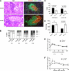
B7-H4.Ig treatment does not block insulitis but alters the aggressiveness of insulitis at later stages. A and B: Representative sections of pancreata from 12-week-old NOD mice treated with B7-H4.Ig (group 1) or mouse Ig (group 2) were stained with hematoxylin and eosin (H&E) and insulin plus CD45 to determine the infiltrate scores and loss of islet cells. Insulitis scores were calculated according to hematoxylin and eosin staining of the pancreata. Scores were given as follows: free of infiltrates, score 1; <25, 25–50, and >50% infiltrates per islet (scores 2, 3, and 4, respectively). C: Inflammation and loss of islets in the pancreata from mice at the age of 16 weeks were compared in groups 1 and 2. Percentages of inflammation and loss of islets were calculated based on the severe intraislet infiltrates and insulin staining, respectively. Both inflammation and loss of islets were significantly lower in B7-H4.Ig treatment group 1 compared with group 2 (inflammation, P = 0.0003; loss of islets, P = 0.0006). D: Total insulin content extracted from each pancreas was detected using a sensitive mouse insulin kit. The pancreas was homogenized in acid-ethanol (0.1 N HCl in 100% ethanol), and the supernatant was diluted in 10 mM Tris-HCl, PH7.5, 1 mM EDTA. The amount of insulin was measured by ELISA kit (Crystal Chem Inc., Chicago, IL) according to the manufacturer's instructions. There was no significant difference in the amount of insulin from group 1 or 2 at 12 weeks of age. However, there was a significant loss in the control group at 16 weeks (P = 0.003). An IPGTT was performed at 12 weeks (E) or 16 weeks (F). IPGTT was performed by intraperitoneal injection of 2 g/kg glucose after 5 h fasting. Blood samples were collected for examining insulin secretion. Blood glucose was tested at the indicated times. A similar blood glucose level was detected in both groups at 12 weeks. By contrast, glucose intolerance was detected in the control group compared with the B7-H4–treated group at 16 weeks. Six to eight animals are included in each group. W, weeks. (A high-quality digital representation of this figure is available in the online issue.)
FIG. 4.
FIG. 4.
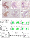
Phenotype of IICs. A: Histology of pancreas from B7-H4.Ig– (group 1) and mouse Ig–treated (group 2) NOD mice at 12–16 weeks of age. Frozen sections were stained with anti-CD4, CD8, or Foxp3 (shown in brown). The number of CD4-, CD8-, or Foxp3-positive cells was blindly counted by microscopy and described as means/field. FACS analysis of IICs (BE). IICs were isolated and stained with anti-CD4, CD8, and CD69, or anti-CD4, CD25, and Foxp3. Representative plots of CD4 vs. CD69, CD8 vs. CD69, and histogram of Foxp3+ gated on CD4+ T-cell subset (B and D). Quantitative data (C and E). F: mRNA expression in IICs. mRNA was isolated from IICs and subjected to real-time PCR analysis. Expression level of mRNA in control group was considered as 1. Total RNA was extracted using RNeasy Mini kits (QIAGEN, Mississauga, ON, Canada). All RNA samples were digested with RNase-free DNase I (QIAGEN). First-strand cDNA was then synthesized from this RNA by Superscript II reverse transcriptase (GIBCO, Burlington, ON, Canada) using oligo(dT). Mouse glyceraldehyde-3-phosphate dehydrogenase mRNA was used as an internal control to confirm equivalent loading of the total RNA. Quantitative real-time PCR was done in duplicate using 25 ng cDNA with 0.4 μmol/L of each primer in a final reaction volume of 20 μL containing 10 μL of 2 SYBR PCR Master Mix (QIAGEN). PCR parameters were as follows: 50°C for 2 min and 95°C for 10 min, followed by 45 cycles of 95°C for 15 s, 55°C for 20 s, and 72°C for 30 s. Relative expression level was expressed as 2–(CTubiquitin–CTgene) (where CT is cycling threshold) with ubiquitin RNA as the endogenous control for normalization. Each group represents four to six mice. Gzmb, granzyme B. G: Representative plots of intracellular staining of IFN-γ among CD4+ or CD8+. H: Expression levels of IFN-γ were generated from (G). Each dot represents one mouse (controls, ▲; B7-H4 treated, □). (A high-quality digital representation of this figure is available in the online issue.)
FIG. 5.
FIG. 5.
B7-H4.Ig transiently upregulates Foxp3+ T cells in the draining lymph nodes. A: Representative plots of Foxp3+CD4+ subsets and pool percentage (B) in two treatment groups at 4 weeks of age in the PLN are shown. Control mouse Ig– and B7-H4.Ig–treated groups are shown in ▲ and □, respectively. Each dot represents one individual mouse; n = 8–11 per group. C: Lymphocytes from PLNs of B7-H4.Ig–treated NOD mice protected against the development of diabetes in NOD SCID mice. A mixture of single cells from PLNs of B7-H4.Ig– or control Ig–treated, 4-week-old NOD mice (10 × 106 cells) and splenic leukocytes from diabetic NOD mice (15 × 106 cells) was adoptively transferred intravenously into NOD SCID recipients. B7-H4.Ig treatment significantly delayed the incidence of diabetes in NOD SCID mice induced by diabetic splenic leukocytes, compared with control Ig treatment (*P = 0.01, B7-H4.Ig vs. control Ig). Lymphocytes from a mixture of 4-week-old NOD mice of either control Ig– (D) or B7-H4.Ig–treated (E) or diabetic NOD mice showed protection against development of hyperglycemia, compared with lymphocytes from diabetic NOD mice alone in the NOD SCID mice (*P = 0.01, mouse Ig vs. diabetic NOD alone; **P = 0.0001, B7-H4.Ig vs. diabetic NOD alone). Data are pooled from two independent experiments, with n = 10 per group. (A high-quality color representation of this figure is available in the online issue.)
FIG. 6.
FIG. 6.
A reduced autoimmune response in the periphery occurs after B7-H4 treatment. A: Purified CD3+ T cells from B7-H4– or mouse Ig–treated (group 1 and group 2, respectively) NOD mice were cultured with irradiated splenocytes and GAD p35 at indicated concentrations for 4 days and pulsed with 1 μCi of [3H]-thymidine in the last 18 h. [3H] incorporation is shown on the y-axis. (▲, group 1; ■, group 2; △, group 1 depleted with anti-CD25; □, group 2 depleted with anti-CD25). B: Purified CD3+ T cells from groups 1 and 2 were cultured with irradiated splenocytes, GAD p35 (20 μg/mL), and 20 u/mL rIL-2 for 4 days and restimulated with phorbol myristic acid and ionomycin for 4 h for intracellular staining of IFN-γ or replated on a 24-well plate precoated with anti-CD3 (1 μg/mL) for cytokine detection. Supernatants were collected at 48 h and subjected to analysis for cytokine production. The production of IFN-γ and IL-10 protein was measured by sandwich ELISA using pair antibodies from eBioscience (clone numbers of capture and detection antibodies for IFN-γ: XMG1.2 and R4-6A2; for IL-10: JES5-16E3 and JES5-2A5). Representative plots and quantitative analysis of IFN-γ expression (CE). Each dot represents one individual mouse at the age of 8–12 weeks; n = 5–9 per group. (A high-quality color representation of this figure is available in the online issue.)

VSports最新版本 - References

    1. Tisch R, McDevitt H. Insulin-dependent diabetes mellitus. Cell 1996;85:291–297 - PubMed
    1. Solomon M, Sarvetnick N. The pathogenesis of diabetes in the NOD mouse. Adv Immunol 2004;84:239–264 - PubMed
    1. Miyazaki A, Hanafusa T, Yamada K, et al. Predominance of T lymphocytes in pancreatic islets and spleen of pre-diabetic non-obese diabetic (NOD) mice: a longitudinal study. Clin Exp Immunol 1985;60:622–630 - V体育安卓版 - PMC - PubMed
    1. Gorsuch AN, Spencer KM, Lister J, et al. Evidence for a long prediabetic period in type I (insulin-dependent) diabetes mellitus. Lancet 1981;2:1363–1365 - "VSports" PubMed
    1. Anderson MS, Bluestone JA. The NOD mouse: a model of immune dysregulation. Annu Rev Immunol 2005;23:447–485 - "VSports" PubMed

Publication types

MeSH terms

VSports最新版本 - Substances