Skip to main page content
U.S. flag

An official website of the United States government

Dot gov

The . gov means it’s official. Federal government websites often end in VSports app下载. gov or . mil. Before sharing sensitive information, make sure you’re on a federal government site. .

Https

The site is secure V体育官网. The https:// ensures that you are connecting to the official website and that any information you provide is encrypted and transmitted securely. .

. 2011 Nov 15;71(22):7091-102.
doi: 10.1158/0008-5472.CAN-11-1367. Epub 2011 Sep 26.

AGR2 is a novel surface antigen that promotes the dissemination of pancreatic cancer cells through regulation of cathepsins B and D

Affiliations

AGR2 is a novel surface antigen that promotes the dissemination of pancreatic cancer cells through regulation of cathepsins B and D

Laurent Dumartin et al. Cancer Res. .

Abstract

Pancreatic ductal adenocarcinoma (PDAC) remains one of the most lethal cancers largely due to disseminated disease at the time of presentation. Here, we investigated the role and mechanism of action of the metastasis-associated protein anterior gradient 2 (AGR2) in the pathogenesis of pancreatic cancer. AGR2 was induced in all sporadic and familial pancreatic intraepithelial precursor lesions (PanIN), PDACs, circulating tumor cells, and metastases studied. Confocal microscopy and flow cytometric analyses indicated that AGR2 localized to the endoplasmic reticulum (ER) and the external surface of tumor cells. Furthermore, induction of AGR2 in tumor cells regulated the expression of several ER chaperones (PDI, CALU, RCN1), proteins of the ubiquitin-proteasome degradation pathway (HIP2, PSMB2, PSMA3, PSMC3, and PSMB4), and lysosomal proteases [cathepsin B (CTSB) and cathepsin D (CTSD)], in addition to promoting the secretion of the precursor form pro-CTSD. Importantly, the invasiveness of pancreatic cancer cells was proportional to the level of AGR2 expression. Functional downstream targets of the proinvasive activity of AGR2 included CTSB and CTSD in vitro, and AGR2, CTSB, and CTSD were essential for the dissemination of pancreatic cancer cells in vivo. Taken together, the results suggest that AGR2 promotes dissemination of pancreatic cancer and that its cell surface targeting may permit new strategies for early detection as well as therapeutic management VSports手机版. .


Conflict of interest statement

Disclosure of Potential Conflicts of Interest

No potential conflicts of interest were disclosed.

Figures

Figure 1
Figure 1
AGR2 protein expression in pancreatic tissues. Immunohistochemical analysis of AGR2 expression in normal pancreas (A), familial PanIN1, PanIN2, PanIN3, and PDAC (B–E, respectively); perineural invasion, circulating tumor cells, lymph node, and liver metastasis from sporadic pancreatic cancer samples (F–I, respectively). Scale bars, 50 μm.
Figure 2
Figure 2
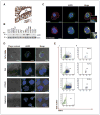
Localization of AGR2 in pancreatic cancer cells. A, immunohistochemical analysis of AGR2 in a representative human PDAC sample showing both cytoplasmic and membranous (arrows) immunoreactivity. B, Western blot analysis showing AGR2 expression in a panel of pancreatic cancer and normal HPDE cells. C, coimmunostaining of AGR2 (green) and actin or calreticulin (red) in permeabilized PaTu 8988s cells showing that AGR2 is situated in the endoplasmic reticulum. D, cell surface immunostaining for AGR2 (green) on nonpermeabilized PaTu 8988s cells. Scale bars, 10 μm. E, i, flow cytometric analysis of AGR2 cell surface expression on intact pancreatic cells in the same order (as in D) and (ii) sorting of AGR2-positive–gated cells. MiaPaCa2 cells that do not endogenously express AGR2 were used as a negative control.
Figure 3
Figure 3
Functional roles of AGR2. A, Western blot analysis of AGR2 expression in (i) MiaPaCa2 cells stably transfected with empty vector control (pCEP) or pCEP-AGR2 vector and (ii) in FA6 cells 48 hours after transfection with specific AGR2 siRNA or nontargeting (NT) control siRNA. Actin was used as a loading control. B, invasion assays using (i) stably transfected MiaPaCa2 cells and (ii) siRNA-transfected FA6 cells. Mean values of triplicate experiments are shown. *, P < 0.05.
Figure 4
Figure 4
Proteomic profiling of MiaPaCa2 AGR2-expressing cells. A, Representative gel image displaying protein spots with AGR2-dependent changes in expression is shown. Magnified regions showing several differentially expressed proteins. CALU, calumenin; RCN1, reticulocalbin 1.
Figure 5
Figure 5
CTSB and CTSD are functional downstream targets of AGR2. A, Western blot analysis (i) and densitometry quantification (ii) showing increased expression of precursor and mature CTSB and CTSD in MiaPaCa2-pCEP4 and pCEP4 AGR2 lysates. Actin was used as a loading control. III, semiquantitative real-time PCR analysis of CTSB and CTSD gene expression in MiaPaCa2-pCEP4 and pCEP4 AGR2 showing no change in their transcript levels. S16 was used as a reference gene. B, Western blot (i and ii) and densitometry (iii and iv) showing increased protein levels of precursor and mature CTSB and CTSD in FA6 pancreatic cancer cells in comparison with HPDE cells and in metastatic PaTu 8988s in comparison with nonmetastatic PaTu 8988t. C, levels of pro-CTSD in the culture supernatant of MiaPaCa2-pCEP4 and pCEP4 AGR2 and in FA6 cells 48 hours after transfection with AGR2 or nontargeting (NT) siRNA. D, after siRNA silencing of CTSB and CTSD for 48 hours (i and iii), decreased invasion of MiaPaCa2-pCEP4 AGR2 cells (ii) and FA6 cells (iv) was seen. Mean values of triplicate experiments are shown. *, P < 0.05; **, P < 0.005; ***, P < 0.0001.
Figure 6
Figure 6
AGR2 and CTSB/CTSD are major regulators of pancreatic cancer cell dissemination in vivo. A, fifty to 100 PaTu 8988s cells stained with CMTMR or CMFDA dyes were injected into the yolk sack of 48-hour-old embryos and assessed by epifluorescence microscopy (i) of the whole embryo and confocal microscopy (ii) of the injection area. B, twenty-four hours after injection, disseminated cancer cells were observed in the tail of the embryo (i) or in the yolk sack (ii). Elongated cells imaged by confocal microscopy in the tail (iii) and the yolk sack (iv) of the embryo. Scale bars, 50 μm. C, labeled PaTu 8988s cells transfected with AGR2 or nontargeting (NT) siRNA (i) were assessed by epifluorescence microscopy 24 hours after injection. The number of disseminated cells was statistically significantly decreased after AGR2 silencing (ii and iii). D, representative image of decreased tumor cell dissemination in zebrafish tail after AGR2 silencing (i). The enlarged respective images of the 2 marked areas in tail are shown on ii and iii. E and F, after silencing of CTSB/CTSD (i), a significant decrease in the number of disseminated cells was also seen (ii and iii), respectively. **, P < 0.005; ***, P < 0.0001.

References

    1. Jemal A, Siegel R, Xu J, Ward E. Cancer statistics, 2010. CA Cancer J Clin. 2010;60:277–300. - "V体育官网" PubMed
    1. Barraclough DL, Platt-Higgins A, de Silva Rudland S, Barraclough R, Winstanley J, West CR, et al. The metastasis-associated anterior gradient 2 protein is correlated with poor survival of breast cancer patients. Am J Pathol. 2009;175:1848–57. - PMC - PubMed
    1. Liu D, Rudland PS, Sibson DR, Platt-Higgins A, Barraclough R. Human homologue of cement gland protein, a novel metastasis inducer associated with breast carcinomas. Cancer Res. 2005;65:3796–805. - PubMed
    1. Fritzsche FR, Dahl E, Dankof A, Burkhardt M, Pahl S, Petersen I, et al. Expression of AGR2 in non small cell lung cancer. Histol Histopathol. 2007;22:703–8. - PubMed
    1. Park K, Chung YJ, So H, Kim K, Park J, Oh M, et al. AGR2, a mucinous ovarian cancer marker, promotes cell proliferation and migration. Exp Mol Med. 2011;43:91–100. - PMC - PubMed

Publication types

MeSH terms