VSports手机版 - Skip to main page content
U.S. flag

An official website of the United States government

Dot gov

The . gov means it’s official. Federal government websites often end in . gov or . mil VSports app下载. Before sharing sensitive information, make sure you’re on a federal government site. .

Https

The site is secure. The https:// ensures that you are connecting to the official website and that any information you provide is encrypted and transmitted securely. V体育官网.

. 2011 Sep 11;13(10):1214-23.
doi: 10.1038/ncb2332.

Midbody accumulation through evasion of autophagy contributes to cellular reprogramming and tumorigenicity

Affiliations

Midbody accumulation through evasion of autophagy contributes to cellular reprogramming and tumorigenicity

Tse-Chun Kuo et al. Nat Cell Biol. .

"VSports" Erratum in

  • Nat Cell Biol. 2011 Dec;13(12):1467

Abstract

The midbody is a singular organelle formed between daughter cells during cytokinesis and required for their final separation. Midbodies persist in cells long after division as midbody derivatives (MB(d)s), but their fate is unclear. Here we show that MB(d)s are inherited asymmetrically by the daughter cell with the older centrosome. They selectively accumulate in stem cells, induced pluripotent stem cells and potential cancer 'stem cells' in vivo and in vitro. MB(d) loss accompanies stem-cell differentiation, and involves autophagic degradation mediated by binding of the autophagic receptor NBR1 to the midbody protein CEP55. Differentiating cells and normal dividing cells do not accumulate MB(d)s and possess high autophagic activity. Stem cells and cancer cells accumulate MB(d)s by evading autophagosome encapsulation and exhibit low autophagic activity. MB(d) enrichment enhances reprogramming to induced pluripotent stem cells and increases the in vitro tumorigenicity of cancer cells. These results indicate unexpected roles for MB(d)s in stem cells and cancer 'stem cells' VSports手机版. .


Figures
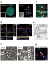
Figure 1
Figure 1
MBds accumulate within cells. (a, b) Multiple MBds associate with a PC3 cell (a) and a B-lymphoblast (b). Insets (a) MBd labeling and (b) merged phase-contrast image with MBd labeling to show cell boundaries. MKLP1, MBd marker (a, b; red); CD44, membrane (a; green); DAPI, DNA (a; blue). Bar, 5 μm (a) and 2 μm (b). (c, d) Three-dimensional reconstruction of polarized cells in a monolayer (c) and a HeLa cell (d) show intracellular MBds. (c) ZO-1, tight junction; MKLP1, MBds. Bar, 2 μm. Enlargement (c, bottom) of box (c, top) shows five MBds (arrows). (d) Wheat germ agglutinin, plasma membrane (red); MKLP1-GFP, MBds (green); DAPI, DNA (blue). Bar, 5 μm. (e) Electron micrograph of a MBd in a permeabilized MCF-7 cell showing immungold labeling with MKLP1 antibodies. Inset, lower magnification of the MBd (boxed) in cell; nucleus, right. Bar, 200 nm. (f) Time-lapse images during extracellular trypsin treatment of HeLa cells show retention of most MBds (MKLP1-GFP, red). Two MBds (yellow arrows) are lost upon treatment, suggesting digestion and/or dissociation. Time (hr:min) post-trypsin. Bar, 5 μm. (g) Two-day co-cultures of HeLa cell expressing either MKLP1-GFP (MBd marker) or cytosolic RFP. Green MBds (arrows) associated with red cells (asterisk) indicate post-mitotic transfer of MBds between cells. Bar, 10 μm.
Figure 2
Figure 2
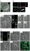
MBds are preferentially inherited by the cell with the older centrosome. (a) CETN1-GFP signal is brighter in upper centrosome/spindle pole of a mitotic spindle. The merged DIC image with CETN1-GFP labeling at two centrosomes shows metaphase chromosome. Insets (lower left, upper right), enlargement and semi-quantitative integrated intensity profile of centrioles. Bar, 5 μm. (b) The brighter CETN1-GFP signal represents the older centrosome as it co-stains more intensely for hCenexin1 and remains more intense throughout cell division (supplementary information, Fig. S1a). Bar, 5 μm. Lower left, merge. (c, d) Time-lapse images show that the mitotic MB is preferentially inherited by the daughter cell with the older centrosome in HeLa cells (c) and hESCs (d). Cells were imaged at the indicated times (hr:min) from telophase by phase-contrast microscopy (c) and from metaphase by DIC microscopy (d). Middle panel of (c) and left panel of (d), CETN1-GFP at centrosomes; enlargements and integrated intensity profiles show the daughter cell having the older centrosome (c, upper; d, lower) inherits the MBd (Time-lapse images: 9:59 in c; lower right image in d). Mitotic MB and MBds (c, d; arrows). MKLP1, MBd marker (red); α-tubulin, mitotic MB and cell boundary marker (green); DAPI, DNA (blue). Bars, 10 μm (c, d).
Figure 3
Figure 3
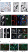
MBds accumulate in stem cells in vivo and in vitro. (a) Histological section through mouse seminiferous tubules labeled for MKLP1 shows several MKLP1+ puncta in cells of the basal layer where stem cells reside. Bar, 20 μm. Inset, enlargement of the cell (arrow) (b, c) Electron micrographs of mitotic MB (b, arrow) and multiple MB-like structures in interphase cells with similar shape and size in a juxtanuclear position (c, arrows) in basal cells of mouse seminiferous tubules. N, nucleus. Bars, 1 μm. (d) Representative planes of a neural progenitor cell in the ventricular zone (Sox2+, left-bottom panel) of an E13.5 mouse brain show that an intracellular MBd (asterisk) is associated with the ventricle-facing daughter in the asymmetrically dividing cell (top row). The bottom row emphasizes the position of paired chromosomes in a dividing anaphase cell. CD133, MB/MBd marker (green); Na-K-ATPase, cell-border marker (red); DRAQ5, DNA (blue); DAPI, DNA. Ventricle (V). Bar, 5 μm. Note that abscission occurs apically in these cells. (e) A histological section through a hair follicle (left, phase-contrast microscopy) stained for the stem cell marker keratin 15 to identify the bulge region (dotted box), the stem cell niche. DNA stain (DAPI) and the phase-contrast image show full follicle architecture. (f) Upper panels show MBd-accumulating cells in the bulge region (boxed) colabeled with K15 and MKLP1. Enlargements (lower panels) of the boxed region highlight a cell with four MBds (asterisks). N, nucleus. Bar, 5 μm. (g-i) Quantitative analysis and representative images show a decrease in MBd-accumulating cells upon the differentiation of pluripotent stem cells (g, H1-OGN) to fibroblast-like cells (h, dH1f), and an increase in MBd-accumulating cells after reprogramming differentiated cells (h) to induced pluripotent stem cells (i, dH1f-iPS). (g-i) numbers refer to mean ± s.d., n=3. MKLP1, MBds; ZO-1, tight junctions; α-tubulin, microtubules; Aurora B, MBs. Bar, 10 μm.
Figure 4
Figure 4
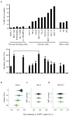
MBd-accumulation is high in stem cells and subpopulations of cancer cells and does not correlate with cell doubling time. (a) Percent of cells that accumulate MBds (>1) in a range of different cell types, as indicated. Below, doubling-times of representative cell lines aligned with MBd-accumulation data. Data are presented as mean ± s.d.; Cell lines are examined in triplicate (MCF-10A, DLD-1, MCF-10AT, MCF-7, H1, and H9), or quadruplicate (e.v. B6 MEFs, HeLa, SAOS-2, and MCF-10CA1a), except hRPE-1 (n=6), U2OS (n=7) and NCC-IT (n=8). Horizontal line, cell lines with different MBd-accumulation potential (14-fold) but similar doubling time. (b) Cells pulse-chased with EdU show a decrease in EdU intensity (x-axis) over time (y-axis), reflecting dilution of dye after cell divisions. (c, d) After a 96-hr chase period, EdU levels were compared between cells with MBd numbers of >1, 1, and 0 (y-axis) in HeLa (c) and SAOS-2 cells (d). In both cases, no significant differences were noted (c, p=0.2101; d, p=0.5609, one-way ANOVA, with at least 800 cells analyzed for each experiment, n=3), indicating similar cycling rates among different subpopulations of cells. (b-d) Each graph is a representative experiment. Cells analyzed shown by green points, median depicted by vertical red lines, and horizontal red lines with ticks illustrate the interquartile range.
Figure 5
Figure 5
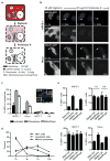
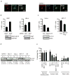
MBds in stem and cancer cells evade membrane encapsulation and lysosomal degradation. (a) Depiction of fluorescence protease protection (FPP) assay. Digitonin selectively permeabilizes the plasma membrane but not internal membranes. Proteinase K degrades cytoplasmic components but membranous compartments remain intact. Under these conditions, MKLP1-GFP-labeled MBds (blue circle) in the cytoplasm will be degraded whereas those inside membrane-bound compartments (MBCs) will not. (b) MBds in MBd-poor hRPE-1 cells are largely protected (~90% in membranous compartments, cells analyzed=10), whereas most MBds in HeLa cells are not (~27%, cells analyzed: 11), and are thus degraded in cytoplasm. Bar, 5 μm. (c) Graph depicting the presence of MBds in lysosomes upon chloroquine or E64d/pepstatin A (E64d/PepA) inhibition in hRPE-1 and HeLa cells, but not in MCF-7 and H9 hESCs. Chloroquine treatment of H9 hESCs is not included as it caused differentiation and cell death. A representative image of hRPE-1 cells inhibited by chloroquine is shown depicting two MBds inside lysosomes. MKLP1 and LAMP2 are used as MBd (red) and lysosome (green) markers, respectively. DAPI, DNA (blue). n=100 MBds/treatment in each of the biological triplicates. Bar, 5 μm. (d) Graph showing the percent of MBd+ cells (MBd levels), the percent of MBds within lysosomes, and the percent of cells exiting cytokinesis following synchronization. MKLP1 and LAMP2 are used as markers as in (c). Note that MBds are transferred into only one of the two nascent daughter cells after abscission (Fig. 2d), so a 50% maximum will be expected for MBd+ cells. The peak of MBds transferred to cells is 3 hours after plating followed by a peak of MBds entering lysosomes at 7 hours. (e) Both chloroquine and E64d/PepA treatments increase the percent of MBd+ cells in hRPE-1 cells and HeLa cells (chloroquine: p=0.0021 and p=0.0187, respectively; E64d/PepA: p=0.0022 and p=0.0043, respectively; n=3 for all experiments). In contrast, lysosomal inhibition has no detectable effect on hESCs (H1, H9) and MCF-7 cancer cells. Data are presented as mean ± s.d. (c-e), except mean ± s.e.m. in hESCs (e).
Figure 6
Figure 6
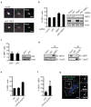
Autophagy controls intracellular MBd levels. (a) Single-plane confocal images of MBds within LC3-positive autophagosomes in MEFs expressing GFP-LC3 (left) and in hRPE-1 cells stained for endogenous LC3 (right). MBd markers: Cep55, MKLP1, or mgcRACGAP. Autophagosomes: GFP-LC3 or LC3. Note that MKLP1 (blue) and mgcRACGAP (red) are co-localized (magenta) in the autophagosome (green), suggesting that MBds are sorted into autophagosomes. Bars, 2 μm. (b) Decreasing autophagy levels by deletion of Atg5 gene (left, MEFs) or depletion of Atg7 by siRNA (right, HeLa) significantly increases the percent of MBd+ cells (p=0.0019 and p=0.021, respectively, n=3). Immunoblots confirm loss of the Atg5-Atg12 conjugation in mutant cells and depletion of Atg7 (asterisk). (c) Rapamycin (Rapa) and lithium chloride (LiCl) co-treatment induces autophagy and decreases the percent of MBd+ cells (left, HeLa; p=0.0056, n=3). Immunoblots showing increased LC3-II levels confirm autophagy induction. Induction of autophagy by over-expression of Flag-tagged BECN1 reduces the percent of MBd+ cells (right, MCF-7; p=0.0008, n=4) (d) Representative immunoblots showing high autophagy levels in normal cells and low levels in stem cells and cancer cells. Autophagic flux (autophagic activity) was measured by changes in the levels of LC3-II, in the presence or absence of lysosomal inhibitors E64d/PepA. U, uninhibited. I, inhibited. Below, the average of the percent change in LC3-II levels after lysosomal inhibition from 3 experiments. α-tubulin, loading control. (e) Quantification of autophagic flux from 3 experiments in different cell lines. Normal dividing cells (MBd-poor) typically have high autophagic flux, whereas stem and cancer cells (MBd-rich) have low autophagic flux. The data are presented as mean ± s.d. (b-e).
Figure 7
Figure 7
NBR1 is a receptor for targeting MBds to the autophagy pathway. (a) Single-plane confocal images showing co-localization of the MBd and the autophagic receptor, NBR1, in U2OS cells and p62-deleted MEFs. MBd markers: MKLP1 or Cep55. Bar, 2 μm. (b) The percent of MBd+ cells is significantly increased following the depletion of NBR1 (p=0.022, n=3), but not another autophagic receptor, p62. Co-depletion of NBR1 and p62 does not further increase MBd levels over NBR1 depletion alone. (c) Deletion of the p62 gene does not affect the percent of MBd+ cells. For (b) and (c), immunoblots verify protein loss. (d) Co-immunoprecipitation reveals Cep55 and NBR1 form a complex. Precipitated proteins and 5% of the input material (Input) were analyzed by immunoblotting with antibodies against NBR1 or Cep55. (e-g) Over-expression of CEP55-EGFP increases the percent of MBd+ cells (e; p=0.0007, n=3) and the percent of NBR1-negative MBds (f; p=0.0568, n=3), presumably by sequestering NBR1 (red) away from MBds in cells expressing CEP55-EGFP (green) as shown in (g), and consequently preventing MBd degradation. The dotted box in (g) is enlarged (top right panel), and the labeling of NBR1 and CEP55-EGFP (middle and bottom right panel) are also presented. DAPI, DNA (blue). Bar, 5 μm. The data are presented as mean ± s.d. (b, c, e, and f).
Figure 8
Figure 8
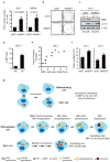
MBd enrichment increases reprogramming efficiency and enhances in vitro tumorigenicity. (a-c) Reprogramming is more efficient after MBd enrichment. Differentiated cells (dH1f) and embryonic fibroblasts (IMR90) are reprogrammed after stable expression of either NBR1-specific shRNA (shNBR1) or non-targeting shRNA (shNT). Emerging iPSC colonies are scored based on Tra-1-60 expression. (a, b) Cells depleted of NBR1 to increase MBd levels show an increase in iPSC colony formation (a, dH1f: 3.1±0.5-fold, n=15, p=0.00035; IMR90: 3.4±0.8-fold, n=3, p=0.02; data are mean ± s.e.m.) but insignificant changes in autophagic activity (c) over shNT control. (b) Representative plates with Tra-1-60-immunostained iPSC colonies. Immunoblot (c, top) and densitometry (c, bottom; percent of autophagic flux) show representative result (n=3); α-tubulin, loading control. (d) MCF-7 side-population (SP) cells have a significantly higher percentage of MBd+ cells over the non-SP population (MP; p=0.0015, n=3; data are mean ± s.d.). (e, f) MBd enrichment in cancer cells leads to increased anchorage-independent growth. MKLP1-GFP-expressing HeLa cells are separated into “MBd high” and “MBd low” subpopulations. An increase in the “MBd high” over “MBd low” ratio is associated with an increase in soft-agar colony formation (e). No significant difference was observed when the enrichment of MBd high subpopulation was less than 3-fold. More soft-agar colonies are formed when MBds are enriched by NBR1-depletion (shNBR1) in HeLa (f, left; p=0.0012, n=3) and mouse 134-4 cells (f, right; p=0.0086, n=3); control, shNT. Data are mean ± s.d., and the colony number (e, f) is the sum of INT-violet-stained colonies from 10 random fields. (g) Model for MBd fate in cells. The newly-formed MBd is preferentially inherited by the daughter cell with the older centrosome (top panel). The inherited MBd (black ring) is recognized by binding of the NBR1 autophagic receptor (grey circle) with the MB protein Cep55 (magenta). The MBd is then encapsulated by the autophagosome (yellow circle), and degraded after fusion of autophagosome and lysosome (red circle) in differentiated cells. This pathway prevents MBd-accumulation. In contrast, stem cells efficiently accumulate MBds through successive divisions and evasion of NBR1-mediated autophagy. Additionally, differentiated and stem cells possess overall high and low autophagic activity, respectively.

Comment in

References

    1. Eggert US, Mitchison TJ, Field CM. Animal cytokinesis: from parts list to mechanisms. Annu Rev Biochem. 2006;75:543–66. - PubMed
    1. Neumüller RA, Knoblich JA. Dividing cellular asymmetry: asymmetric cell division and its implications for stem cells and cancer. Genes Dev. 2009;23:2675–99. - "VSports在线直播" PMC - PubMed
    1. Doxsey S, McCollum D, Theurkauf W. Centrosomes in cellular regulation. Annu Rev Cell Dev Biol. 2005;21:411–34. - VSports app下载 - PubMed
    1. Yamashita YM, Mahowald AP, Perlin JR, Fuller MT. Asymmetric inheritance of mother versus daughter centrosome in stem cell division. Science. 2007;315:518–21. - PMC - PubMed
    1. Wang X, et al. Asymmetric centrosome inheritance maintains neural progenitors in the neocortex. Nature. 2009;461:947–55. - PMC (V体育官网) - PubMed

"V体育平台登录" Publication types

MeSH terms