Single-molecule detection of H₂O₂ mediating angiogenic redox signaling on fluorescent single-walled carbon nanotube array
- PMID: 21899329
- PMCID: PMC3850173
- DOI: 10.1021/nn201904t
Single-molecule detection of H₂O₂ mediating angiogenic redox signaling on fluorescent single-walled carbon nanotube array
Abstract
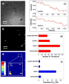
Reactive oxygen species, specifically hydrogen peroxide (H(2)O(2)), activate signal transduction pathways during angiogenesis and therefore play an important role in physiological development as well as various pathophysiologies. Herein, we utilize a near-infrared fluorescent single-walled carbon nanotube (SWNT) sensor array to measure the single-molecule efflux of H(2)O(2) from human umbilical vein endothelial cells (HUVEC) in response to angiogenic stimulation. Two angiogenic agents were investigated: the pro-angiogenic cytokine, vascular endothelial growth factor A (VEGF-A) and the recently identified inorganic pro-angiogenic factor, europium(III) hydroxide in nanorod form. The nanosensor array consists ofa SWNT embedded within a collagen matrix that exhibits high selectivity and sensitivity to single molecules of H(2)O(2). A calibration from 12. 5 to 400 nM quantifies the production of H(2)O(2) at nanomolar concentration in HUVEC with 1 s temporal and 300 nm spatial resolutions VSports手机版. We find that the production of H(2)O(2) following VEGF stimulation is elevated outside of HUVEC, but not for stimulation via nanorods, while increased generation is observed in the cytoplasm for both cases, suggesting two distinct signaling pathways. .
Figures


References
-
- Folkman J. Angiogenesis in Cancer, Vascular, Rheumatoid and Other Disease. Nature Medicine. 1995;1:27–31. - PubMed
-
- Risau W. Mechanisms of angiogenesis. Nature. 1997;386:671–674. - PubMed
-
- Basu S, et al. The neurotransmitter dopamine inhibits angiogenesis induced by vascular permeability factor/vascular endothelial growth factor. Nature Medicine. 2001;7:569–574. - PubMed
-
- Cross MJ, Dixelius J, Matsumoto T, Claesson-Welsh L. VEGF-receptor signal transduction. Trends in Biochemical Sciences. 2003;28:488–494. - PubMed
-
- Coultas L, Chawengsaksophak K, Rossant J. Endothelial cells and VEGF in vascular development. Nature. 2005;438:937–945. - PubMed
Publication types
- "VSports最新版本" Actions
MeSH terms
- "V体育官网" Actions
- Actions (V体育2025版)
- V体育官网 - Actions
- Actions (VSports)
- Actions (V体育官网入口)
- Actions (VSports手机版)
- "V体育2025版" Actions
- Actions (VSports手机版)
- Actions (VSports手机版)
- Actions (V体育ios版)
- "VSports最新版本" Actions
- "VSports手机版" Actions
- VSports app下载 - Actions
V体育官网 - Substances
- "VSports手机版" Actions
Grants and funding
LinkOut - more resources
VSports - Full Text Sources
Other Literature Sources

