"VSports app下载" Skip to main page content
U.S. flag

An official website of the United States government

Dot gov

The . gov means it’s official. Federal government websites often end in . gov or . mil. Before sharing sensitive information, make sure you’re on a federal government site VSports app下载. .

Https

The site is secure V体育官网. The https:// ensures that you are connecting to the official website and that any information you provide is encrypted and transmitted securely. .

. 2011 Aug 26;35(2):299-311.
doi: 10.1016/j.immuni.2011.08.007.

V体育安卓版 - Genome-wide analyses of transcription factor GATA3-mediated gene regulation in distinct T cell types

Affiliations

Genome-wide analyses of transcription factor GATA3-mediated gene regulation in distinct T cell types

Gang Wei (V体育官网) et al. Immunity. .

Abstract

The transcription factor GATA3 plays an essential role during T cell development and T helper 2 (Th2) cell differentiation VSports手机版. To understand GATA3-mediated gene regulation, we identified genome-wide GATA3 binding sites in ten well-defined developmental and effector T lymphocyte lineages. In the thymus, GATA3 directly regulated many critical factors, including Th-POK, Notch1, and T cell receptor subunits. In the periphery, GATA3 induced a large number of Th2 cell-specific as well as Th2 cell-nonspecific genes, including several transcription factors. Our data also indicate that GATA3 regulates both active and repressive histone modifications of many target genes at their regulatory elements near GATA3 binding sites. Overall, although GATA3 binding exhibited both shared and cell-specific patterns among various T cell lineages, many genes were either positively or negatively regulated by GATA3 in a cell type-specific manner, suggesting that GATA3-mediated gene regulation depends strongly on cofactors existing in different T cells. .


Figures

Figure 1
Figure 1. Genome-wide GATA3 binding patterns in various T cell lineages
A. Cartoon showing various development and differentiation stages of T cells analyzed in this study. GATA3-mediated steps are highlighted in red. B. Overlaps of GATA3 binding sites in various T cell types. The number of binding sites identified in each cell type is indicated on the right of the heatmap. Number within each cell (i, j) in the heatmap and the corresponding intensity indicate the percentage of sites in cell type i (row) overlapping those in cell type j (column). The cluster pattern shown on the left was generated based on the overall binding similarity. C. GATA3 binding pattern at the genomic region containing Ctla4 and Icos genes. Blue arrowheads indicate the peaks identified by MACS with a P value of ≤10−13. D. Overlap of GATA3-bound genes in Th1, Th2, Th17 and iTreg cells. GATA3 was considered associated with a gene if a MACS peak was located anywhere from 10 kb upstream of transcription start site to 3 kb downstream of transcription end site.
Figure 2
Figure 2. GATA3 regulates distinct transcription programs in different cells
A. Number of genes that undergo change in expression and are bound by GATA3. “Affected genes” indicates all genes that show changed expression in a specific T cell stage-lineage by deleting the Gata3 gene. “P-reg” indicates the genes that are positively regulated by GATA3. “P & bound” indicates the genes that are positively regulated and bound by GATA3. “N-reg” indicates the genes that are negatively regulated by GATA3. “N & bound” indicates the genes that are negatively regulated and bound by GATA3. B. Deletion of Gata3 results in changes in expression of cell-specific genes. The expression pattern of a set of genes characteristic of each cell type (indicated on the right) is shown. Heatmap intensity denotes the normalized expression (log2 RPKM values). C. Overlap of GATA3-regulated genes among all Th cells. D. Overlap of GATA3-regulated genes among both DP subsets and Th2 cells. Genes regulated by GATA3 in each cell type were determined by the overlaps of two independent biological replicates.
Figure 3
Figure 3. GATA3 binding is associated with primary and various secondary motifs
A. Motif enrichment analysis of GATA3 binding sites in various T cells. The co-occurring motifs associated with the primary WGATAA motif in the GATA3 binding sites were also analyzed and the top two secondary motifs (indicated at the bottom) from each cell were displayed. B. Genome Browser image showing that Fli1 but not Ets1 co-localized with GATA3 at the Th2 cytokine locus in Th2 cells. C. Genome-wide co-localization of GATA3 and Fli1 binding sites in Th2 cells. D. Genome Browser image showing that Gata3 deletion abrogated Fli1 binding to the Th2 cytokine locus in CD8+ T cells.
Figure 4
Figure 4. GATA3 regulates critical components of signaling and transcription during T cell development in the thymus
A–C. Genome Browser image showing that the Thpok gene (zbtb7b) (A), the Notch1 gene (B) and Cd3d and Cd3e (C) were bound by GATA3 in DN, DP and naïve CD4+ T cells (left panel). Gata3 deletion abolished their expression in CD69+CD3hiCD4+CD8int-hi DP cells (right panel). D. Gata3 deletion resulted in a decrease of surface T cell receptor complex in CD4+CD8int cells by FACS analysis.
Figure 5
Figure 5. GATA3 binding is correlated with both gene activation and repression in Th2 cells
A. Venn diagrams showing the overlaps of Th2 cell-specific genes, GATA3-bound genes and the genes positively regulated by GATA3. The locations of some interesting genes are indicated. All the affected and bound genes were determined by the overlaps of two independent biological repeats. B. The percentages of GATA3-bound genes among various classes of genes in Th2 cells. C. Differential distribution of genes positively and negatively regulated by GATA3. A total of 2,039 GATA3-bound genes were binned into 10 equal-sized groups according to their expression in wild-type Th2 cells (X axis), and the percentage of differentially expressed genes resulting from the Gata3 deletion was plotted (Y-axis). D. GATA3 binding is correlated with both activation and repression. Y-axis indicates the fraction of genes that are either positively or negatively regulated by GATA3 in each group of genes separated by the number of GATA3 binding sites in a gene.
Figure 6
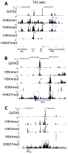
Figure 6. Active and repressive activities of GATA3 are associated with distinct epigenetic modification patterns
A–C. Genome Browser image showing the patterns of GATA3 binding and H3K4me1, H3K4me2, H3K4me3 and H3K27me3 modifications at the Il4 and Il13 gene locus (A), at the Tbx21 gene (B) and at the Ifng gene (C) in Th2 cells. The histone H4K4 methylation peaks co-localized with GATA3 peaks are indicated by arrowheads and the regions enriched with H3K27me3 are highlighted by the blue box.
Figure 7
Figure 7. GATA3 regulates transcription by modulating H3K4 and H3K27 methylations of enhancers
A–D. Genome Browser image showing that deletion of Gata3 resulted in changes in H3K4me2 (indicated by arrowheads) and/or H3K27me3 (highlighted in blue box) at or near the GATA3 sites of the Il4/Il13 locus (A), of the Il10 locus (B), of the Tbx21 locus (C) and of the Ifng locus (D) in Th2 cells. E. Global analysis indicated that deletion of Gata3 resulted in decreases in H3K4me2 signals over the distal GATA3 sites in Th2 cells. Upper panel: the H3K4me2 tag densities (Y-axis) surrounding the total GATA3 sites (left), promoter-proximal (middle) and distal (right) GATA3 sites were plotted for a region of 100kb on each side of the GATA3 binding site. Lower panel: Y-axis shows the ratio of the H3K4me2 signals in Gata3-deficient cells to that in wild type cells surrounding the GATA3 binding sites across the same region as in the upper panel. The P-values for the histone modification differences between the Gata3-deficient and wild type cells are obtained using the Kolmogorov-Smirnoff test and shown in the panels. F. Gata3 deletion-induced changes in histone methylation correlate with changes in gene expression in Th2 cells. Upper panel: plotted are the ratios of H3K4me2 signals between Gata3-deficient cells and wild type cells surrounding the GATA3 binding sites associated with the genes positively regulated (green), negatively regulated (red) and not regulated (black) by GATA3; lower panel: the same are plotted for H3K27me3. The P-values for the histone modification differences between the regulated genes and non-regulated genes are obtained using the Kolmogorov-Smirnoff test and shown either in red or green. G. GATA3 regulates both H3K4me2 and H3K27me3 at numerous target genes. The changes in H3K4me2 and H3K27me3 modifications (P<10−5; ratio≥2 or ≤0.5) after Gata3 deletion) and expression level at each individual GATA3-bound gene were summarized. The arrows indicate the direction of changes in histone modification and gene expression after Gata3 deletion. The P values of either increased or reduced gene expression correlated with a change in histone modification are indicated.

References

    1. Ansel KM, Djuretic I, Tanasa B, Rao A. Regulation of Th2 differentiation and Il4 locus accessibility. Annu Rev Immunol. 2006;24:607–656. - PubMed
    1. Barski A, Cuddapah S, Cui K, Roh TY, Schones DE, Wang Z, Wei G, Chepelev I, Zhao K. High-resolution profiling of histone methylations in the human genome. Cell. 2007;129:823–837. - PubMed
    1. Cheng Y, Wu W, Kumar SA, Yu D, Deng W, Tripic T, King DC, Chen KB, Zhang Y, Drautz D, et al. Erythroid GATA1 function revealed by genome-wide analysis of transcription factor occupancy, histone modifications, and mRNA expression. Genome Res. 2009;19:2172–2184. - PMC - PubMed
    1. Collins A, Littman DR, Taniuchi I. RUNX proteins in transcription factor networks that regulate T-cell lineage choice. Nat Rev Immunol. 2009;9:106–115. - V体育ios版 - PMC - PubMed
    1. Eisbacher M, Holmes ML, Newton A, Hogg PJ, Khachigian LM, Crossley M, Chong BH. Protein-protein interaction between Fli-1 and GATA-1 mediates synergistic expression of megakaryocyte-specific genes through cooperative DNA binding. Mol Cell Biol. 2003;23:3427–3441. - PMC - PubMed

Publication types

MeSH terms

VSports注册入口 - Associated data