VSports手机版 - Skip to main page content
U.S. flag

An official website of the United States government

Dot gov

The . gov means it’s official VSports app下载. Federal government websites often end in . gov or . mil. Before sharing sensitive information, make sure you’re on a federal government site. .

Https

The site is secure V体育官网. The https:// ensures that you are connecting to the official website and that any information you provide is encrypted and transmitted securely. .

. 2011;6(8):e23132.
doi: 10.1371/journal.pone.0023132. Epub 2011 Aug 1.

TIRAP, an adaptor protein for TLR2/4, transduces a signal from RAGE phosphorylated upon ligand binding

Affiliations

TIRAP, an adaptor protein for TLR2/4, transduces a signal from RAGE phosphorylated upon ligand binding

Masakiyo Sakaguchi (V体育安卓版) et al. PLoS One. 2011.

Abstract

The receptor for advanced glycation end products (RAGE) is thought to be involved in the pathogenesis of a broad range of inflammatory, degenerative and hyperproliferative diseases. It binds to diverse ligands and activates multiple intracellular signaling pathways. Despite these pivotal functions, molecular events just downstream of ligand-activated RAGE have been surprisingly unknown. Here we show that the cytoplasmic domain of RAGE is phosphorylated at Ser391 by PKCζ upon binding of ligands VSports手机版. TIRAP and MyD88, which are known to be adaptor proteins for Toll-like receptor-2 and -4 (TLR2/4), bound to the phosphorylated RAGE and transduced a signal to downstream molecules. Blocking of the function of TIRAP and MyD88 largely abrogated intracellular signaling from ligand-activated RAGE. Our findings indicate that functional interaction between RAGE and TLRs coordinately regulates inflammation, immune response and other cellular functions. .


Conflict of interest statement

Competing Interests: Akira Motoyama and Toshihiko Hibino are employees of the Shiseido Research Center. There are no patents, products in development or marketed products to declare V体育安卓版. This does not alter the authors' adherence to all the PLoS ONE policies on sharing data and materials.

VSports注册入口 - Figures
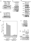
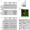
Figure 1
Figure 1. RAGE is phosphorylated at Ser391 by PKCζ upon ligand binding.
(A) Phosphorylation of full-length RAGE (wt, closed arrowhead) but not cytoplasmic domain-deleted RAGE (ΔCyt: open arrowhead). HEK293 cells were transfected with epitope-tagged RAGE constructs (RAGE-Myc-HA-Flag-6His), [32P]phosphate-labeled, and treated with the ligands (10 ng/ml, 1 h), and then RAGE proteins were pulled down with anti-HA antibody (clone HA-7)-agarose before Western blotting and autoradiography. The results were confirmed by five similar experiments. (B and C) Phosphorylation of RAGE(Cyt) in vitro. 6His-2HA-RAGE(Cyt) purified from transfected HEK293 cells by anti-HA antibody (clone HA-7)-agarose was incubated with constitutively active recombinant kinases of commercial source. Western blotting with anti-HA and anti-6His antibodies was performed for determination of amounts of RAGE(Cyt) and kinase proteins, respectively. The results were confirmed by five similar experiments (B) or by an independent and four related experiments (C). (D) Down-regulation of endogenous PKCζ but not of other isoforms by siRNA abrogated S100A11 (10 ng/ml, 6 h)-induced phosphorylation of co-transfected RAGE-Myc-HA-Flag-6His in HEK293 cells. The results are representative of three independent experiments. (E) Activation of endogenous PKCζ by RAGE ligands. HEK293 cells transfected with RAGE-Myc-HA-Flag-6His (wt or ΔCyt) were treated with 10 ng/ml of GST, S100A11, S100A12, and HMGB1 or 100 µg/ml of AGE for 1 h. Cell extracts were incubated with a specific PKCζ-substrate in the presence of [γ-32P]-ATP. The experiment was performed in quadruplicate, and the results were confirmed by three related experiments. (F) Determination of phosphorylation site of RAGE(Cyt). Recombinant PKCζ-6His (top panel) was incubated with isolated variants of 6His-2HA-RAGE(Cyt) [SSST (wild-type), ASST (Ser391 to Ala), SAST (Ser399 to Ala), SSAT (Ser400 to Ala), SSSA (Thr401 to Ala)] and assayed as for (B) and (C). The results were confirmed by an independent and two related experiments. (G) S100A11 (10 ng/ml)-dependent phosphorylation of transfected 6His-2HA-RAGE(wt) at Ser391 in HEK293 cells. [32P]Phosphate-labeled cell extracts were pulled down with anti-HA agarose and immunoblotted with anti-6His antibody (top) or analyzed by autoradiography (middle). Co-precipitated endogenous PKCζ was detected with anti-PKCζ antibody (bottom). The results were confirmed by an independent and five related experiments.
Figure 2
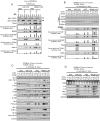
Figure 2. TIRAP and MyD88 function as adaptor proteins for RAGE.
(A) Co-precipitation of endogenous TIRAP and MyD88 but not TRAM with recombinant RAGE(wt). HEK293 cells were transfected with RAGE(wt) and RAGE(ΔCyt) tagged with -Myc-HA-Flag-6His, [32P]phosphate-labeled, and treated with AGE at 100 µg/ml or with other ligands at 10 ng/ml for 1 h. TLR2/4 blocking mixture (Mixture, see Materials and Methods) was preincubated with the ligands for 30 min at room temperature. Cell extracts were analyzed by Western blotting with or without prior immunoprecipitation and by autoradiography. The results were confirmed by 7 related experiments. (B and C) AGE (100 µg/ml)-induced phosphorylation of RAGE at Ser391 is essential for recruitment of endogenous TIRAP and MyD88 (B: The results were confirmed by an independent and two related experiments.) and downstream signal transduction (C: The results were confirmed by an independent and two related experiments.) in HEK293 cells. Full-length 6His-2HA-RAGE constructs were used. SSST, wild-type; ASST, Ser391 to Ala. (D and E) Abortive signal transduction from activated endogenous RAGE in MyD88-/- or TIRAP-down regulated mouse fibroblasts was rescued by forced expression of the corresponding human genes. Mouse TIRAP siRNA and expression vectors were transfected 48 h before treatment with AGE (100 µg/ml, 1 h). Experiments were performed under the conditions similar to those described in (A) except for immunoprecipitation using a biotinylated antibody against endogenous RAGE. 32P shows the results of autoradiography of the immunoprecipitated RAGE. The results were confirmed by an independent and two related experiments.
Figure 3
Figure 3. Specific activation of endogenous RAGE by AGE induced cytokines in HUVECs.
(A and B) Activation of downstream signaling molecules by specific activation of endogenous RAGE. Experiments were performed under the conditions similar to those described in Fig. 2A except for down regulation of RAGE with siRNA, immunoprecipitation using an antibody against RAGE, and type of cells used. The results were confirmed by an independent and two related experiments (A) or by an independent and three related experiments (B). (C) Induction of IL-6 by AGE (100 µg/ml for 12 h) demonstrated by Northern blot analysis in HUVECs in serum- and supplement-free conditions. Cell-permeable inhibitory peptides (100 µM) were added 12 h prior to the addition of AGE. The results were confirmed by two independent and one related experiments. (D) Induction of IL-6 in HUVECs but not in HASMCs by treatment with AGE. HUVECs co-cultured with HASMCs pre-infected with an adenovirus carrying GFP were treated under conditions similar to those described in (C). Fixed cells were immunostained with anti-IL-6 antibody. Bars, 20 µm. The results were confirmed by three independent experiments. (E) Detection of cytokines secreted into the medium by HUVECs treated with AGE using an antibody array [Human cytokine Antibody Array VI & 6.1 (60), RayBiotech, Norcross GA]. Dotted squares indicate antibodies against corresponding cytokines. Cells were treated under conditions similar to those described in (C). The results were confirmed by an independent experiment.
Figure 4
Figure 4. Behavior of adaptor proteins and signal transduction upon differential stimulation of RAGE and TLR4.
(A) Co-precipitation of endogenous adaptor proteins with full-length 6His-2HA-RAGE(wt) and TLR4(wt)-Myc transfected into 293-hMD2-CD14 cells. The cells were treated with control BSA (100 µg/ml), LBP-LPS (100 ng/ml each, preincubated for 2 h), AGE (100 µg/ml), and HMGB1-LPS (100 ng/ml each, preincubated for 2 h) for 30 min. Cell extracts were pulled down with anti-Myc + anti-HA, anti-Myc, or anti-HA agarose and immunoblotted with indicated antibodies. Open triangle, TLR4; closed triangle, RAGE. The results were confirmed by two independent and two related experiments. (B) Co-precipitation of endogenous adaptor proteins with RAGE-Myc-HA-Flag-6His and TLR4-Myc (w, wild-type; D, cytoplasmic domain-deleted variant). Experiments were performed under conditions similar to those described in (A). The results were confirmed by two independent and two related experiments. (C) Downstream signal transduction from RAGE and TLR4. Experiments were performed under conditions similar to those described in (B). The results were confirmed by two independent and two related experiments. (D) Induction of cytokines by differential stimulation of RAGE and TLR4 as assayed by Northern blot analysis. Experiments were performed under conditions similar to those described in (B) except for treatment time (12 h). The results were confirmed by an independent and four related experiments.
Figure 5
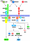
Figure 5. Schematic diagram of signal transduction from RAGE and TLR4.
Both receptors partially share ligands and adaptor proteins. TLR4 transduces a signal to NFκB via TIRAP, MyD88 and IRAK4 and to IRF3 via TRAM and TRIF. RAGE phosphorylated by PKCζ upon ligand binding also uses TIRAP, MyD88 and IRAK4 for signal transduction to NFκB. Presence of yet unknown signaling pathway from RAGE is still possible. Simultaneous activation of both receptors resulted in enhanced activation of NFκB. The signaling machinery is also linked to Akt and caspase-8, and possibly Rac1.

VSports app下载 - References

    1. Neeper M, Schmidt AM, Brett J, Yan SD, Wang F, et al. Cloning and expression of a cell surface receptor for advanced glycosylation end products of proteins. J Biol Chem. 1992;267:14998–15004. - VSports在线直播 - PubMed
    1. Schmidt AM, Hori O, Cao R, Yan SD, Brett J, et al. RAGE: a novel cellular receptor for advanced glycation end products. Diabetes. 1996;45(Suppl 3):S77–80. - PubMed
    1. Yan SF, Ramasamy R, Schmidt AM. Mechanisms of disease: advanced glycation end-products and their receptor in inflammation and diabetes complications. Nat Clin Pract Endocrinol Metab. 2008;4:285–293. - PubMed
    1. Lin L, Park S, Lakatta EG. RAGE signaling in inflammation and arterial aging. Front Biosci. 2009;14:1403–1413. - PMC - PubMed
    1. Sims GP, Rowe DC, Rietdijk ST, Herbst R, Coyle AJ. HMGB1 and RAGE in inflammation and cancer. Annu Rev Immunol. 2010;28:367–388. - PubMed (VSports)

Publication types

MeSH terms