V体育官网入口 - Skip to main page content
U.S. flag

An official website of the United States government

Dot gov

The . gov means it’s official. Federal government websites often end in . gov or VSports app下载. mil. Before sharing sensitive information, make sure you’re on a federal government site. .

Https

The site is secure. The https:// ensures that you are connecting to the official website and that any information you provide is encrypted and transmitted securely. V体育官网.

. 2011;6(7):e21924.
doi: 10.1371/journal.pone.0021924. Epub 2011 Jul 18.

Exploiting mitochondrial dysfunction for effective elimination of imatinib-resistant leukemic cells (V体育2025版)

Affiliations

Exploiting mitochondrial dysfunction for effective elimination of imatinib-resistant leukemic cells (VSports在线直播)

Jérome Kluza et al. PLoS One. 2011.

"VSports app下载" Abstract

Challenges today concern chronic myeloid leukemia (CML) patients resistant to imatinib. There is growing evidence that imatinib-resistant leukemic cells present abnormal glucose metabolism but the impact on mitochondria has been neglected VSports手机版. Our work aimed to better understand and exploit the metabolic alterations of imatinib-resistant leukemic cells. Imatinib-resistant cells presented high glycolysis as compared to sensitive cells. Consistently, expression of key glycolytic enzymes, at least partly mediated by HIF-1α, was modified in imatinib-resistant cells suggesting that imatinib-resistant cells uncouple glycolytic flux from pyruvate oxidation. Interestingly, mitochondria of imatinib-resistant cells exhibited accumulation of TCA cycle intermediates, increased NADH and low oxygen consumption. These mitochondrial alterations due to the partial failure of ETC were further confirmed in leukemic cells isolated from some imatinib-resistant CML patients. As a consequence, mitochondria generated more ROS than those of imatinib-sensitive cells. This, in turn, resulted in increased death of imatinib-resistant leukemic cells following in vitro or in vivo treatment with the pro-oxidants, PEITC and Trisenox, in a syngeneic mouse tumor model. Conversely, inhibition of glycolysis caused derepression of respiration leading to lower cellular ROS. In conclusion, these findings indicate that imatinib-resistant leukemic cells have an unexpected mitochondrial dysfunction that could be exploited for selective therapeutic intervention. .


Conflict of interest statement

Competing Interests: The authors have declared that no competing interests exist.

V体育安卓版 - Figures

Figure 1
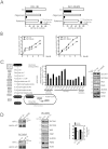
Figure 1. Characterization of glucose metabolism in imatinib-sensitive and -resistant cells.
(A) DA1-3b (imatimib-sensitive) and DA1-3b/M2 (imatinib-resistant) leukemic cells were cultured in DMEM (containing 25 mM of glucose) (□) or in medium containing 1 mM glucose and 10 mM 2-Deoxyglucose (2-DG) (▪) for 18 h, then treated with 1 µg/ml oligomycin or 1 µM rotenone for 4 h. Cells were then collected for total ATP measurement as described in Materials and Methods. Data shown are means +/− SD of four separate cell preparations. (B) Exponentially growing DA1-3b and DA1-3b/M2 cells were resuspended in fresh DMEM medium at a density of 2×10⁶ cells/ml. At indicated times, glucose (left) and lactate (right) were measured in the supernatants. Data are means +/− SD of three independent experiments (C) (left) Schematic overview of glucose metabolism to point out the position of studied genes in metabolism. (middle) Quantitative PCR analysis of relative transcript levels of glucose transporters, glycolytic enzymes and enzymes related to lactate metabolism in DA1-3b/M2 cells compared with DA1-3b cells. Data are means +/− SD of four independent experiments. (right) Immunoblot analysis of protein abundance in DA1-3b and DA1-3b/M2 cells. Densitometric values of proteins are normalized on the basis of actin expression and expressed as fold induction in DA1-3b/M2 cells over DA1-3b cells (n = 2). (D) (left) Expression of HIF-1α protein in DA1-3b and DA1-3b/M2 cells. Nuclear extracts were analyzed by immunoblotting against HIF-1α. Lamin A/C was used as a loading control. Alternatively, DA1-3b/M2 cells were treated with either HIF-1α siRNA or control non-targeting siRNA. Knock down of HIF-1α was confirmed by immunoblot at 48 h post transfection. Data are representative of 3 different experiments. (middle). The effect of HIF-1α knock-down in DA-13b/M2 cells on expression of glycolysis-related proteins. Protein expression was measured by immunoblot densitometry and normalized to actin. The values in HIF-1α silenced DA1-3b/M2 cells are reported as the fold decrease over control (n = 2). (right) Effects of loss of HIF-1α expression in DA-13b/M2 cells on glucose utilization and lactate production. Exponentially growing DA1-3b/M2 cells transfected with siControl or si HIF-1α were incubated in fresh DMEM medium at a density of 2×10⁶ cells/ml. At 24 h, glucose and lactate were measured in the supernatants. Data are means of two independent experiments.
Figure 2
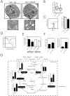
Figure 2. Characterization of mitochondrial alterations in imatinib-resistant cells.
(A) Transmission electron microscopic images of DA1-3b (imatimib-sensitive), DA1-3b/M2 (imatinib-resistant) cells. Typical representative morphologic features of cells (magnification, ×7,000) and mitochondria (magnification, ×20,000); (B) Size of mitochondria per DA1-3b cells or DA1-3b/M2 cells. A total of 500 cells were examined in transmission electronic microscopy and mitochondrial size were evaluated. Values are medians (horizontal bars) with 25–75% interquartile ranges (boxes) and minimum–maximum values (I) (*P = 0.02 between DA1-3b and DA1-3b/M2 cells); (C) (left) Representative flow cytometric measurement of mitochondrial mass. DA1-3b and DA1-3b/M2 cells were labeled with the MTG fluorescent dye. (right) Flow cytometric measurement of ΔΨm. DA1-3b and DA1-3b/M2 cells were labeled with JC-1. JC-1 fluorescence (mean fluorescence intensity) ratio is normalized to that of depolarized cells (cells incubated with 20 µM ClCCP used as a positive control for loss of ΔΨm). Values are means ± S.D of five measurements; (D) Comparison of oxygen consumption in DA1-3b and DA1-3b/M2 cells. DA1-3b or DA1-3b/M2 cells (2×10⁶/ml) in DMEM were added to a closed chamber with an oxygen electrode at 37°C and oxygen levels were monitored over time. Data are representative of five independent experiments; (E) Proportions of mitochondrial oxygen consumption due to proton leak and ATP turnover reactions in DA1-3b and DA1-3b/M2 cells. Proton leak corresponds to the respiration that is not modified by the inhibitor of the ATP synthase, oligomycin whereas ATP turnover constitutes the respiration that is inhibited by oligomycin. Oxygen consumption was determined as in (D). Data are means +/− SD of three separate experiments; (F) Determination of cytoplasmic NADH/NAD ratio (left) and mitochondrial NADH in DA1-3b and DA1-3b/M2 cells. Data are means +/− SD of three independent experiments in triplicates; (G) Quantified levels of mitochondrial TCA cycle related metabolites in DA1-3b and DA1-3b/M2. Metabolites detected (black boxes) are represented on a comprehensive metabolic pathway map. Results are means +/− SD of fifteen 1H NMR spectra of the metabolome for each of the individual cell cultures.
Figure 3
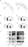
Figure 3. Impairment of the mitochondrial electron chain complex activity in imatinib resistant cells.
(A) Traces of oxygen consumption of digitonin-permeabilized DA1-3b vs. DA1-3b/M2 cells (left), and K562 vs. K562-IM, K562-NI cells (right). Respiration in response to complex I (glutamate+malate, 5 mM each) or complex II (succinate, 5 mM) substrates was determined in the presence or absence of 1 mM ADP. Mitochondrial substrates, N,N,N′,N′-tetramethyl p-phenylenediamine (TMPD 0.5 mM) + ascorbate (1 mM) and respiratory inhibitors, rotenone (2 µM), antimycin A (1 µM) and potassium cyanide (KCN, 100 µM) were used as indicated: As expected, KCN used as control, blocked mitochondrial respiration. Values for oxygen consumption are represented on the curves as nmol oxygen/min; One representative experiment of five performed (B–E) Mitochondrial respiratory chain complex activities (left axes) and protein expression levels of complexes (right axes) in mitochondria isolated from DA1-3b and DA1-3b/M2 cells. (B) NADH ubiquinone reductase (Complex I), (C) Succinate cytochrome c reductase (Complex II+III), (D) Ubiquinone cytochrome c reductase (Complex III) and (E) cytochrome c oxidase (Complex IV activity) activities were measured by spectrophotometry as described in Material and Methods. Rotenone, antimycin A and sodium azide, (prototypic inhibitors of complex activities) were used as control. Quantitative densitometric analysis of mitochondrial repiratory chain proteins was evaluated on Western blot as depicted in (F). The results are expressed as a ratio of actin density following reprobing of the membrane. Results are representative of three independent experiments. (*P<0.05 between DA1-3b and DA1-3b/M2 cells); (F) Western blot analysis of mitochondrial respiratory chain complex proteins such as complex I (20 kDa subunit), complex II (SDHB), III (core 2 protein), IV (Cox II) and V (F1α ATPase). Protein expression patterns were assessed in DA1-3b and DA1-3b/M2 cells, in human K562 (sensitive to imatinib), K562-NI (resistant to nilotinib and imatinib) and K562-IM (resistant to imatinib) CML cell lines, in normal CD34+ cells (2 separate specimen) and in cells isolated from four CML patients. BCR-ABL (+) CML cells were isolated from 4 patients: one patient with primary resistance to imatinib (patient 1), two patients with secondary resistance to imatinib in accelerated/blastic phase (patients 3 and 4) of the disease and one patient (Patient 2) had only a partial response.
Figure 4
Figure 4. Regulation of mitochondrial dysfunction in imatinib resistant cells.
(A) Effect of dichloroactetate (DCA) on lactate production. DA1-3b and DA1-3b/M2 cells were incubated in the presence of 6 mM DCA then at the indicated time points, lactate production was determined. Values are means of two independent experiments in triplicates; (B) The oxygen consumption was measured as described in Materials and Methods following the sequential addition of DCA (1.5 mM every 2 min, arrows) in DA1-3b and DA1-3b/M2 cells. Typical result out of two independent experiments; (C) Whole-cell oxygen consumption measurements in DA1-3b/M2 cells treated with 10 mM 2-DG for 18 h or grown in galactose DMEM medium for 48 h relative to untreated cells (Control, Co.). Data are means +/− SD of four independent experiments in duplicate; * indicates p<0.05 compared to control (D) Typical western blot analysis of mitochondrial respiratory chain complex proteins in DA1-3b/M2 cells treated as in (C). Actin was used as a loading control. Data are representative of three independent experiments.
Figure 5
Figure 5. Mitochondrial ROS production in imatinib- sensitive and -resistant cell lines.
(A) Cytofluorometric analysis of mitochondrial ROS production in DA1-3b and DA1-3b/M2. Cells were kept untreated (Control) or treated with the mitochondrial redox cycling promoter menadione (100 µm, 1 h) used as positive control then labeled with the fluorescent probe MitoSox as described in Materials and Methods. Alternatively, cells were treated with menadione in the presence of 10 mM NAC (inset). Data represent typical results of one out of three independent experiments; (B) NADPH oxidase activity in the plasma membrane of DA1-3b and DA1-3b/M2 cells. When indicated, cells were treated with the flavoprotein inhibitor diphenyleneiodonium (DPI), used as control. Lucigenin chemiluninescence assay was used as described in Material and methods. Results are means +/− SD of two independent experiments carried out in triplicates; (C) Effects of various inhibitors of the mitochondrial respiratory chain on ROS production in DA1-3b cells. Cells were kept untreated (Control) or treated with 1 µM rotenone (rot, a know inhibitor of mitochondrial electron transport complex I), 1 µM 2-thenoyltrifluoroacetone (TTFA, a conventional complex II inhibitor), 1 µM stigmatellin or 1 µM antimycin A (AA, two specific inhibitors of mitochondrial complex III) in the presence or absence of 10 mM N-acetyl cystein (NAC) for 18 h then the levels of ROS were determined by flow cytometry after Mitosox staining. Inhibitors were used at sub-toxic concentrations which induce moderate inhibition of cell respiration (around 20%). Data are means +/− SD of four independent experiments; * indicate significant differences from untreated cells at p<0.05 (D) DA1-3b/M2 cells were incubated in the presence of the indicated doses of DCA for 1 h, or with 10 mM 2-DG for 18 h, or grown in galactose medium for 48 h then the levels of ROS were determined by flow cytometry after Mitosox staining. Data are means +/− SD of four independent experiments. * indicate p<0.05 compared to control.
Figure 6
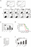
Figure 6. In vitro and in vivo antitumor activity of PEITC and Trisenox in imatinib resistant cells.
(A) DA1-3b and DA1-3b/M2 cells were treated with 1, 3, 5, 10, 15 and 20 µM of the pro-oxidant phenethyl isothiocyanate (PEITC) for 18 h (left) or with 1, 5, 10 and 20 µM of arsenic trioxide (Trisenox) for 4 h then mitochondrial ROS production was assessed by flow cytometry using MitoSox. When indicated, cells were pre-treated with 10 mM NAC followed by incubation with 20 µM PEITC for 18 h or with 20 µM PEITC for 4 h. MitoSox fluorescence intensity was presented in arbitrary units (A.U.). Data are means of three independent experiments (SD<10%); (B) DA1-3b and DA1-3b/M2 cells were treated with PEITC for 18 h or Trisenox for 4 h in the presence or absence of pretreatment with the antioxidant NAC (10 mM for 1 h). Apoptotic cell death was then determined by flow cytometry after annexin V–FITC and propidium iodide staining. Flow cytometric profiles shown are representative of two replicate experiments; (C) Effect of loss of HIF-1α expression in DA-13b/M2 cells on ROS production and cell death. DA1-3b/M2 cells were treated with either HIF-1α siRNA or control non-targeting siRNA. At 24 h post transfection, cells were treated with 20 µM Trisenox for 4 h, 20 µM PEITC for 18 h or 300 µM Menadione for 1 h, then ROS production and cell death were determined by flow cytometry after MitoSox or PI staining, respectively. Results are presented as the percentage of inhibition of ROS production or cell death in siRNA HIF-1α transfected cells relative to siControl. (D) In vivo therapeutic activity of PEITC and Trisenox Survival of mice after injection of 1×10⁶ GFP-tagged DA1-3b/M2 cells. Cumulative survival data were compiled from two separate experiments and presented by a Kaplan–Meier survival analysis. GFP+ DA1-3b/M2 bearing mice were treated with Trisenox (4 mg/kg ip 5days/week) or PEITC (50 mg/kg ip 5days/week). Survival of mice treated with Trisenox (n = 10) or with PEITC (n = 10) in comparison to control animals (n = 20) was significant (P = 0.006 and P = 0.0008, respectively); (E) Decrease in percentage GFP+ DA1-3b/M2 cells in spleen after Trisenox and PEITC treatment. Untreated mice, PEITC- or Trisenox- treated mice were killed 37 days after inoculation of GFP+ DA1-3b/M2 cells. The percentage of GFP+ cells in spleen was evaluated by flow cytometry. Data are means +/− SD of three independent experiments; (F) Mitochondrial ROS levels in GFP-tagged DA1-3b/M2 cells after Trisenox and PEITC treatment in vivo. Untreated mice, PEITC- or Trisenox- treated mice were killed 37 days after inoculation of GFP+ DA1-3b/M2 cells. Spleen cells were stained with MitoSox to determine relative levels of ROS. The experiment was performed twice. Results are fold change in Mitosox fluorescence intensity of GFP and + spleen cells. The values for untreated cells were normalized to 1 for the analysis. As control, cells were treated with 100 µM menadione for 1 h in vitro.
Figure 7
Figure 7. Suggested sequence of metabolic alterations leading to cell death in imatinib-resistant cells.
Imatinib-resistant cells presented high glycolytic activity controlling mitochondrial oxidative phosphorylation. As a result, mitochondrial dysfunction generated more ROS spontaneously and after treatment with the pro-oxidants PEITC and Trisenox enhancing cell death sensitivity. Metabolic organization in imatinib-resistant cells is under control of HIF-1α (see text for details).

References

    1. Walz C, Sattler M. Novel targeted therapies to overcome imatinib mesylate resistance in chronic myeloid leukemia (CML). Crit Rev Oncol Hematol. 2006;57:145–164. - PubMed
    1. Shah NP, Nicoll JM, Nagar B, Gorre ME, Paquette RL, et al. Multiple BCR-ABL kinase domain mutations confer polyclonal resistance to the tyrosine kinase inhibitor imatinib (STI571) in chronic phase and blast crisis chronic myeloid leukemia. Cancer Cell. 2002;2:117–125. - PubMed
    1. Druker BJ, Guilhot F, O'Brien SG, Gathmann I, Kantarjian H, et al. Five-year follow-up of patients receiving imatinib for chronic myeloid leukemia. N Engl J Med. 2006;355:2408–2417. - "VSports在线直播" PubMed
    1. Wei Y, Hardling M, Olsson B, Hezaveh R, Ricksten A, et al. Not all imatinib resistance in CML are BCR-ABL kinase domain mutations 1. Ann Hematol. 2006;85:841–847. - PubMed
    1. Barnes K, McIntosh E, Whetton AD, Daley GQ, Bentley J, et al. Chronic myeloid leukaemia: an investigation into the role of Bcr-Abl-induced abnormalities in glucose transport regulation. Oncogene. 2005;24:3257–3267. - PubMed

"VSports" Publication types

MeSH terms

"V体育官网入口" LinkOut - more resources