<big dropzone="aygyi"><noframes id="JLUYOsrM"> Skip to main page content
U.S. flag

An official website of the United States government

Dot gov

The . gov means it’s official. Federal government websites often end in . gov or . mil. Before sharing sensitive information, make sure you’re on a federal government site VSports app下载. .

Https

The site is secure. The https:// ensures that you are connecting to the official website and that any information you provide is encrypted and transmitted securely. V体育官网.

. 2011 Nov;141(5):1897-906.
doi: 10.1053/j.gastro.2011.06.051. Epub 2011 Jun 25.

"V体育2025版" A proinflammatory role for interleukin-22 in the immune response to hepatitis B virus

Affiliations

A proinflammatory role for interleukin-22 in the immune response to hepatitis B virus

Ye Zhang et al. Gastroenterology. 2011 Nov.

"VSports" Abstract

Background & aims: T-helper (Th)17 cells that secrete interleukin (IL)-22 have immunomodulatory and protective properties in the liver and other tissues. IL-22 induces expression of proinflammatory genes but is also mitogenic and antiapoptotic in hepatocytes. Therefore, it could have multiple functions in the immune response to hepatitis B virus (HBV). VSports手机版.

Methods: We examined the role of IL-22 in regulating liver inflammation in HBV transgenic mice and measured levels of IL-22 in HBV-infected patients. V体育安卓版.

Results: In HBV transgenic mice, injection of a single dose of IL-22 increased hepatic expression of proinflammatory genes but did not directly inhibit virus replication. When splenocytes from HBV-immunized mice were transferred into HBV transgenic mice, the severity of the subsequent liver damage was ameliorated by neutralization of IL-22. In this model, IL-22 depletion did not affect interferon gamma-mediated noncytopathic inhibition of virus replication initiated by HBV-specific cytotoxic T cells, but it significantly inhibited recruitment of antigen-nonspecific inflammatory cells into the liver V体育ios版. In patients with acute HBV infections, the percentage of Th17 cells in peripheral blood and concentration of IL-22 in serum were significantly increased. .

Conclusions: IL-22 appears to be an important mediator of the inflammatory response following recognition of HBV by T cells in the liver. These findings might be relevant to the development of cytokine-based therapies for patients with HBV infection VSports最新版本. .


Conflict of interest statement

Potential conflict of interest: Nothing to report.

Figures

Figure 1
Figure 1. IL-22 induces an acute-phase like response in the liver of HBV Tg mice but does not inhibit virus replication
(A) Groups of age, sex, and serum HBeAg matched transgenic mice (3 mice per group) were injected intravenously or intraperitoneally with a single dose of 25 μg of IL-22, and analyzed 1 day post injection. HBV replication in the liver was measured by Southern blot (SB) analysis of relaxed-circle (RC) and single-stranded (SS) DNA replication forms, and compared to levels in control animals that did not receive the cytokine. HBV 3.5 kb and 2.1+2.4 kb mRNA expression from total liver RNA was examined by northern blot (NB) analysis, and compared to the housekeeping gene GAPDH. (B) Intrahepatic expression of amyloid A (AA), haptoglobin (HB), and anti-chymotrypsin (α-CT) were measured by quantitative real-time RT-PCR (qRT-PCR). Results are displayed as fold differences relative to one control mouse, and normalized to GAPDH expression. (C) Serum amyloid A (SAA) levels from the same mice were measured by ELISA. (D) The level of serum alanine aminotransferase (sALT) was measured before and after injection. The data represent mean ± SD of three mice.
Figure 2
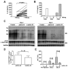
Figure 2. IL-22 depletion does not affect IFN-γ mediated inhibition of HBV replication, but ameliorates subsequent liver damage following transfer of immunized splenocytes
(A) IL-22 expression in immunized splenocytes was measured by ELISA after a 4-day incubation in vitro with or without HBsAg stimulation. Matched data points indicate splenocytes from an individual animal. (B) IL-22 expression from pooled total, CD4-depleted (Miltenyi Biotec; CD4−), or CD4-purified (CD4+) splenocytes. (C) HBV.CB6F1 mice (4 per group) were injected with 2×10⁸ immunized splenocytes with or without anti-IL-22 Ab. Mice were euthanized at days 2 and 5 postinjection, and total hepatic DNA was analyzed for HBV replication by Southern blot (SB). Bands corresponding to integrated transgene (Tg), relaxed-circular (RC), and single-stranded (SS) HBV DNA replication intermediates are indicated. Northern blot (NB) was used for analysis of HBV 3.5 kb and 2.1+2.4 kb mRNA expression, normalized to GAPDH. Results were compared to control livers from another four matched transgenic littermates injected with unimmunized splenocytes. (D) HBV.CB6F1 mice (4 per group) were injected with 2×10⁸ CFSE-stained immunized splenocytes with or without anti-IL-22 Ab. The livers and spleens were harvested at day 2 after splenocyte transfer, and the percentages of CFSE+CD8+ cells in the liver and CFSE+ cells in the spleen were analyzed by flow cytometry. The data represent mean ± SD of four mice. (E) The mean sALT activity (± SD), measured 1 day pre-injection and at euthanasia 2 or 5 days post transfer, is indicated for each group.
Figure 3
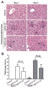
Figure 3. IL-22 neutralization reduces liver inflammation and pathology following transfer of immunized splenocytes
(A) Representative sections of liver from control HBV Tg mice given splenocytes from non-immunized mice (upper panels), immunized splenocytes alone (middle panels) and with anti-IL-22 Ab (lower panels) at day 2 (left) and day 5 (right) post administration. The overall morphology of control livers (upper panels) was normal. Mice with HBV-specific splenocyte induced hepatic injury alone had more frequent foci of necrotic hepatocytes (arrowheads) and inflammation (asterisks) at day 5 (middle right panel) than day 2 (middle left panel). Mice with HBV-specific splenocyte induced hepatic injury given anti-IL-22 Ab had fewer foci of necrosis and inflammation (lower panels) than mice not given anti-IL-22 Ab. Scale Bars = 50 microns. (B) Histopathology scores for necrosis and inflammation were significantly reduced at day 5 in anti-IL-22 treated mice.
Figure 4
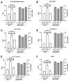
Figure 4. Depletion of IL-22 blocks the recruitment of antigen-nonspecific cells into the liver
Livers from HBV Tg mice receiving splenocytes from non-immunized mice (control) or immunized mice in the absence (-anti-IL-22) or presence (+anti-IL-22) of IL-22 Ab were weighed at the time of euthanasia, and IHLs were isolated from two liver lobes of a similar weight and analyzed by flow cytometry. The indicated numbers of total IHLs or splenocytes and cell subsets represent the absolute numbers in the liver or spleen, respectively. (A) total IHL and splenocyte number, (B) CD4 T cells, (C) CD8 T cells, (D) NK cells, (E) neutrophils, and (F) B cells. Similar patterns were observed for NKT cells, lymphoid dendritic cells, myeloid dendritic cells, and macrophages (not shown).
Figure 5
Figure 5. IL-22 depletion reduces chemokine expression in the liver
Expression of CXCL9 and CXCL10 in the liver of HBV Tg mice 2 days after receiving splenocytes from non-immunized mice (control; n=6) or immunized mice in the absence (−anti-IL-22; n=8) or presence (+anti-IL-22; n=6) of IL-22 Ab. RNA expression was measured by qRT-PCR, and results are displayed as fold differences relative to the control group, normalized to GAPDH.
Figure 6
Figure 6. Th17 cells and IL-22 expression in HBV-infected patients
The percentage of circulating Th17 cells (A) and concentration of IL-22 in serum (B) were measured by flow cytometry or ELISA in healthy controls (n=16), patients with acute hepatitis B (AHB, n=16), chronic hepatitis B (CHB, n=41), and asymptomatic HBV carriers (AsC, n=20). Horizontal bars indicate the mean value of each subset. The individual frequency for each subject is shown. Significance was calculated using the Dunn’s multiple comparison test. (C and D) Spearman correlation analysis of Th17 percentage with serum AST and ALT in sixteen AHB patients.

References

    1. Eyerich S, Eyerich K, Pennino D, Carbone T, Nasorri F, Pallotta S, Cianfarani F, Odorisio T, Traidl-Hoffmann C, Behrendt H, Durham SR, Schmidt-Weber CB, Cavani A. Th22 cells represent a distinct human T cell subset involved in epidermal immunity and remodeling. J Clin Invest. 2009;119:3573–85. - PMC - PubMed
    1. Hughes T, Becknell B, McClory S, Briercheck E, Freud AG, Zhang X, Mao H, Nuovo G, Yu J, Caligiuri MA. Stage 3 immature human natural killer cells found in secondary lymphoid tissue constitutively and selectively express the TH 17 cytokine interleukin–22. Blood. 2009;113:4008–10. - PMC (VSports在线直播) - PubMed
    1. Liang SC, Tan XY, Luxenberg DP, Karim R, Dunussi-Joannopoulos K, Collins M, Fouser LA. Interleukin (IL)-22 and IL-17 are coexpressed by Th17 cells and cooperatively enhance expression of antimicrobial peptides. J Exp Med. 2006;203:2271–9. - PMC - PubMed
    1. Xie MH, Aggarwal S, Ho WH, Foster J, Zhang Z, Stinson J, Wood WI, Goddard AD, Gurney AL. Interleukin (IL)-22, a novel human cytokine that signals through the interferon receptor-related proteins CRF2-4 and IL-22R. J Biol Chem. 2000;275:31335–9. - VSports最新版本 - PubMed
    1. Wolk K, Kunz S, Witte E, Friedrich M, Asadullah K, Sabat R. IL-22 increases the innate immunity of tissues. Immunity. 2004;21:241–54. - PubMed

Publication types

MeSH terms (VSports app下载)

Substances