Skip to main page content
U.S. flag

An official website of the United States government

Dot gov

The . gov means it’s official. Federal government websites often end in . gov or . mil VSports app下载. Before sharing sensitive information, make sure you’re on a federal government site. .

Https

The site is secure V体育官网. The https:// ensures that you are connecting to the official website and that any information you provide is encrypted and transmitted securely. .

. 2011 Jun 22;3(88):88ra55.
doi: 10.1126/scitranslmed.3002194.

The role of Nogo and the mitochondria-endoplasmic reticulum unit in pulmonary hypertension

Affiliations

The role of Nogo and the mitochondria-endoplasmic reticulum unit in pulmonary hypertension

Gopinath Sutendra et al. Sci Transl Med. .

Abstract

Pulmonary arterial hypertension (PAH) is caused by excessive proliferation of vascular cells, which occlude the lumen of pulmonary arteries (PAs) and lead to right ventricular failure. The cause of the vascular remodeling in PAH remains unknown, and the prognosis of PAH remains poor. Abnormal mitochondria in PAH PA smooth muscle cells (SMCs) suppress mitochondria-dependent apoptosis and contribute to the vascular remodeling. We hypothesized that early endoplasmic reticulum (ER) stress, which is associated with clinical triggers of PAH including hypoxia, bone morphogenetic protein receptor II mutations, and HIV/herpes simplex virus infections, explains the mitochondrial abnormalities and has a causal role in PAH VSports手机版. We showed in SMCs from mice that Nogo-B, a regulator of ER structure, was induced by hypoxia in SMCs of the PAs but not the systemic vasculature through activation of the ER stress-sensitive transcription factor ATF6. Nogo-B induction increased the distance between the ER and mitochondria and decreased ER-to-mitochondria phospholipid transfer and intramitochondrial calcium. In addition, we noted inhibition of calcium-sensitive mitochondrial enzymes, increased mitochondrial membrane potential, decreased mitochondrial reactive oxygen species, and decreased mitochondria-dependent apoptosis. Lack of Nogo-B in PASMCs from Nogo-A/B-/- mice prevented these hypoxia-induced changes in vitro and in vivo, resulting in complete resistance to PAH. Nogo-B in the serum and PAs of PAH patients was also increased. Therefore, triggers of PAH may induce Nogo-B, which disrupts the ER-mitochondria unit and suppresses apoptosis. This could rescue PASMCs from death during ER stress but enable the development of PAH through overproliferation. The disruption of the ER-mitochondria unit may be relevant to other diseases in which Nogo is implicated, such as cancer or neurodegeneration. .


VSports最新版本 - Figures

Fig. 1
Fig. 1
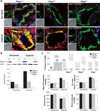
Nogo-B and ATF6 are increased in pulmonary arteries (PAs), pulmonary arterial smooth muscle cells (PASMCs), and serum from patients with PAH. (A) Resistance PAs in the lungs of five patients with idiopathic PAH had increased levels of Nogo-B protein (red) compared to normal PAs from three transplant donors, as shown by immunofluorescence and confocal microscopy (n = 7 PAs per patient). *P < 0.01, unpaired Student’s t test. Results from one patient are shown here; see fig. S1A for the remaining results. Although the antibody detects both Nogo-A and Nogo-B, Nogo-A is not expressed in the lungs (27). Resistance PAs were co-stained with an antibody to smooth muscle actin (SMA; green). PA morphology is shown in the differential interference contrast (DIC) panel. In the merged panel, Nogo-B and SMA colocalization is shown (yellow), along with nuclei stained blue with 4′,6-diamidino-2-phenylindole (DAPI). Mean data were calculated for all patients (n = 5 PAH, n = 3 normal; seven PAs per patient were analyzed) and expressed as arbitrary fluorescence units (AFUs). (B) Serum levels of Nogo-A/B in PAH patients are increased compared to normal and secondary pulmonary hypertension (thromboembolic disease) (Sec) patients. Sample sizes are shown in parentheses. *P < 0.01 versus normal; #P < 0.01 versus secondary PAH using ANOVA with Fisher’s least significant difference (FLSD) post hoc analysis. A previously described ELISA method was used (51). Individual patient values are shown in fig. S2. (C) PASMCs isolated from resistance PAs taken from patients with idiopathic PAH have higher levels of Nogo-B protein (45 kD) than do PASMCs from normal PAs as shown by immunoblot (n = 4 experiments per group). *P < 0.01 using unpaired Student’s t test. Although the antibody detects both Nogo-A and Nogo-B isoforms, Nogo-A is absent from vascular and lung tissues; in addition, Nogo-B (45 kD) is separated from Nogo-A (180 kD) by molecular weight. (D) PASMCs from normal donor patients exposed to ER stress inducers, hypoxia, and thapsigargin (Tg) and PASMCs from PAH patients have increased Nogo-B mRNA values compared to normal vehicle-treated PASMCs, as shown by qRT-PCR. Thapsigargin, but not hypoxia, increased Nogo-B mRNA in CASMCs from transplant donors. An ATF6 inhibitor [4-(2-aminoethyl)benzenesulfonyl fluoride hydrochloride (AEBSF)] and ATF6 siRNA decrease Nogo-B mRNA in both hypoxia- and thapsigargin-treated PASMCs and in PAH PASMCs (n = 3 experiments per group). *P < 0.05 versus normoxia; P < 0.05 versus hypoxia; #P < 0.05 versus thapsigargin; **P < 0.05 versus PAH normoxia by ANOVA with FLSD post hoc analysis. (E) PASMCs from normal donor patients exposed to hypoxia and PASMCs from PAH patients have increased nuclear ATF6 (red) and Nogo-B (green) levels as shown by immunocytochemistry. ATF6 colocalization with the nuclei, stained with DAPI (blue), is shown in pink in the merged panel. Nuclear ATF6 and Nogo-B were decreased by the ATF6 inhibitor AEBSF in hypoxia-treated normal donor PASMCs and PAH PASMCs. Hypoxia did not increase nuclear ATF6 or Nogo-B levels in CASMCs (n = 70 cells per group). *P < 0.05 versus normoxia PASMC; P < 0.05 versus hypoxia PASMC; **P < 0.05 versus normoxia PAH PASMC using ANOVA with Tukey’s post hoc analysis. Mean data are presented in AFUs.
Fig. 2
Fig. 2
In mice, chronic hypoxia-induced PAH is dependent on Nogo-B. (A) Resistance PAs in the lungs of control (Nogo+/+) and heterozygote (Nogo+/−) littermates exposed to chronic hypoxia have increased levels of Nogo-B protein (red) compared to normoxia-treated controls, as shown by immunofluorescence and confocal microscopy. Nogo-deficient (Nogo−/−) mice had no detectable Nogo-B protein in either normoxic or chronic hypoxic PAs. Although the antibody detects both Nogo-A and Nogo-B, Nogo-A is not expressed in the lungs (27). Resistance PAs were co-stained with an antibody to SMA (green). PA morphology is shown in the differential interference contrast (DIC) panel. Nogo-B and SMA colocalization (yellow) is shown along with the nuclear stain DAPI (blue) in the merged panel. (B) Lungs of Nogo+/+ and Nogo+/−, but not Nogo−/−, mice exposed to chronic hypoxia had increased levels of Nogo-B protein when compared to those from normoxic mice, as shown by immunoblot (n = 5 mice per group). *P < 0.05 versus normoxia controls using ANOVA with FLSD post hoc analysis. (C) (Top) Representative pressure traces obtained by closed chest, right heart catheterization of anesthetized mice with a Millar catheter advanced through the internal jugular vein into the right atrium (RA), right ventricle (RV), and PA. (Bottom) Nogo+/+ and Nogo+/−, but not Nogo−/−, mice exposed to chronic hypoxia showed increased mean PA pressure, RV mass [RV/LV (left ventricle) + septum weight ratio], and decreased functional capacity (distance run on rodent treadmill). Systemic blood pressure was not different among groups (n = 8 mice per group). *P < 0.05 versus normoxia controls using ANOVA with FLSD post hoc analysis.
Fig. 3
Fig. 3
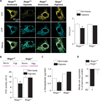
Hypoxia-induced Nogo-B disrupts the mitochondria-ER unit in mice PASMC. (A) Nogo+/+ PASMCs exposed to hypoxia had increased nuclear ATF6 (red) and Nogo-B (green) levels as shown by immunofluorescence. ATF6 colocalization, with the nuclear stain DAPI (blue), is shown in pink in the merged panel. Nuclear ATF6 and Nogo-B were decreased by the ATF6 inhibitor AEBSF in hypoxia-treated Nogo+/+ PASMCs. Hypoxia-treated Nogo−/− PASMCs had increased nuclear ATF6 levels but no detectable Nogo-B. Hypoxia did not increase nuclear ATF6 and Nogo-B levels in Nogo+/+ CASMCs (n = 70 cells per group). *P < 0.05 versus normoxia; #P < 0.05 versus hypoxia using ANOVA with Tukey’s post hoc analysis. Mean data are presented in AFUs. (B) Nogo+/+ PASMCs, but not Nogo+/+ CASMCs or RASMCs, exposed to hypoxia had increased Nogo-B and GRP-78 mRNA values, as shown by qRT-PCR., Nogo−/− PASMCs exposed to hypoxia had increased GRP-78 mRNA, whereas no detectable Nogo-B mRNA was observed (n = 3 experiments per group). *P < 0.001 versus normoxia using unpaired Student’s t test. (C) Nogo+/+, but not Nogo−/−, PASMCs exposed to hypoxia had structurally disrupted mitochondria-ER units as shown by the increase in the minimal distance between the mitochondria and ER in electron microscopy photomicrographs (arrows) (n = 40 contact sites in PASMCs isolated from three mice per group). The distance between the outer mitochondrial membrane and the nearest ER membrane was measured on magnified photomicrographs using ImageJ software. *P < 0.001 using unpaired Student’s t test. (D) Nogo+/+, but not Nogo−/−, PASMCs exposed to hypoxia had functionally disrupted mitochondria-ER units as shown by decreased mitochondrial synthesis of phosphatidylethanolamine (PtdEtn) (n = 6 per group). *P < 0.01 versus normoxia using unpaired Student’s t test. Human PAH PASMCs had decreased mitochondrial synthesis of PtdEtn compared to normal donor PASMCs (n = 3 per group). *P < 0.01 using unpaired Student’s t test. Data presented as ×10−3 disintegrations per minute (dpm) per microgram of protein. PSS, PtdSer synthase; PSD, PtdSer decarboxylase.
Fig. 4
Fig. 4
Hypoxia induction of mitochondrial Ca2+, pyruvate dehydrogenase (PDH), and isocitrate dehydrogenase (IDH) depends on Nogo-B. (A) Nogo+/+, but not Nogo−/−, PASMCs exposed to hypoxia had decreased mitochondrial Ca2+ compared to normoxic controls, as shown by the ratio of bound mitochondrial Ca2+ [yellow fluorescent protein (YFP), yellow] to unbound mitochondrial Ca2+ [cyan fluorescent protein (CFP) signal, blue] with the fluorescence resonance energy transfer (FRET) and confocal microscopy. Mean data are presented as YFP/CFP signal ratio (n = 25 cells per group). *P< 0.05 versus normoxia using unpaired Student’s t test. (B)Nogo+/+, but not Nogo−/−, PASMCs exposed to hypoxia had decreased PDH activity compared to normoxic controls (n = 3 experiments per group). *P < 0.01 versus normoxia using unpaired Student’s t test. Mean data presented in AFUs. (C) Nogo+/+, but not Nogo−/−, PASMCs exposed to hypoxia had decreased α-ketoglutarate (α-KG) levels, a direct index of IDH activity, compared to normoxic controls (n = 4 experiments per group). *P < 0.05 versus normoxia using unpaired Student’s t test. (D)Nogo+/+, but not Nogo−/−, PASMCs exposed to hypoxia had decreased whole-cell respiration rates (n = 5 experiments per group). *P < 0.01 versus normoxia using an independent one-sample t test. Mean data are presented as percent decrease of hypoxia-treated PASMCs to normoxic control.
Fig. 5
Fig. 5
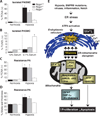
Hypoxia disruption of the mitochondria–mROS–Kv channel axis depends on Nogo-B. (A) Nogo+/+ and Nogo+/− PASMCs exposed to hypoxia had hyperpolarized mitochondria as assessed by tetramethylrhodamine methyl ester (TMRM) and decreased mROS production as assessed by MitoSOX compared to normoxic controls. In contrast, Nogo−/− PASMCs exposed to hypoxia had similar mitochondrial potential (ΔΨm) and mROS production as normoxic PASMCs (n = 100 cells per group). *P < 0.05 versus normoxic control using ANOVA with Tukey’s post hoc analysis. (B) Nogo+/+, but not Nogo−/−, PASMCs exposed to hypoxia had lower outward K+ current density compared to normoxic controls, measured by whole-cell patch clamping. The K+ current in normoxic Nogo+/+ and Nogo−/− PASMCs was sensitive to 4-aminopyridine (4-AP) (n = 8 to 10 cells per group). *P < 0.05 versus normoxia using ANOVA with FLSD post hoc analysis. Mean data are shown as current density (pA/pF) over membrane potential (mV). (C) Nogo+/+ and Nogo+/− PASMCs exposed to hypoxia and phenylephrine had increased intracellular Ca2+, as assessed by FLUO-3, compared to normoxic controls., Nogo−/− PASMCs showed increased intracellular Ca2+ to phenylephrine but not hypoxia (n= 100 cells per group). *P < 0.05 versus normoxic control using ANOVA with Tukey’s post hoc analysis. (D) Nogo+/+ PASMCs exposed to hypoxia had increased nuclear HIF-1α (red) and Nogo-B (green) levels, as shown by immunofluorescence. HIF-1α colocalization with the nuclear stain DAPI (blue) is shown in pink in the merged panel. Nuclear HIF-1α and Nogo-B levels were decreased by the ATF6 inhibitor AEBSF in hypoxia-treated Nogo+/+ PASMCs., Nogo−/− PASMCs exposed to hypoxia had similar HIF-1α levels compared to normoxic controls and no detectable Nogo-B levels (n = 70 cells per group). *P < 0.05 versus normoxia; #P < 0.05 versus hypoxia using ANOVA with Tukey’s post hoc analysis. Mean data are presented in AFUs.
Fig. 6
Fig. 6
Nogo-B levels correlate positively with the degree of proliferation under hypoxia and the degree of resistance to apoptosis under starving conditions. (A) Nogo+/+ and Nogo+/−, but not Nogo−/−, PASMCs exposed to hypoxia had increased proliferation (% PCNA-positive cells) compared to normoxic controls using immunofluorescence microscopy (n = 100 cells per group). *P < 0.05 using ANOVA with Tukey’s post hoc analysis. (B) Nogo−/−, but not Nogo+/+ and Nogo+/−, PASMCs exposed to 0.1% serum had increased apoptosis (% TUNEL-positive cells) compared to controls treated with 10% serum using immunofluorescence microscopy (n = 100 cells per group). *P < 0.05 versus controls, using ANOVA with Tukey’s post hoc analysis. (C) Nogo+/+ and Nogo+/−, but not Nogo−/−, mice exposed to chronic hypoxia had increased cell proliferation in resistance PAs compared to normoxic controls, as shown by % PCNA-positive cells in resistance PAs, using immunofluorescence microscopy (n = 100 cells from 15 PAs per group). *P < 0.05 versus normoxic control using ANOVA with Tukey’s post hoc analysis. (D) Nogo+/+ and Nogo+/−, but not Nogo−/−, mice exposed to chronic hypoxia had increased % media wall thickness in resistance PAs (<300 µm) compared to normoxic controls, as assessed by hematoxylin and eosin staining and light microscopy. These lungs were obtained from the same mice that underwent hemodynamic assessment in Fig. 2 (n = 40 PAs per group). *P < 0.05 versus normoxic controls, using ANOVA with Tukey’s post hoc analysis. (E) A schematic diagram showing how Nogo links ER stress to mitochondrial function. Many PAH-associated triggers, including hypoxia, bone morphogenetic protein receptor II (BMPRII) mutations, viruses, inflammation, or induction of Notch, are known triggers of endoplasmic reticulum (ER) stress. ER stress activates ATF6 in a pulmonary circulation–specific manner, which increases Nogo expression. This in turn disrupts the mitochondria-ER unit, resulting in a suppression of mitochondrial function, explaining the metabolic remodeling and several molecular responses in PAH. IDH, isocitrate dehydrogenase; αKG, α-ketoglutarate; mROS, mitochondrial-reactive oxygen species; MTP, mitochondrial transition pore; NFAT, nuclear factor of activated T cells; PDH, pyruvate dehydrogenase.

Comment in

V体育安卓版 - References

    1. McLaughlin VV, Archer SL, Badesch DB, Barst RJ, Farber HW, Lindner JR, Mathier MA, McGoon MD, Park MH, Rosenson RS, Rubin LJ, Tapson VF, Varga J, Harrington RA, Anderson JL, Bates ER, Bridges CR, Eisenberg MJ, Ferrari VA, Grines CL, Hlatky MA, Jacobs AK, Kaul S, Lichtenberg RC, Moliterno DJ, Mukherjee D, Pohost GM, Schofield RS, Shubrooks SJ, Stein JH, Tracy CM, Weitz HH, Wesley DJ. ACCF/AHA, ACCF/AHA 2009 expert consensus document on pulmonary hypertension: A report of the American College of Cardiology Foundation Task Force on Expert Consensus Documents and the American Heart Association: Developed in collaboration with the American College of Chest Physicians, American Thoracic Society, Inc., and the Pulmonary Hypertension Association. Circulation. 2009;119:2250–2294. - PubMed
    1. Macchia A, Marchioli R, Marfisi R, Scarano M, Levantesi G, Tavazzi L, Tognoni G. A meta-analysis of trials of pulmonary hypertension: A clinical condition looking for drugs and research methodology. Am. Heart J. 2007;153:1037–1047. - PubMed (V体育平台登录)
    1. Macchia A, Marchioli R, Tognoni G, Scarano M, Marfisi R, Tavazzi L, Rich S. Systematic review of trials using vasodilators in pulmonary arterial hypertension: Why a new approach is needed. Am. Heart J. 2010;159:245–257. - VSports手机版 - PubMed
    1. Michelakis ED, Wilkins MR, Rabinovitch M. Emerging concepts and translational priorities in pulmonary arterial hypertension. Circulation. 2008;118:1486–1495. - PubMed
    1. Dromparis P, Sutendra G, Michelakis ED. The role of mitochondria in pulmonary vascular remodeling. J. Mol. Med. 2010;88:1003–1010. - PubMed

Publication types

MeSH terms