<b dir="2nD17N"></b> "V体育官网" Skip to main page content
U.S. flag

An official website of the United States government

Dot gov

The . gov means it’s official VSports app下载. Federal government websites often end in . gov or . mil. Before sharing sensitive information, make sure you’re on a federal government site. .

Https

The site is secure. The https:// ensures that you are connecting to the official website and that any information you provide is encrypted and transmitted securely V体育官网. .

Clinical Trial
. 2011 Sep;34(9):2026-32.
doi: 10.2337/dc11-0472. Epub 2011 Jun 16.

Phase I (safety) study of autologous tolerogenic dendritic cells in type 1 diabetic patients

Affiliations
Clinical Trial

Phase I (safety) study of autologous tolerogenic dendritic cells in type 1 diabetic patients

Nick Giannoukakis et al. Diabetes Care. 2011 Sep.

Abstract

Objective: The safety of dendritic cells to selectively suppress autoimmunity, especially in type 1 diabetes, has never been ascertained. We investigated the safety of autologous dendritic cells, stabilized into an immunosuppressive state, in established adult type 1 diabetic patients VSports手机版. .

Research design and methods: A randomized, double-blind, phase I study was conducted V体育安卓版. A total of 10, otherwise generally healthy, insulin-requiring type 1 diabetic patients between 18 and 60 years of age, without any other known or suspected health conditions, received autologous dendritic cells, unmanipulated or engineered ex vivo toward an immunosuppressive state. Ten million cells were administered intradermally in the abdomen once every 2 weeks for a total of four administrations. The primary end point determined the proportion of patients with adverse events on the basis of the physician's global assessment, hematology, biochemistry, and immune monitoring for a period of 12 months. .

Results: The dendritic cells were safely tolerated. There were no discernible adverse events in any patient throughout the study. Other than a significant increase in the frequency of peripheral B220+ CD11c- B cells, mainly seen in the recipients of engineered dendritic cells during the dendritic cell administration period, there were no statistically relevant differences in other immune populations or biochemical, hematological, and immune biomarkers compared with baseline V体育ios版. .

Conclusions: Treatment with autologous dendritic cells, in a native state or directed ex vivo toward a tolerogenic immunosuppressive state, is safe and well tolerated VSports最新版本. Dendritic cells upregulated the frequency of a potentially beneficial B220+ CD11c- B-cell population, at least in type 1 diabetes autoimmunity. .


Figures
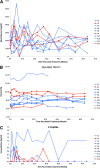
Figure 1
Figure 1
Effects of control and immunosuppressive dendritic cells on nonfasting blood glucose levels (A), nonfasting glycated HbA1c levels (B), and nonfasting, nonstimulated C-peptide levels in serum (C). Week 0 represents pretreatment/baseline levels. The symbols and lines in blue represent the immunosuppressive dendritic cell recipients and those in red represent the control dendritic cell recipients. The legend to the right of each graph shows the symbols that correspond to each individual patient (P). The values were measured in freshly obtained blood and serum at each of the weeks of the trial, shown on the x-axis in the graph. For C-peptide levels, only values >0.5 ng/mL are shown based on standard clinical reference.
Figure 2
Figure 2
A: Serum levels of IL-4 and IL-10 in dendritic cell recipients. The presence and the concentration of serum cytokines was measured by Luminex multianalyte assay systems (LincoPLEX and Beadlyte). Serum IL-4 and IL-10 were reproducibly detectable. The points and lines in red indicate cytokine concentration in the serum of control dendritic cell recipients and the blue points and lines show the concentration in immunosuppressive dendritic cell recipients. The graph at the top shows the levels of IL-4 and that on the bottom shows IL-10. The legend to the right shows the symbols that correspond to each individual patient (P). B: Dendritic cell administration does not confer systemic immunosuppression. Proliferation of dendritic cell-recipient T cells in allogeneic MLR in vitro. 2 × 10⁵ PBMCs from the freshly obtained blood of the dendritic cell recipients were cultured alone or mixed with an equal number of allogeneic irradiated PBMCs in standard one-way MLR. The cells were cultured for 4 days before BrdU addition. BrdU incorporation into proliferating cells was measured on day 5 by flow cytometry. The graph shows the ratio of BrdU+ cells in the presence of allogeneic stimulators to BrdU+ cells in the absence of stimulators at each of the indicated weeks of the trial in seven individual patients compared with baseline (visit 2). Patients 6, 8, 10, 12, and 13 received immunosuppressive dendritic cells; patients 9 and 14 received control dendritic cells. The volumes of blood (and hence cell number) obtained from the remainder of the patients were not permissive to properly conduct biologically meaningful MLR. The legend to the right shows the symbols that correspond to each individual patient (P). C: The ELISPOT response of dendritic cell-recipient T cells to vaccination antigens in vitro. PBMCs from freshly isolated blood were frozen and, for this assay, thawed at a later time. The thawed cells were plated on interferon-γ ELISPOT strips (Human Interferon ELISPOT Pro; Mabtech) at densities of 1 × 10⁵ or 3 × 10⁵ cells. PBMCs were plated alone, with a viral peptide pool (CEF Peptide Pool; Mabtech) at 2 µg/mL, or with a kit-supplied CD3 agonistic antibody. An ELISPOT assay was conducted as recommended by the manufacturer, and the spots were counted electronically (KS ELISPOT; Zeiss) in an Axioplan 2 microscope with ELISPOT adapter/module. The data in the graph are shown for each patient where frozen PBMCs from each of the indicated weeks of the trial were available. The data are represented as the ratio of interferon-γ spots in viral peptide–stimulated PBMCs to the spot number in unstimulated cells. Viability of frozen PBMCs after thawing from the remainder of the patients precluded the conduct of a biologically meaningful MLR assay. The legend to the right shows the symbols that correspond to each individual patient (P).

References

    1. Eisenbarth GS. Insulin autoimmunity: immunogenetics/immunopathogenesis of type 1A diabetes. Ann N Y Acad Sci 2003;1005:109–118 - PubMed
    1. Fan Y, Rudert WA, Grupillo M, He J, Sisino G, Trucco M. Thymus-specific deletion of insulin induces autoimmune diabetes. EMBO J 2009;28:2812–2824 - PMC - PubMed
    1. Lo J, Clare-Salzler MJ. Dendritic cell subsets and type I diabetes: focus upon DC-based therapy. Autoimmun Rev 2006;5:419–423 - PubMed
    1. Steinman RM, Nussenzweig MC. Avoiding horror autotoxicus: the importance of dendritic cells in peripheral T cell tolerance. Proc Natl Acad Sci USA 2002;99:351–358 - PMC - PubMed
    1. McCurry KR, Colvin BL, Zahorchak AF, Thomson AW. Regulatory dendritic cell therapy in organ transplantation. Transpl Int 2006;19:525–538 - PubMed

"V体育ios版" Publication types

MeSH terms

Substances (VSports注册入口)