Skip to main page content
U.S. flag

An official website of the United States government

Dot gov

The . gov means it’s official. Federal government websites often end in . gov or . mil VSports app下载. Before sharing sensitive information, make sure you’re on a federal government site. .

Https

The site is secure. The https:// ensures that you are connecting to the official website and that any information you provide is encrypted and transmitted securely V体育官网. .

. 2011 Jun 12;18(7):769-76.
doi: 10.1038/nsmb.2062.

ATRX ADD domain links an atypical histone methylation recognition mechanism to human mental-retardation syndrome

Affiliations

ATRX ADD domain links an atypical histone methylation recognition mechanism to human mental-retardation syndrome

Shigeki Iwase et al. Nat Struct Mol Biol. .

"V体育官网入口" Abstract

ATR-X (alpha-thalassemia/mental retardation, X-linked) syndrome is a human congenital disorder that causes severe intellectual disabilities. Mutations in the ATRX gene, which encodes an ATP-dependent chromatin-remodeler, are responsible for the syndrome. Approximately 50% of the missense mutations in affected persons are clustered in a cysteine-rich domain termed ADD (ATRX-DNMT3-DNMT3L, ADD(ATRX)), whose function has remained elusive. Here we identify ADD(ATRX) as a previously unknown histone H3-binding module, whose binding is promoted by lysine 9 trimethylation (H3K9me3) but inhibited by lysine 4 trimethylation (H3K4me3) VSports手机版. The cocrystal structure of ADD(ATRX) bound to H3(1-15)K9me3 peptide reveals an atypical composite H3K9me3-binding pocket, which is distinct from the conventional trimethyllysine-binding aromatic cage. Notably, H3K9me3-pocket mutants and ATR-X syndrome mutants are defective in both H3K9me3 binding and localization at pericentromeric heterochromatin; thus, we have discovered a unique histone-recognition mechanism underlying the ATR-X etiology. .


Conflict of interest statement

COMPETING FINANCIAL INTERESTS

Y. S is a co-founder of Constellation Pharmaceuticals. D. J. Patel is on the Epigenetics Advisory Board of Epinova-Glaxo V体育安卓版. The remaining authors declare no competing financial interests.

Figures
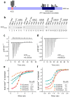
Figure 1
Figure 1. Methylation at K4 and K9 inversely regulates interaction between AD-DATRX and histone H3
(a) ATRX patient mutations (circles) are predominantly found in either the ADD or the SNF2 (Sucrose Non Fermenting)-type ATP-dependent chromatin remodeling domain. (b) ADDATRX interacts with the unmodified H3 peptide (1–21 aa) (lane 3), and the interaction was enhanced by K9 methylation (lanes 4–6). (c–f) Experimental ITC titration curves are displayed in panel c for unmodified H3 (peptide to buffer control shown in the top), and panel d for H3K9me3 peptide. The fitting curves and the calculated binding affinities are listed in panels e and f. Methylation at K4 (panel e) and K9 (panel f) displayed entirely different effects on the binding affinity. Detailed peptide sequences and complete ITC fitting parameters are summarized in Supplementary Table 1.
Figure 2
Figure 2. Atrx is recruited to PCH by Suv39h1/2-mediated H3K9 trimethylation
(a) Atrx fails to locate on PCH in Suv39h1/h2 double knock-out (DKO) cells. (b and c) Suv39h1-WT but not a catalytically inactive mutant Suv39h1-H324L restores the PCH localization of Atrx in DKO cells. Suv39h1-WT (panels a to d) and Suv39h1-H324L (panels e to h) were expressed in DKO cells. (c) Quantification of presence or absence of H3K9me3 signal in the Suv39h1-positive cells (left panel), and punctate PCH localization or diffused distribution of Atrx (right panel). (d) Mecp2 is localized at PCH in both WT- (panels a to c) and DKO- (panels d to f) MEF cells. (e) Schematic diagram of ATRX proteins used for testing their localization. (f) Deletion of PxVxL motif resulted in partial loss of PCH localization. Subnuclear distribution of ATRX proteins represented in (Fig. 4f) was quantified. (g) Targeting of Atrx (panels b and f) to Daxx-positive PML bodies (panels a and e) is not affected in Suv39h DKO cells (panels e to h).
Figure 3
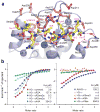
Figure 3. Molecular basis for H3K9me3 recognition by ADDATRX
(a) Ribbon representation of ADDATRX domain in complex with H31–15K9me3 peptide. ADDATRX domain is a hybrid of a GATA-like zinc finger (cyan) and a PHD finger (magenta). Three zinc ions are depicted as spheres and the H3 peptide is colored yellow with key residues Lys4 and K9me3 shown in stick representation. (b) Surface electrostatic view of ADDATRX domain in complex with H31–15K9me3 peptide, color-coded with red representing negatively-charged and blue positively-charged potentials. The Fo-Fc omit map was contoured at 3.5 σ level for the bound H3 peptide. (c) Stereo view of the K9me3-binding pocket. The GATA-like segment Tyr203–Ser210 and the PHD finger segment Gln219–-Glu225 are color-coded in cyan and magenta, respectively. Blue dotted lines refer to nonconventional C-H:O hydrogen bonds. (d) A snug fit of the bulky trimethyllysine group inserted into the reader pocket of ADDATRX.
Figure 4
Figure 4. Effect of H3 sequence context on ADDATRX binding
(a) Details of H3-ADDATRX interaction. Histone H3 Ala1–Ser10 containing K9me3 modification is depicted as yellow sticks. ADDATRX is shown as light blue ribbons with key residues that interact with H3 peptide shown as pink sticks. Red dotted lines denote hydrogen bonds. Three zinc ions and water molecules are depicted as large blue and small red spheres, respectively. (b) ITC titration fitting curves and the binding constants of synthetic H3 peptide variants to WT-ADDATRX. R2A, K4A, K9A: Arg2, Lys4, Lys9 to alanine substitution, respectively; AAH3: H3 N-terminal double alanine extension; R2me2s: Arg2 symmetrical dimethylation; R2me2a: Arg2 asymmetrical dimethylation; K9me3: Lys9 trimethylation; K9ac: Lys9 acetylation; n.d., not determined. Detailed peptide sequences and complete ITC fitting parameters are summarized in Supplementary Table 1.
Figure 5
Figure 5. Mutations in ADDATRX compromise histone binding and targeting of ATRX onto PCH
(a) Alignment of the ADD domains. Hs: Homo sapiens (human), Dr: Danio rario (zebrafish), Xt: Xenopus tropicalis (frog). Conserved cysteines are presented as red letters. (b) Mutations in the K9me3-pocket limit the binding of ADDATRX to H3 peptides methylated at Lys9. (c) Mutations in the K9me3-pocket disrupt binding to nucleosomes. GST alone (lane 1) or GST-ADDATRX WT (lane 2) as well as mutants (lanes 3 to 6) were incubated with nucleosomes. (d and e) Four patient mutations resulted in defective H3 peptide binding (d) and nucleosome binding (e). (f) Representative images of subnuclear distribution of recombinant ATRX proteins in NIH3T3 cells. (g and h) Quantification of the distribution patterns is shown for the four K9me3-pocket mutants (g) and additional four syndrome-associated mutants (h).
Figure 6
Figure 6. Model of the mechanism underlying ATR-X syndrome caused by the mutations in ADDATRX
Atrx is recruited on PCH via interaction between ADD and H3K9me3 generated by Suv39h HMTs. Physical associations of Hp1/Atrx and Mecp2/Atrx support the PCH anchoring of Atrx. Patient mutations in ADD disrupt H3K9me3 binding; then Atrx falls off from PCH. Loss of Atrx from PCH may leads to compromised higher-order structure of PCH, followed by chromosome missegregation and apoptosis in neuroprogenitor cells.

References

    1. Li H, et al. Molecular basis for site-specific read-out of histone H3K4me3 by the BPTF PHD finger of NURF. Nature. 2006;442:91–5. - PMC - PubMed
    1. Wysocka J, et al. A PHD finger of NURF couples histone H3 lysine 4 trimethylation with chromatin remodelling. Nature. 2006;442:86–90. - PubMed
    1. Shi X, et al. ING2 PHD domain links histone H3 lysine 4 methylation to active gene repression. Nature. 2006;442:96–9. - PMC - PubMed
    1. Pena PV, et al. Molecular mechanism of histone H3K4me3 recognition by plant homeodomain of ING2. Nature. 2006;442:100–3. - VSports在线直播 - PMC - PubMed
    1. Bannister AJ, et al. Selective recognition of methylated lysine 9 on histone H3 by the HP1 chromo domain. Nature. 2001;410:120–4. - PubMed

Publication types

MeSH terms