"VSports注册入口" Skip to main page content
U.S. flag

An official website of the United States government

Dot gov

The . gov means it’s official. Federal government websites often end in . gov or . mil. Before sharing sensitive information, make sure you’re on a federal government site VSports app下载. .

Https

The site is secure V体育官网. The https:// ensures that you are connecting to the official website and that any information you provide is encrypted and transmitted securely. .

. 2011 Jun 8;13(6):701-11.
doi: 10.1016/j.cmet.2011.04.008.

High-mobility group box 1 is essential for mitochondrial quality control

Affiliations

High-mobility group box 1 is essential for mitochondrial quality control (V体育ios版)

Daolin Tang et al. Cell Metab. .

Abstract

Mitochondria are organelles centrally important for bioenergetics as well as regulation of apoptotic death in eukaryotic cells. High-mobility group box 1 (HMGB1), an evolutionarily conserved chromatin-associated protein which maintains nuclear homeostasis, is also a critical regulator of mitochondrial function and morphology VSports手机版. We show that heat shock protein beta-1 (HSPB1 or HSP27) is the downstream mediator of this effect. Disruption of the HSPB1 gene in embryonic fibroblasts with wild-type HMGB1 recapitulates the mitochondrial fragmentation, deficits in mitochondrial respiration, and adenosine triphosphate (ATP) synthesis observed with targeted deletion of HMGB1. Forced expression of HSPB1 reverses this phenotype in HMGB1 knockout cells. Mitochondrial effects mediated by HMGB1 regulation of HSPB1 expression serve as a defense against mitochondrial abnormality, enabling clearance and autophagy in the setting of cellular stress. Our findings reveal an essential role for HMGB1 in autophagic surveillance with important effects on mitochondrial quality control. .


VSports注册入口 - Figures

Figure 1
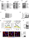
Figure 1. HMGB1 sustains cellular bioenergetics and normal mitochondrial morphology in MEFs
(A) Transient transfection of HMGB1 (pEGFPN1-HMGB1) or empty vector (pEGFPN1) into Hmgb1−/− MEFs increased HMGB1 protein expression after 48 h by western blot assay “+/+”: Hmgb1+/+ MEFs; “−/−”: Hmgb1−/− MEFs. (B) Expression of HMGB1 restores normal cellular bioenergetics in Hmgb1−/− MEFs. Cells were treated sequentially as indicated with oligomycin (“Olig”, 1 uM), p-trifluoromethoxy carbonyl cyanide phenyl hydrazone (“FCCP”, 0.3 uM), 2-deoxyglucose (“2DG”, 100 mM) and rotenone (“Rote”, 1 uM) in sequence as shown. Oxygen consumption rates (OCR, indicative of OXPHOS) and extracellular acidification rates (ECAR, indicative of glycolysis) were monitored using the Seahorse Bioscience Extracellular Flux Analyzer in real time (mean ±SD, n = 3). “+/+”: Hmgb1+/+ MEFs, “−/−”: Hmgb1−/− MEFs. (C) ATP levels were assessed using an ATP Assay Kit (PerkinElmer Life Sciences) in Hmgb1−/− (“−/−“) MEFs with or without pEGFPN1 empty vector or pEGFPN1-HMGB1 transfection. Data represent relative ATP levels, with Hmgb1+/+ (“+/+”) MEFs set as 100% (mean ±SD, n=3, *p < 0.01, “ns”: not significant). (D) Knockout or knockdown of HMGB1 decreases cellular proliferation in MEFs and NIH3T3 cells. Data are expressed as means ± SD. (E) Mitochondrial (“Mit”) morphology was evaluated using antibodies against mitochondrial Complex I subunit GRIM-19 (red) with or without pUNO1 empty vector or pUNO1-HMGB1 transfection. HMGB1 and the nucleus were stained by HMGB1 antibody (green) or Hoechst 33342 (blue), respectively. Mitochondrial fragmentation (%) in Hmgb1+/+ (“+/+”) and Hmgb1−/− (“−/−“) MEFs were quantified from 15–20 random fields. In parallel, the mitochondrial membrane potential was assayed for JC-1 expression by flow cytometry (mean ±SD, n=3, *p < 0.01, ns: not significant, Hmgb1+/+ group set as 1).
Figure 2
Figure 2. HMGB1 is required to sustain cellular bioenergetics and mitochondrial morphology in MEFs and NIH3T3 cells
(A) Western blot assay for HMGB1 expression in the indicated cells after transfection for 48h with control shRNA or HMGB1 shRNA. (B) Knockdown of HMGB1 in MEFs or NIH3T3 cells decreased OXPHOS. Cells were sequentially treated as indicated with oligomycin (“Olig”, 1 uM), p-trifluoromethoxy carbonyl cyanide phenyl hydrazone (“FCCP”, 0.3 uM), 2-deoxyglucose (“2DG”, 100 mM), and rotenone (“Rote”, 1 uM). Oxygen consumption rates (OCR, indicative of OXPHOS) and extracellular acidification rates (ECR, indicative of glycolysis) were monitored using the Seahorse Bioscience Extracellular Flux Analyzer in real time (mean ±SD, n = 3). (C) ATP levels were assessed using an ATP Assay Kit (PerkinElmer Life Sciences) in control shRNA or HMGB1 shRNA transfected cells. Data represent relative ATP levels, with control shRNA group set as 100% (mean ±SD, n=3, *p < 0.01). (D) Mitochondrial morphology was evaluated using antibody against the mitochondrial Complex I subunit GRIM-19 (red) in control shRNA or HMGB1 shRNA transfected cells. Images are representative of 15–20 random fields. All data are representative of two or three experiments. Note in particular the fragmented mitochondria and larger nuclei in Hmgb1−/− cells.
Figure 3
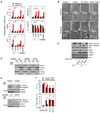
Figure 3. HMGB1 and HSPB1 are required to sustain OXPHOS in pancreatic and colon tumor cells
(A) Western blot assay for HMGB1 expression in the indicated cells after transfection for 48h with control shRNA or HMGB1 shRNA or HSPB1 shRNA. (B) Knockdown of HMGB1 or HSPB1 in pancreatic and colon tumor cells decreased OXPHOS (coincident curves below control). Cells were sequentially treated as indicated with oligomycin (“Olig”, 1 uM), p-trifluoromethoxy carbonyl cyanide phenyl hydrazone (“FCCP”, 0.3 uM), 2-deoxyglucose (“2DG”, 100 mM), and rotenone (“Rote”, 1 uM). Oxygen consumption rates (OCR, indicative of OXPHOS) and extracellular acidification rates (ECR, indicative of glycolysis) were monitored using the Seahorse Bioscience Extracellular Flux Analyzer in real time (mean ±SD, n = 3). (C) ATP levels were assessed using an ATP Assay Kit (PerkinElmer Life Sciences) in control shRNA or HMGB1 shRNA or HSPB1 shRNA transfected cells. Data represent relative ATP levels, with control shRNA group set as 100% (mean ±SD, n=3, *p < 0.01 vs. control shRNA group). All data are representative of two or three experiments.
Figure 4
Figure 4. Knockdown of HSPB1 recapitulates the HMGB1-deficient metabolic phenotype and mitochondrial morphology
(A) HMGB1 regulates HSPB1 but not other HSP protein levels. Western blot analysis of HSPB1 and other proteins implicated in mitochondrial morphology in Hmgb1+/+ (“+/+”) and Hmgb1−/− (“−/−“) MEFs, and MEFs or NIH3T3 cells transfected with control shRNA or HMGB1 shRNA are shown. (B) Dependence of HSPB1 protein and mRNA transactivation levels on HMGB1. Transient transfection of pUNO1-HMGB1 (“HMGB1”) into Hmgb1−/− MEFs restored HSPB1 protein expression by western blot assay (top panel) and HSPB1 mRNA expression by reverse transcription polymerase chain reaction (bottom panel). “+/+”: Hmgb1+/+ MEFs, “−/−”: Hmgb1−/− MEFs. (C) Western blot analysis of HSPB1 and HSP70 expression in Hmgb1+/+ and Hmgb1−/− MEFs after heat shock (60 min at 42.5°C water bath, then 12 h recovery). (D) In parallel, heat shock element (HSE) activity was evaluated in a luciferase reporter assay (“AU”: arbitrary unit). Data are expressed as means ± SD (n=3). (E) Knockdown of HSPB1 by shRNA reproduces the bioenergetic phenotype observed in HMGB1-deficient cells. The indicated cells were treated sequentially with oligomycin (“Olig”, 1 uM), p-trifluoromethoxy carbonyl cyanide phenyl hydrazone (“FCCP”, 0.3 uM), 2-deoxyglucose (“2DG”, 100 mM) and rotenone (“Rote”, 1 uM). Oxygen consumption rates (OCR, indicative of OXPHOS) and extracellular acidification rates (ECAR, indicative of glycolysis) were monitored using the Seahorse Bioscience Extracellular Flux Analyzer in real time (mean ±SD, n = 3). “+/+”: Hmgb1+/+ MEFs, “−/−”: Hmgb1−/− MEFs. (F) ATP levels were assessed in MEFs transfected with HSPB1 shRNA. Data represent relative ATP levels, with control shRNA MEFs set as 100% (mean ±SD, n=3, *p < 0.01 vs. control shRNA group). (G) Mitochondrial morphology was evaluated using an antibody against the mitochondrial Complex I subunit GRIM-19 (red). Mitochondrial fragmentation numbers (%) in cells were quantified from 15–20 random fields (mean ±SD, *p < 0.01 vs. control shRNA group). All data are representative of two or three experiments.
Figure 5
Figure 5. HSPB1 expression restores cellular bioenergetics and mitochondrial morphology in Hmgb1−/− MEFs
(A) Transient transfection of pcDNA4-HisMaxC-HSPB1 (“HSPB1 cDNA”) into Hmgb1−/− MEFs increased HSPB1 protein expression after 48h as determined by western blot assay. “+/+”: Hmgb1+/+ MEFs, “−/−”: Hmgb1−/− MEFs. Control cDNA=pcDNA4-HisMaxC. (B) Expression of HSPB1 rescues alterations in cellular bioenergetics in Hmgb1−/− MEFs. Cells were sequentially treated as indicated with oligomycin (“Olig”, 1 uM), p-trifluoromethoxy carbonyl cyanide phenyl hydrazone (“FCCP”, 0.3 uM), 2-deoxyglucose (“2DG”, 100 mM), and rotenone (“Rote”, 1 uM). Oxygen consumption rates (OCR, indicative of OXPHOS) and extracellular acidification rates (ECAR, indicative of glycolysis) were monitored using the Seahorse Bioscience Extracellular Flux Analyzer in real time (mean ±SD, n = 3). “+/+”: Hmgb1+/+ MEFs, “−/−”: Hmgb1−/− MEFs. (C) ATP levels were assessed in Hmgb1−/− MEFs with or without HSPB1 cDNA transfection. Data represent relative ATP levels, with Hmgb1+/+ MEFs set as 100% (mean ±SD, n=3, *p < 0.01. ns: not significant). “+/+”: Hmgb1+/+ MEFs, “−/−”: Hmgb1−/− MEFs. (D) Mitochondrial morphology was evaluated using an antibody against the mitochondrial Complex I subunit, GRIM-19 (red). Mitochondrial fragmentation as a percent of cells was quantified in 15–20 random fields (mean ±SD, *p < 0.01, ns: not significant). “+/+”: Hmgb1+/+ MEFs, “−/−”: Hmgb1−/− MEFs. All data are representative of two or three experiments.
Figure 6
Figure 6. HMGB1 and HSPB1 regulate mitophagy following rotenone-induced disruption of oxidative phosphorylation
(A) Confocal microscopyana colocalizes mitochondria with LC3, LAMP2 and actin in Hmgb1+/+ (“+/+”), Hmgb1−/− (“−/−”), control shRNA or HSPB1 shRNA MEFs with or without rotenone (2 uM, 6 h). Cytochalasin D (“CytD”, 1 uM, 6h) was used to inhibit actin polymerization. Images were acquired digitally from a randomly selected pool of 10–15 fields under each condition. Quantitative analysis of colocalization (%) was performed. Representative images are showed in Figure S4. In parallel, apoptosis was analyzed by the Annexin V-FITC apoptosis detection kit and mitochondrial membrane potential depolarization (Hmgb1+/+ control group seat as 1) was measured by JC-1 fluorescence. Data are expressed as mean ± SD (*p < 0.01, n=3). (B) Ultrastructural features assessed by electron microscopy in Hmgb1+/+, Hmgb1−/−, control shRNA and HSPB1 shRNA MEFs with or without ATP depletion by rotenone (2 uM, 4h) (arrows point to mitochondria in autophagosomes or autolysosomes which are not apparent in Hmgb1−/− or HSPB1 knockdowns). (C) ATP depletion by rotenone (2 uM, 6 h) increased the expression of p-HSPB1 (Ser15 and Ser86) in Hmgb1+/+ (“+/+”) cells but not Hmgb1−/− (“−/−”) cells. HSPB1 mutants (S15A, S86A) cannot restore the decreased LC3-II and increased p62 expression observed in Hmgb1−/− cells following rotenone treatment. (D) Cells as indicated were treated with rotenone (2 uM) for 6 h, and then cytoplasmic (Cyt), and mitochondrial (Mit) extracts from cells were separated by SDS-PAGE. The blots were probed with antibody against Parkin, Cox IV, tubulin, VDAC1 and Pink1. (E) Cells as indicated were treated with rotenone (2 uM) for 6 h, and then VDAC1 ubiquitylation was assayed by immunoprecipitation (IP) and western blot (IB). (F) Cellular ATP levels and mitochondrial fragmentation was assayed in Hmgb1+/+ (“+/+”) and Hmgb1−/− (“−/−”) cells transfected with HSPB1 vectors with or without Pink1 or Parkin shRNA (mean ±SD, *p < 0.01, ns: not significant).
Figure 7
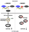
Figure 7. Schematic of the mechanism by which HMGB1 modulates mitochondrial respiration and morphology by sustaining mitophagy
In wild type cells, HMGB1 functions as a transcriptional regulator of HSPB1 gene expression. Phosphorylation of HSPB1 is necessary to regulate the actin cytoskeleton, which affects the cellular transport of autophagy/mitophagy in response to mitochondrial injury. Loss of either HMGB1 or HSPB1 results in phenotypically similar deficient mitophagy typified by mitochondrial fragmentation, decreased aerobic respiration and subsequent ATP production. OXPHOS: oxidative phosphorylation.

References (VSports app下载)

    1. Arrigo AP. The cellular "networking" of mammalian Hsp27 and its functions in the control of protein folding, redox state and apoptosis. Adv Exp Med Biol. 2007;594:14–26. - PubMed
    1. Benndorf R, Hayess K, Ryazantsev S, Wieske M, Behlke J, Lutsch G. Phosphorylation and supramolecular organization of murine small heat shock protein HSP25 abolish its actin polymerization-inhibiting activity. J Biol Chem. 1994;269:20780–20784. - PubMed
    1. Bjorkoy G, Lamark T, Brech A, Outzen H, Perander M, Overvatn A, Stenmark H, Johansen T. p62/SQSTM1 forms protein aggregates degraded by autophagy and has a protective effect on huntingtin-induced cell death. J Cell Biol. 2005;171:603–614. - "VSports最新版本" PMC - PubMed
    1. Boldogh IR, Pon LA. Interactions of mitochondria with the actin cytoskeleton. Biochim Biophys Acta. 2006;1763:450–462. - V体育平台登录 - PubMed
    1. Brooks C, Wei Q, Cho SG, Dong Z. Regulation of mitochondrial dynamics in acute kidney injury in cell culture and rodent models. J Clin Invest. 2009;119:1275–1285. - PMC - PubMed

VSports手机版 - Publication types

MeSH terms

Substances