Skip to main page content
U.S. flag

An official website of the United States government

Dot gov

The . gov means it’s official. Federal government websites often end in VSports app下载. gov or . mil. Before sharing sensitive information, make sure you’re on a federal government site. .

Https

The site is secure V体育官网. The https:// ensures that you are connecting to the official website and that any information you provide is encrypted and transmitted securely. .

Comparative Study
. 2011 Jun;13(6):591-9.
doi: 10.1093/neuonc/nor042.

Myeloid-derived suppressor cell accumulation and function in patients with newly diagnosed glioblastoma

Affiliations
Comparative Study

Myeloid-derived suppressor cell accumulation and function in patients with newly diagnosed glioblastoma

Baisakhi Raychaudhuri et al. Neuro Oncol. 2011 Jun.

Abstract (V体育ios版)

To assess the accumulation of myeloid-derived suppressor cells (MDSCs) in the peripheral blood of patients with glioma and to define their heterogeneity and their immunosuppressive function. Peripheral blood mononuclear cells (PBMCs) from healthy control subjects and from patients with newly diagnosed glioma were stimulated with anti-CD3/anti-CD28 and T cells assessed for intracellular expression of interferon (IFN)-γ. Antibody staining of PBMCs from glioma patients and healthy donors (CD33, HLADR, CD15, and CD14) followed by 4-color flow cytometry analysis-defined MDSC levels in the peripheral blood. To assess the role of MDSCs in suppressing T cell IFNγ production, PBMCs were depleted of MDSCs using anti-CD33 and anti-CD15 antibody-coated beads prior to T cell stimulation. Enzyme-linked immunosorbent assays were used to assess plasma arginase activity and the level of granulocyte colony-stimulating factor (G-CSF). Patients with glioblastoma have increased MDSC counts (CD33+HLADR-) in their blood that are composed of neutrophilic (CD15(+); >60%), lineage-negative (CD15(-)CD14(-); 31%), and monocytic (CD14(+); 6%) subsets. After stimulation, T cells from patients with glioblastoma had suppressed IFN-γ production when compared with healthy, age-matched donor T cells. Removal of MDSCs from the PBMCs with anti-CD33/CD15-coated beads significantly restored T cell function. Significant increases in arginase activity and G-CSF levels were observed in plasma specimens obtained from patients with glioblastoma. The accumulation of MDSCs in peripheral blood in patients with glioma likely promotes T cell immune suppression that is observed in this patient population. Increased plasma levels of arginase and G-CSF may relate to MDSC suppressor function and MDSC expansion, respectively, in patients with glioma VSports手机版. .


Figures

Fig. 1.
Fig. 1.
Patients with glioblastoma (GBM) have elevated levels of myeloid-derived suppressor cells (MDSCs) compared with age-matched healthy donors and other cancer patients. Peripheral blood mononuclear cells (PBMCs) from patients with newly diagnosed GBM, healthy age-matched donors, and other patients with cancer were isolated and stored in liquid nitrogen. Cells were thawed, washed, and blocked with human immunoglobulin G and then immediately stained at 4°C with monoclonal antibodies to CD14, CD15, CD33, and HLA-DR. Cells were fixed with 1% paraformaldehyde, then acquired and analyzed by multicolor flow cytometry.
Fig. 2.
Fig. 2.
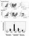
Neutrophilic subsets are prevalent in patients with glioblastoma (GBM). (A) The difference in FACS forward and side scatter profile shows that peripheral blood mononuclear cells (PBMCs) from the patients with GBM have a significant population of cells that is not detectable in PBMCs from healthy donors. Dot plots represent live gated events. The forward and side scatter gate was analyzed for CD3+HLADR cells. Then the CD33+HLADR gate was analyzed for cells expressing CD15 and CD14. (B) Columns represent mean percentages of myeloid-derived suppressor cells (MDSCs) in total PBMCs from healthy, age-matched donors, patients with GBM, and patients with other cancers. The majority of the MDSCs in patients with GBM are neutrophilic CD15+ CD14 (82%), followed by lineage-negative (lyn15%) and monocytic (3%) MDSCs.
Fig. 3.
Fig. 3.
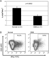
Patients with glioblastoma (GBM) are deficient in IFN-γ production. Peripheral blood mononuclear cells from patients with GBM or healthy donors were stored in liquid N2, thawed, and rested overnight in complete media, and equal numbers of nonadherent cells were stimulated the following day with anti-CD3/CD28 antibody–coated beads and interleukin-2 for 72 h. CD3+ gated cells were analyzed for intracellular cytokine (ICC) staining by FACS. Columns in top panel represent mean percentage of interferon (IFN)–γ-positive cells. Live gated events are shown in the bottom panel.
Fig. 4.
Fig. 4.
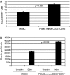
In vitro depletion of patient myeloid-derived suppressor cells (MDSCs) partially restores patient T cell production and proliferation of interferon (IFN)-γ. (A) PBMCs from patients with glioblastoma (GBM) (n = 3) were thawed and immediately stained for the expression of myeloid surface markers with or without the depletion of MDSC by anti-CD15 magnetic microbeads and magnetic column. Cells were then plated and stimulated with anti-CD3/CD28 antibodies, and after 72 h, IFN-γ was detected in CD3+ cells by FACS. Columns represent mean percentage of CD3+ cells staining positive for IFN-γ. (B) Patients’ PBMCs were left alone or CD33/CD15 magnetic beads were used to remove MDSCs. The cells were stimulated or not with anti-CD3/CD28 for 72 h. Then, 3H-thymidine was added 18 h before the harvest of the cells. Proliferation was measured by 3H incorporation in DNA.
Fig. 5.
Fig. 5.
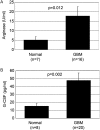
Patients with glioblastoma (GBM) have elevated levels of plasma G-CSF and arginase activity. (A) Serum arginase levels for healthy, age-matched donors (n = 7) and patients with GBM (n = 16) were measured using an arginase kit (Bio Assay Systems). Serum samples from patients had a significantly higher level of arginase than did control plasma samples (P = .012). (B) Serum samples from patients with GBM (n = 20) and healthy donors (n = 8) were thawed, and the G-CSF level was measured using an enzyme-linked immunosorbent assay (R&D Systems) in accordance with the instructions supplied by the vendor. Data represent mean ± standard error of the mean of 3 different experiments (P = .002).

References

    1. Ostrand-Rosenberg S. Myeloid-derived suppressor cells: more mechanisms for inhibiting antitumor immunity. Cancer Immunol Immunother. 2010;59(10):1593–1600. doi:10.1007/s00262-010-0855-8. - DOI - PMC - PubMed
    1. Gabrilovich DI, Nagaraj S. Myeloid-derived suppressor cells as regulators of the immune system. Nat Rev Immunol. 2009;9(3):162–174. V体育安卓版 - doi:10.1038/nri2506. - DOI - PMC - PubMed
    1. Bronte V, Serafini P, Apolloni E, Zanovello P. Tumor-induced immune dysfunctions caused by myeloid suppressor cells. J Immunother. 2001;24(6):431–446. VSports手机版 - doi:10.1097/00002371-200111000-00001. - DOI - PubMed
    1. Ugel S, Delpozzo F, Desantis G, et al. Therapeutic targeting of myeloid-derived suppressor cells. Curr Opin Pharmacol. 2009;9(4):470–481. doi:10.1016/j.coph.2009.06.014. - DOI - PubMed
    1. Almand B, Clark JI, Nikitina E, et al. Increased production of immature myeloid cells in cancer patients: a mechanism of immunosuppression in cancer. J Immunol. 2001;166(1):678–689. - V体育平台登录 - PubMed

MeSH terms

LinkOut - more resources