<b dir="LhCfEX0h"></b> Skip to main page content
U.S. flag

An official website of the United States government

Dot gov

The . gov means it’s official VSports app下载. Federal government websites often end in . gov or . mil. Before sharing sensitive information, make sure you’re on a federal government site. .

Https

The site is secure. The https:// ensures that you are connecting to the official website and that any information you provide is encrypted and transmitted securely. V体育官网.

. 2011 Jun 1;29(7):607-14.
doi: 10.1038/nbt.1873.

"VSports最新版本" Mapping in vivo protein-RNA interactions at single-nucleotide resolution from HITS-CLIP data

Affiliations

Mapping in vivo protein-RNA interactions at single-nucleotide resolution from HITS-CLIP data

Chaolin Zhang (VSports最新版本) et al. Nat Biotechnol. .

Abstract

Mammalian RNA complexity is regulated through interactions of RNA-binding proteins (RBPs) with their target transcripts. High-throughput sequencing together with UV-crosslinking and immunoprecipitation (HITS-CLIP) is able to globally map RBP-binding footprint regions at a resolution of ~30-60 nucleotides. Here we describe a systematic way to analyze HITS-CLIP data to identify exact crosslink sites, and thereby determine protein-RNA interactions at single-nucleotide resolution. We found that reverse transcriptase used in CLIP frequently skips the crosslinked amino-acid-RNA adduct, resulting in a nucleotide deletion. Genome-wide analysis of these crosslinking-induced mutation sites (CIMS) in HITS-CLIP data for Nova and Argonaute (Ago) proteins in mouse brain tissue revealed deletions in ~8-20% of mRNA tags, which mapped to Nova and Ago binding sites on mRNA or miRNA. CIMS analysis provides a general and more precise means of mapping protein-RNA interactions than currently available methods and insight into the biochemical properties of such interactions in living tissues. VSports手机版.


Figures

Figure 1
Figure 1. Overview of CIMS analysis
a. Schematic representation of HITS-CLIP and cross-linking induced mutations. Protein-RNA complexes are purified by immunoprecipitation and stringent washing, followed by proteinase K treatment, a broad-specificity enzyme which cleaves peptide bonds. Remaining cross-linked amino acid(s) attached to RNA, as indicated by the red cross, impose an obstacle for RT, so that a mutation may be induced (~8–20% frequency; see below) during reverse transcription of RNA to cDNA. CLIP tags are then PCR amplified and read-out by high-throughput sequencing. b. Schematic representation of the CIMS analysis method. Mutations detected during alignment, indicated by blue triangles, are clustered into discrete sites according to their genomic coordinates. Each site (cluster) is characterized by the total number of overlapping unique tags k and the number of tags with particular types of mutations m at the position. A permutation-based approach, which preserves the distribution of CLIP tags in the genome and also the positional bias of mutations relative to the 5′ end of reads, is used to evaluate the statistical significance of clustering for each given k and to estimate the FDR (see Online Methods for more details).
Figure 2
Figure 2. Cross-linking induces deletions, but not substitutions that precisely map Nova-mRNA interactions
a. The positional profiles of deletions (top panels, blue) and substitutions (bottom panels, green) relative to the 5′ end of reads are shown. Analysis of CLIP data is shown on the left whereas analysis of non-cross-linked mRNA-Seq data is shown on the right as a control. b. Distribution of deletion sites or substitution sites is shown as a function of the number of tags supporting the mutation (m). c. Enrichment of YCAY around clustered deletion sites (blue curve) is calculated from the number of YCAY starting at each position relative to the deletion sites, normalized by the frequency in flanking sequences. The same CLIP clusters are re-anchored at the CLIP tag cluster peak to calculate the enrichment of YCAY shown in the yellow curve. d. Similar to (c) but the enrichment of YCAY around clustered substitution sites (green) or around CLIP cluster peaks of the corresponding CLIP clusters (yellow curve) is shown.
Figure 3
Figure 3. Frequency of CIMS in CLIP tag clusters and association of CIMS with Nova regulated alternative exons
a. The proportion of CLIP tag clusters in genic or extended 3′ UTR regions (10k nt downstream of transcript termination) which harbor CIMS defined with varying stringency is shown as a function of CLIP tag cluster peak height (PH). b. Similar to (a) but CIMS are defined with FDR<0.001 and CLIP tag clusters in different genomic regions are shown separately for comparison. c. The breakdown of 325 non-redundant Nova-regulated cassette exons is shown, according to whether they have CIMS in the alternative exon, or upstream or downstream introns that are important for Nova-dependent alternative splicing regulation. CIMS are defined with varying stringency, similar to (a). d. An example (Nova1 exon 4) of CIMS that precisely maps Nova-RNA interactions. Top panel: the Nova1 gene locus, with the number of CLIP tags and frequency of deletions shown in blue and red, respectively. Inclusion or exclusion of exon 4 is autoregulated by Nova . Middle panel: a zoom-in view of exon4 and flanking intronic sequences. In addition to CLIP tags and deletions, positions of YCAY elements, scores of bioinformatically predicted YCAY clusters , and cross-species sequence conservation in mammals are shown. Bottom panel: A further zoom-in view of sequences around the CIMS. YCAY elements and the nucleotides with deletions are highlighted. Note that although the alignment algorithm assigned the deletion to the first of the three uridines shown in red, the actual location could be any of the three nucleotides.
Figure 4
Figure 4. CIMS analysis refines the Nova binding motif
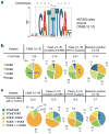
a. A dimeric Nova binding motif with sites identified in 487 of 500 21-nt sequences (−10 to +10 nt) around the top CIMS by de novo motif analysis. The two YCAY elements (highlighted) are separated by a spacer region (shaded), which is variable in length, but predominantly 1 nt as depicted in the motif logo. The consensus of the motif is shown above the logo. b. U is preferred over C in the first or last position of the Nova binding YCAY element around CIMS. The overall frequency (top) and composition (bottom) of the four tetramers conforming to the YCAY consensus are shown for 11-nt sequences around CIMS (−5 to +5 nt around deletion sites, FDR<0.001), 11-nt sequences around CLIP tag peaks of the same set of clusters with CIMS, 11-nt sequences around CLIP tag peaks of the most robust clusters independent of CIMS, or random positions in transcripts. c. A single-nucleotide spacer between the two YCAY elements is preferred for dimeric Nova binding sites around CIMS. The overall frequency (top) and composition of dimeric motif sites with a spacer of different sizes (bottom) are shown for 11-nt sequences around CIMS or sequences around control groups, as in (b).
Figure 5
Figure 5. Cross-linking induces deletions, but not substitutions, that precisely map Ago-mRNA and Ago-miRNA interaction sites
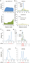
a. The positional profiles of deletions (top panel, blue) and substitutions (bottom panel, green) on Ago mRNA CLIP tags relative to 5′ end of reads. b. Top panel: frequency of miRNA seed matches starting at each position around clustered deletion sites is shown for four top miRNAs (miR-124, miR-9, let-7, and miR-26) with the most seed enrichment and abundant in the brain (blue curve). The same CLIP clusters are re-anchored at the CLIP tag cluster peak to calculate the positional frequency of seed matches shown in the yellow curve. Bottom panel: similar to the top panel, but the frequency of miRNA seed matches around clustered substitution sites (green) or around CLIP tag cluster peak of the corresponding CLIP clusters (yellow curve) is shown. c–f. Positional frequency of deletions for representative individual miRNAs abundant in brain: miR-124 (c), let-7i, b, and c (d), miR-9 (e), and miR-26a (f). For each miRNA, the sequence is shown at the bottom, with inferred crosslink sites highlighted in red.

References

    1. Nilsen TW, Graveley BR. Expansion of the eukaryotic proteome by alternative splicing. Nature. 2010;463:457–463. - PMC (VSports在线直播) - PubMed
    1. Licatalosi DD, Darnell RB. RNA processing and its regulation: global insights into biological networks. Nat Rev Genet. 2010;11:75–87. - PMC - PubMed
    1. Cooper TA, Wan L, Dreyfuss G. RNA and disease. Cell. 2009;136:777–793. - PMC - PubMed
    1. Dredge BK, Darnell RB. Nova regulates GABAA receptor γ2 alternative splicing via a distal downstream UCAU-rich intronic splicing enhancer. Mol Cell Biol. 2003;23:4687–4700. - VSports - PMC - PubMed
    1. Dredge BK, Stefani G, Engelhard CC, Darnell RB. Nova autoregulation reveals dual functions in neuronal splicing. EMBO J. 2005;24:1608–1620. - V体育平台登录 - PMC - PubMed

Publication types

MeSH terms

"V体育安卓版" Substances