Skip to main page content
U.S. flag

An official website of the United States government

Dot gov

The . gov means it’s official. Federal government websites often end in . gov or VSports app下载. mil. Before sharing sensitive information, make sure you’re on a federal government site. .

Https

The site is secure. The https:// ensures that you are connecting to the official website and that any information you provide is encrypted and transmitted securely. V体育官网.

. 2011 Jul 1;187(1):538-52.
doi: 10.4049/jimmunol.1100450. Epub 2011 May 25.

VSports手机版 - Netting neutrophils induce endothelial damage, infiltrate tissues, and expose immunostimulatory molecules in systemic lupus erythematosus

Affiliations

Netting neutrophils induce endothelial damage, infiltrate tissues, and expose immunostimulatory molecules in systemic lupus erythematosus

V体育官网入口 - Eneida Villanueva et al. J Immunol. .

Abstract (V体育官网)

An abnormal neutrophil subset has been identified in the PBMC fractions from lupus patients. We have proposed that these low-density granulocytes (LDGs) play an important role in lupus pathogenesis by damaging endothelial cells and synthesizing increased levels of proinflammatory cytokines and type I IFNs. To directly establish LDGs as a distinct neutrophil subset, their gene array profiles were compared with those of autologous normal-density neutrophils and control neutrophils. LDGs significantly overexpress mRNA of various immunostimulatory bactericidal proteins and alarmins, relative to lupus and control neutrophils. In contrast, gene profiles of lupus normal-density neutrophils do not differ from those of controls. LDGs have heightened capacity to synthesize neutrophils extracellular traps (NETs), which display increased externalization of bactericidal, immunostimulatory proteins, and autoantigens, including LL-37, IL-17, and dsDNA. Through NETosis, LDGs have increased capacity to kill endothelial cells and to stimulate IFN-α synthesis by plasmacytoid dendritic cells. Affected skin and kidneys from lupus patients are infiltrated by netting neutrophils, which expose LL-37 and dsDNA. Tissue NETosis is associated with increased anti-dsDNA in sera. These results expand the potential pathogenic roles of aberrant lupus neutrophils and suggest that dysregulation of NET formation and its subsequent responses may play a prominent deleterious role. VSports手机版.


"V体育平台登录" Figures
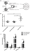
Figure 1
Figure 1. LDGs overexpress defensins and proinflammatory molecules
A. Schematic representation of the sample group and gene list comparisons made using the gene microarray data (q-value <0.01, fold-change ≥1.5 and ≤ 0.7 for the up-regulated and down-regulated genes, respectively). Samples were matched-paired when comparing lupus neutrophils with autologous lupus LDGs (n=10 for each group). B. Lupus LDGs express elevated levels of azurophilic granule genes. Log base 2 mRNA mean expression values of five azurophil genes in control neutrophils (n=9), lupus neutrophils (n=10) and lupus LDGs (n=10): Myeloperoxidase (MPO), Elastase (ELANE), Defensin alpha 4 (DEFA4), Cathepsin G (CTSG) and azurocidin 1 (AZU1). Data are presented as mean ± SD. ***p<0.0001. C. Confirmation of enhanced mRNA expression by real-time PCR of various neutrophils genes in lupus LDGs when compared to autologous lupus neutrophils (n=7–12) and control neutrophils (n=7). Bar graph represents fold mRNA expression (mean ± SEM) after adjusting for housekeeping gene (GAPDH). *p<0.05 LDGs compared to control and/or autologous lupus neutrophils.
Figure 2
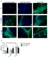
Figure 2. Circulating lupus LDGs undergo increased NETosis
A. Representative images of control neutrophils, lupus neutrophils and lupus LDGs isolated from peripheral blood and analyzed at baseline (T0) or after stimulation for 2 hours with DMSO or PMA. Top panels show immunofluorescent merged images of neutrophil extracellular traps (NETs) which were detected by neutrophil elastase (green) and DNA was labeled with Hoechst 33342 (blue); 40x images, bar graph: 20μm. B. Quantification of the percentage of NETs (elastase-labeled cells over total number of cells) are plotted as mean ± SEM (n = 6 patients/group; * p ≤ 0.05).
Figure 3
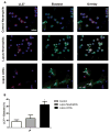
Figure 3. LL-37 externalization in NETs is increased in lupus LDGs
A. Representative images of control neutrophils, lupus neutrophils and lupus LDGs after isolation from peripheral blood. Cells were stained for detection of LL-37 (red), neutrophil elastase (green) and DNA was labeled with Hoechst 33342 (blue). Top panels show images of LL-37 and Hoechst (left), elastase and Hoechst (middle) and merged LL-37, elastase and Hoechst (right); 40x images, bar graph = 20μm. Arrows represent areas of LL-37 localization within the NETs. B. Quantification of the percentage of cells containing LL-37 colocalized with elastase over total number of cells are plotted as mean ± SEM (n ≥ 3 patients; * p ≤ 0.05).
Figure 4
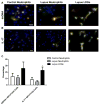
Figure 4. Lupus LDGs externalize ds-DNA and IL-17 through NETosis
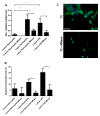
Representative images of control neutrophils, lupus neutrophils and lupus LDGs after isolation from peripheral blood. Cells were stained for detection of neutrophil elastase (green), DNA (Hoechst 33342, blue) and either ds-DNA (red) or IL-17 (red). A. Merged images of ds-DNA, elastase and Hoechst and B. Merged images of IL-17, elastase and Hoechst. C. Quantification of the percentage of cells containing ds-DNA or IL-17 colocalized with elastase over total number of cells are plotted as mean ± SEM (n ≥ 5 patients; * p ≤ 0.05).
Figure 5
Figure 5. Netting neutrophils are present in glomeruli from patients with lupus nephritis
Colocalization of histone H2A (green), MPO (red), and DNA (white) by direct immunofluorescence reveals in vivo evidence of NET formation in a glomerulus from a representative kidney microphotograph from a patient with class IV lupus nephritis. Yellow arrow and inset box highlights intraglomerular NET formation. Original magnification 20X.
Figure 6
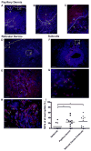
Figure 6. Netting neutrophils are present in affected lupus dermis and subcutis
Direct immunofluorescence staining of DNA (blue) and MPO (red) reveal NETs throughout affected lupus skin. A. Low power view of a punch biopsy from lesional lupus skin. Bar = 200 μm. Arrows highlight perifollicular infiltration of NETs (B, bar = 50 μm), and NETs within the papillary dermis (C, bar = 20 μm). D. Low power view of lupus reticular dermis (bar = 200 μm) with arrows highlighting infiltration by NETs (E, bar = 50 μm). F. Low power view of a large blood vessel in affected lupus subcutis (bar = 200 μm), with arrows highlighting NETs in an area of panniculitis (G, bar = 20 μm). Dotted line delineates the dermal-epidermal junction (in A, C, D) and circumscribes the follicle (“f”) in B. Epidermis is designated “e” and dermis by “d”. H. Cathelicidin (LL-37) is present in NETs within inflamed lupus skin lesions. Expression of LL-37 in neutrophils in lupus tissue was examined by dual-color immunofluorescence staining for LL-37 (green) and MPO (red) with DAPI counterstain (blue). Representative images from one of 3 sections stained with LL-37 and MPO are shown at 600x magnification. Bar = 100 μm. I. Frequency of NETosis in cutaneous lupus lesions. NETs and neutrophils were counted after immunofluorescence staining with MPO and DAPI. Percentage of neutrophils undergoing NETosis of all neutrophils were calculated for the epidermis, papillary dermis, reticular dermis and subcutis. *p<0.05.
Figure 7
Figure 7. IL-17-positive neutrophils infiltrate SLE skin
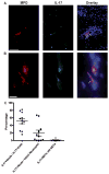
Direct immunofluorescence staining of IL-17 (green) and MPO (red) in affected lupus dermis. A. Arrows highlight intact IL-17+ neutrophils in a blood vessel. Bar = 200 μm. B. IL-17 present in a NET (arrow). Bar = 10μm. C. Frequency of IL-17 expression in intact neutrophils and NETs in SLE skin. Percentages of 1) IL17+ neutrophils of all IL17+ cells, 2) IL17+ neutrophils of all intact neutrophils and 3) IL17+ NETs of all NETs are reported in the graph.
Figure 8
Figure 8. Netting lupus LDGs stimulate IFN-α synthesis by PDCs and kill endothelial cells
A. Gen2.2 pDC cells were stimulated for 16 hours with supernatants from control neutrophils (n=4), lupus neutrophils (n=6) and autologous lupus LDGs (n=6) that had been previously cultured in the presence or absence of micrococcal nuclease (MNase) for 1 hour. Results represent mean ± SEM IFN-α mRNA expression in Gen2.2 cells.*p<0.05, for control neutrophils compared with lupus neutrophils and with LDGs, and for lupus and LDGs compared to lupus or LDGs+MNAse. B. Bar graph represents mean ± SEM percentage of apoptotic HUVECs after exposure to activated LDGs, autologous neutrophils and control neutrophils (n=4–5/group) in the presence or absence of MNase. *p<0.05. C. Representative images of LDGs isolated from peripheral blood, cultured for 2 hours followed by either no MNase treatment (T2) or with MNase (T2+MNase) for 10 min at room temperature and processed for immunofluorescence staining of elastase (green) and DNA (Hoechst 33342, blue); 40X images, Bar= 20μm.

References

    1. Denny MF, Yalavarthi S, Zhao W, Thacker SG, Anderson M, Sandy AR, McCune WJ, Kaplan MJ. A distinct subset of proinflammatory neutrophils isolated from patients with systemic lupus erythematosus induces vascular damage and synthesizes type I IFNs. J Immunol. 184:3284–3297. - PMC - PubMed
    1. Hakkim A, Furnrohr BG, Amann K, Laube B, Abed UA, Brinkmann V, Herrmann M, Voll RE, Zychlinsky A. Impairment of neutrophil extracellular trap degradation is associated with lupus nephritis. Proc Natl Acad Sci U S A. 2010;107:9813–9818. - PMC - PubMed
    1. Bennett L, Palucka AK, Arce E, Cantrell V, Borvak J, Banchereau J, Pascual V. Interferon and granulopoiesis signatures in systemic lupus erythematosus blood. J Exp Med. 2003;197:711–723. - "VSports最新版本" PMC - PubMed
    1. Hacbarth E, Kajdacsy-Balla A. Low density neutrophils in patients with systemic lupus erythematosus, rheumatoid arthritis, and acute rheumatic fever. Arthritis Rheum. 1986;29:1334–1342. - VSports在线直播 - PubMed
    1. Brinkmann V, Reichard U, Goosmann C, Fauler B, Uhlemann Y, Weiss DS, Weinrauch Y, Zychlinsky A. Neutrophil extracellular traps kill bacteria. Science. 2004;303:1532–1535. - PubMed

"V体育2025版" Publication types

MeSH terms

Associated data