Skip to main page content
U.S. flag

An official website of the United States government

Dot gov

The . gov means it’s official. Federal government websites often end in VSports app下载. gov or . mil. Before sharing sensitive information, make sure you’re on a federal government site. .

Https

The site is secure. The https:// ensures that you are connecting to the official website and that any information you provide is encrypted and transmitted securely. V体育官网.

Comparative Study
. 2011 Jul 1;187(1):490-500.
doi: 10.4049/jimmunol.1100123. Epub 2011 May 23.

Mast cells and neutrophils release IL-17 through extracellular trap formation in psoriasis

Affiliations
Comparative Study

Mast cells and neutrophils release IL-17 through extracellular trap formation in psoriasis

Andrew M Lin et al. J Immunol. .

"V体育安卓版" Abstract

IL-17 and IL-23 are known to be absolutely central to psoriasis pathogenesis because drugs targeting either cytokine are highly effective treatments for this disease. The efficacy of these drugs has been attributed to blocking the function of IL-17-producing T cells and their IL-23-induced expansion. However, we demonstrate that mast cells and neutrophils, not T cells, are the predominant cell types that contain IL-17 in human skin. IL-17(+) mast cells and neutrophils are found at higher densities than IL-17(+) T cells in psoriasis lesions and frequently release IL-17 in the process of forming specialized structures called extracellular traps. Furthermore, we find that IL-23 and IL-1β can induce mast cell extracellular trap formation and degranulation of human mast cells. Release of IL-17 from innate immune cells may be central to the pathogenesis of psoriasis, representing a fundamental mechanism by which the IL-23-IL-17 axis mediates host defense and autoimmunity. VSports手机版.


Figures

FIGURE 1
FIGURE 1
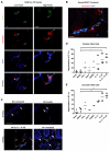
T cells, mast cells, and neutrophils contain IL-17 in psoriasis. Punch biopsies of skin from subjects without psoriasis (NN, n ≥ 10), uninvolved skin from subjects with psoriasis (PN, n ≥ 10), or lesional psoriasis plaques (PP, n ≥ 10, shown) were subjected to dual-color immunofluorescence staining for IL-17 (green) and either CD3 (A), tryptase (B), chymase (C), or MPO (D, red), with DAPI counterstain. These high-powered 600x fields near the superficial dermis illustrate the colocalization of IL-17 with CD3, tryptase, chymase, or MPO (yellow, solid arrows), while simultaneously indicating the presence of other IL-17+ cells (open arrows) in the same field. Bar = 100 μm.
FIGURE 2
FIGURE 2
IL-17+ mast cells and neutrophils are increased in psoriasis. The number of single CD3+, tryptase+, chymase+, or MPO+ cells (A, E, I, M) and their means (bars) in each 200x field were quantified as described in Materials and Methods (each, n ≥ 10). The double CD3+IL-17+, tryptase+IL-17+, chymase+IL-17+, MPO+IL-17+ cells (B, F, J, N) and their means (bars) in each 200x field were also quantified. The proportions of these double positive cells out of all IL-17+ cells and their means (bars) were calculated (C, G, K, M). In addition, the proportions of double positive cells of all CD3+, tryptase+, chymase+, or MPO+ cells and their means (bars) were calculated (D, H, L, P). *P < 0.05, **P < 0.01, ***P < 0.001 (one-way ANOVA, Tukey’s test).
FIGURE 3
FIGURE 3
Neutrophils frequently undergo ETosis in psoriasis. A, Punch biopsies of skin from subjects with lesional psoriasis plaques (PP, n = 12) were subjected to dual-color immunofluorescence staining for IL-17 (green) and MPO (red), with DAPI counterstain (blue). An epidermally-centered field of a mature psoriasis plaque at 200x reveals IL-17-containing neutrophils in a characteristic Munro’s microabscess (MM) and spongiform pustule of Kogoj (SPK). Bar = 100 μm. B, NETs magnified past 1000x were observed to extrude nuclear material (open arrows) and contained both MPO and IL-17. Bar = 10 μm. C, PP skin was also subjected to tri-color immunofluorescence staining (n = 3) for IL-17 (green), LL-37 (red), and MPO (blue), revealing many triple-positive NETs in the dermis (white cells, solid arrow) as well as other non-neutrophil IL-17+ cells (green, open arrow). Bar = 100 μm. D, NETs in a MM were further examined by confocal microscopy at 1000x (solid arrows) and approximately 5000x, showing elaborate distortion of nuclear material. Bar = 10 μm. E, Hematoxylin and eosin stained sections of a MM at 200x and past 1000x also revealed altered nuclear morphology resembling NETs. Bar = 100 μm (top), 10 μm (bottom).
FIGURE 4
FIGURE 4
Neutrophils and LDG from the blood of patients with psoriasis exhibit frequent NET formation. A, Neutrophils isolated from human blood were further sorted by FACSAria with FSC, SSC, CD15, and CD3 gates (red) to >99.5% purity. Flow cytometry plots from one representative sample are shown. B, Western blot analysis of cell lysates from post-sort neutrophils with anti-IL-17A and anti-IL-17F antibodies was performed to confirm the type of IL-17 contained in control neutrophils (n = 3). Staining for β-actin served as the loading control. Abbreviations for lanes: A, recombinant IL-17A; F, recombinant IL-17F; γ, recombinant IFN-γ; N, neutrophils; P, PBMC. C, A higher number of LDG were present in blood from psoriasis patients (n = 15) compared to control blood (n = 6). **P < 0.01 (unpaired two-tailed Student’s t-test). D, Neutrophils and LDG isolated from psoriatic blood and neutrophils from control blood were stained at time 0, 2 hours without stimulation, and at 2 hours after incubation in PMA. In-vitro NETs were examined at 400x magnification using stains for neutrophil elastase (green) and Hoechst 33342 (blue). Bar = 20 μm.
FIGURE 5
FIGURE 5
IL-1β and IL-23 promote mast cell degranulation or MCET formation. A, MCETs observed in PP lesions at 1000x showed extruded nuclear material (open arrows) and contained both chymase (red) and IL-17 (green). Bar = 10 μm. B, MCETs were further observed in the dermis of psoriasis skin by zooming in on a confocal image past 1000x. Bar = 25 μm. C, Punch biopsies of skin from normal controls (NN) were incubated for 3 days in various conditions: media alone, IFN-γ, Substance P, Compound 48/80, IL-1β, IL-23, and IL-1β+IL-23 as described in Materials and Methods. Tissue sections were subjected to dual-color immunofluorescence staining for IL-17 (green) with chymase (red) or tryptase (not shown), with DAPI counterstain (n = 3). Representative images from one NN series stained with chymase and IL-17 are shown at 400x magnification and compared to a typical PP section. Intact cells (solid arrows) and MCETs or degranulated cells (open arrows) are highlighted. Bar = 100 μm. D, The proportion of MCETs or degranulated tryptase+ cells was observed to increase when exposed to IL-23 alone or IL-1β and IL-23. *P < 0.05 for media vs. IL-23 or IL-1β +IL-23 (one-way ANOVA, Dunnett’s test). E, Similarly, chymase+ cells also displayed more frequent MCETs and degranulation when exposed to IL-1β and IL-23. **P < 0.01 for media vs. IL-23 or IL-1β+IL-23.
FIGURE 6
FIGURE 6
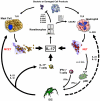
Model: Contributions of innate IL-17 and ETosis to psoriasis.

References

    1. Korn T, Bettelli E, Oukka M, Kuchroo VK. IL-17 and Th17 Cells. Annu Rev Immunol. 2009;27:485–517. - PubMed (V体育平台登录)
    1. Wilson NJ, Boniface K, Chan JR, McKenzie BS, Blumenschein WM, Mattson JD, Basham B, Smith K, Chen T, Morel F, Lecron JC, Kastelein RA, Cua DJ, McClanahan TK, Bowman EP, de Waal Malefyt R. Development, cytokine profile and function of human interleukin 17-producing helper T cells. Nat Immunol. 2007;8:950–957. - PubMed
    1. Yang L, Anderson DE, Baecher-Allan C, Hastings WD, Bettelli E, Oukka M, Kuchroo VK, Hafler DA. IL-21 and TGF-beta are required for differentiation of human T(H)17 cells. Nature. 2008;454:350–352. - PMC - PubMed
    1. Manel N, Unutmaz D, Littman DR. The differentiation of human T(H)-17 cells requires transforming growth factor-beta and induction of the nuclear receptor RORgammat. Nat Immunol. 2008;9:641–649. - PMC - PubMed
    1. Chang YT, Chou CT, Yu CW, Lin MW, Shiao YM, Chen CC, Huang CH, Lee DD, Liu HN, Wang WJ, Tsai SF. Cytokine gene polymorphisms in Chinese patients with psoriasis. Br J Dermatol. 2007;156:899–905. - PubMed

MeSH terms