Skip to main page content (V体育官网入口)
U.S. flag

An official website of the United States government

Dot gov

The VSports app下载. gov means it’s official. Federal government websites often end in . gov or . mil. Before sharing sensitive information, make sure you’re on a federal government site. .

Https

The site is secure V体育官网. The https:// ensures that you are connecting to the official website and that any information you provide is encrypted and transmitted securely. .

. 2011 Jun 1;25(11):1159-72.
doi: 10.1101/gad.2042311. Epub 2011 May 16.

mTOR phosphorylates IMP2 to promote IGF2 mRNA translation by internal ribosomal entry

Affiliations

mTOR phosphorylates IMP2 to promote IGF2 mRNA translation by internal ribosomal entry (VSports最新版本)

Ning Dai et al. Genes Dev. .

Abstract

Variants in the IMP2 (insulin-like growth factor 2 [IGF2] mRNA-binding protein 2) gene are implicated in susceptibility to type 2 diabetes. We describe the ability of mammalian target of rapamycin (mTOR) to regulate the cap-independent translation of IGF2 mRNA through phosphorylation of IMP2, an oncofetal RNA-binding protein. IMP2 is doubly phosphorylated in a rapamycin-inhibitable, amino acid-dependent manner in cells and by mTOR in vitro. Double phosphorylation promotes IMP2 binding to the IGF2 leader 3 mRNA 5' untranslated region, and the translational initiation of this mRNA through eIF-4E- and 5' cap-independent internal ribosomal entry VSports手机版. Unexpectedly, the interaction of IMP2 with mTOR complex 1 occurs through mTOR itself rather than through raptor. Whereas depletion of mTOR strongly inhibits IMP2 phosphorylation in cells, comparable depletion of raptor has no effect; moreover, the ability of mTOR to phosphorylate IMP2 in vitro is unaffected by the elimination of raptor. Dual phosphorylation of IMP2 at the mTOR sites is evident in the mouse embryo, likely coupling nutrient sufficiency to IGF2 expression and fetal growth. Doubly phosphorylated IMP2 is also widely expressed in adult tissues, including islets of Langerhans. .


Figures

Figure 1.
Figure 1.
The rapamycin-sensitive translational initiation of IGF2 L3 mRNA is mediated by its 5′ UTR and is independent of eIF-4E. (A) The effect of rapamycin on the polysomal association of IGF2 L3 and L4 mRNAs in rapidly growing RD cells. Rapamycin (20 nM) was added to RD cells at ∼30% confluency; the cells were harvested at intervals thereafter and extracts were subjected to sucrose density gradient centrifugation. The subpolysomal ([- - -] fractions 1–5, Supplemental Fig. S2) and polysomal ([—] fractions 6–13) regions of the gradient were pooled separately, and each was assayed by qPCR for the content of IGF2 L3 (▪) and L4 (□) mRNAs. The combined results of three experiments are shown ±SEM. (B) The rapamycin sensitivity of IGF2 L3 mRNA translational initiation is conferred by the L3 5′ UTR. The IGF2 L3 and L4 5′ UTR segments were fused to the coding sequences of firefly luciferase (see cartoon) and were transiently transfected into rapidly growing RD cells. After 24 h, cells were treated either with DMSO (filled bars) or rapamycin (20 nM, 3 h.; cross-hatched bars); extracts were separated by sucrose density gradient centrifugation, and the content of endogenous IGF2 L3 (black-filled and hatched bars, right) and L4 (gray-filled and hatched bars, right) mRNAs and of the L3-luciferase (black-filled and hatched bars, left) and L4-luciferase (gray-filled and hatched bars, left) mRNAs in the pooled polysomal fractions was determined by qPCR and is expressed as a percentage of the corresponding total L3 or L4 mRNA in the sample loaded. The combined results of three experiments are shown ±SEM. (C) Overexpression of eIF-4E up-regulates ODC1-luciferase expression but does not alter the expression of L3-luciferase. Plasmids encoding the 5′ UTR of human ODC1 fused to firefly luciferase or L3-firefly luciferase or L4-firefly luciferase, each together with a plasmid encoding Renilla luciferase, were transfected into RD cells that stably express recombinant eIF-4E in a tetracycline-inducible manner. The cells were treated with tetracycline (gray bars) or carrier (black bars) for 24 h. Three hours prior to harvest, cells were treated with DMSO (D) or rapamycin (20 nM, 3 h; R). Thereafter, extracts were assayed for firefly and Renilla luciferase activity and by qPCR for the content of firefly luciferase mRNA. The activity of firefly luciferase was divided by the activity of Renilla luciferase to give a normalized firefly luciferase activity; “translational efficiency” was calculated by dividing the normalized firefly luciferase activity by the measured content of firefly luciferase mRNA, setting to 1 the value of this dividend for the ODC1-luciferase, L3-luciferase, and L4-luciferase conditions in the absence of tetracycline induction or rapamycin treatment (the black bars in ODC1, L3D, and L4D). The unfilled/white bar shows the effect of rapamycin on the expression of ODC1-luciferase in the absence of tetracycline. Each experiment was performed in triplicate, and the combined results of three experiments are shown ±SEM. (D) Overexpression of a nonphosphorylatable mutant of 4E-BP1 suppresses expression of ODC1-luciferase but does not alter expression of L3-luciferase. RD cells were engineered to stably overexpress GST or a GST fused to a nonphosphorylatable 4E-BP1 polypeptide (GST-4E-BP[5Ala]) (Hara et al. 2002). Plasmids encoding ODC1-luciferase, L3-luciferase, or L4-luciferase, each together with a plasmid encoding Renilla luciferase, were transfected into the RD GST (black bars) or GST-4E-BP[5Ala] (gray bars) stable transformants. After 24 h, the cells were treated with DMSO (D) or rapamycin (20 nM, 3 h; R). Translational efficiency was calculated as in C, and set to 1 for the ODC1-luciferase, L3-luciferase, and L4-luciferase conditions in the extracts from cells expressing GST in the absence of rapamycin treatment (the black bars in ODC1, L3D, and L4D). Each experiment was performed in triplicate, and the combined results of three experiments are shown ±SEM.
Figure 2.
Figure 2.
The initiation of IGF2 L3 mRNA translation occurs through internal ribosomal entry. (A) Expression patterns of dicistronic plasmids indicate that translational initiation of mRNAs containing the IGF2 L3 sequences occurs by internal ribosomal entry. Three dicistronic plasmids encoding Renilla luciferase upstream of firefly luciferase, each preceded by the 5′ UTRs shown in the cartoon, were constructed; plasmid 3 is identical to plasmid 1 except for the insertion of a 60-nt hairpin structure (Mauro et al. 2007) as indicated. Twenty-four hours after transfection into RD cells, the activity of Renilla (black bars, black numbers) and firefly (gray bars, gray numbers) luciferase was measured. The results of triplicate transfections are shown. (B) Tetracycline induces a single mRNA and a quantitatively identical fold increase in the Renilla and firefly luciferase activities from a stably expressed dicistronic plasmid containing an internal L3 5′ UTR. RD cells were engineered to stably express the dicistronic plasmid (shown in the cartoon) in a tetracycline-inducible fashion. Extracts were prepared 24 h after the addition of carrier (−) or tetracycline (+). RNA was extracted and luciferase activities were measured. The RNA blot was cut longitudinally in two and hybridized with 32P-labeled cDNA probes against either the Renilla (left) or firefly (right) luciferase coding sequences; an autoradiograph of the reassembled blot and the absolute and relative activities generated from the β-globin–Renilla- and internal L3-firefly-luciferase cDNA are shown. (C) An RNAi against one of the two luciferases encoded by a dicistronic cDNA inhibits the expression of both luciferases to a similar extent. RD cells stably expressing the dicistronic plasmid shown in B were treated with a scramble RNAi or a single RNAi against one luciferase and harvested 48 h later; three different RNAis were employed against each luciferase. The Renilla (or firefly) luciferase activity measured in the extract from the scramble RNAi-treated cells was divided into the Renilla (or firefly) luciferase activity measured from the Renilla (or firefly) RNAi-treated cells. This fraction, subtracted from 1 and multiplied by 100, is shown as the percent inhibition due to the Renilla (or firefly) RNAi. (D) Synthetic monocistronic and dicistronic RNAs containing the IGF2 L3 5′ UTR undergo translational initiation after transient transfection independently of the state of 5′ cap methylation. Capped poly(A) RNA was synthesized in vitro using the mScript mRNA Production system (Epicentre) from dsDNA templates encoding the three constructs indicated. A methylated (N7mG[5′]ppp[2′Om]G; “Me”; solid bars) or unmethylated (G[5′]pppG; “unMe”; hatched bars) 5′ cap was added.One-hundred nanograms of each of the six in vitro transcribed RNA species was transfected in triplicate into 293 cells; 24 h later, extracts were analyzed for the luciferase activities (black bars; black numbers, Renilla; gray bars, gray numbers, firefly) and the abundance of the luciferase RNAs by qPCR (the Ct value is shown). The cap structures, a gel of these six RNAs, and a table containing the actual luciferase measurements are shown in Supplemental Figure S3A–C, respectively.
Figure 3.
Figure 3.
Rapamycin inhibits translational initiation of L3-mRNAs by inhibiting the binding of IMP2 to L3. (A) Rapamycin displaces the IMP polypeptides from polysomes. Extracts of RD cells treated with DMSO (black bars) or rapamycin (20 nM, 3 h; gray bars) were subjected to sucrose gradient centrifugation. The subpolysomal (fractions 1–5) (Supplemental Fig. S2) and polysomal (fractions 6–13) regions of the gradient were pooled separately, and matched aliquots of each were subjected to SDS-PAGE and immunoblot for IMP1, IMP2, and IMP3. For each IMP blot, the intensity of the subpolysomal and polysomal images were added and divided into the value of the polysomal image and multiplied by 100 to give the percent of that IMP polypeptide in the polysomal fractions. The combined results of five experiments are shown. (B) Rapamycin reduces the amount of IGF2 L3 mRNA bound to IMP2 without affecting IMP2 association with IGF2 L4 mRNA. Extracts of RD cells treated with DMSO (D) or rapamycin (20 nM, 3 h; R) were subjected to immunoprecipitation with anti-IMP2 or nonimmune IgG. (Middle panel) RNA was extracted from the washed immunoprecipitates, and coprecipitating IGF2 L3 and IGF2 L4 mRNAs were quantified by qPCR and are shown in the bar graph (combined results of three experiments). (C) The endogenous IMP polypeptides bind selectively and in a rapamycin-sensitive manner to an L3-luciferase mRNA. An aptamer encoding a streptavidin-binding motif (called S1) was inserted into the L3-luciferase (cartoon) and L4-luciferase plasmids immediately after the stop triplet of the coding region. RD cells stably expressing similar amounts of the recombinant L3-S1mRNAs or L4-S1mRNAs were selected (see Supplemental Fig. S4). Aliquots of extracts prepared from rapidly growing S1 transformants, treated with either DMSO or rapamycin (20 nM, 3 h), were subjected to formaldehyde cross-linking and streptavidin affinity purification as described in the Materials and Methods; the amount of S1-containing mRNAs was quantified by qPCR, and comparable recoveries from cells treated with DMSO or rapamycin was observed. After reversal of the cross-links, the released polypeptides were separated by SDS-PAGE and were subjected to immunoblot for IMP1, IMP2, and IMP3 as shown in the lowest panel. (D) Overexpression of IMP2 in RD cells up-regulates L3-luciferase selectively and abolishes its inhibition by rapamycin. Plasmids encoding L3-luciferase or L4-luciferase, each together with a plasmid encoding Renilla luciferase, were transfected into RD cells that stably express a tetracycline-inducible IMP2, and the cells were treated with tetracycline (gray bars) or carrier (black bars) for 24 h. Three hours prior to harvest, cells were treated with DMSO (D) or rapamycin (20 nM; R). Translational efficiency was calculated as in Figure 1C and set to 1 for the L3-luciferase and L4-luciferase conditions in the extracts from cells not exposed to tetracycline or rapamycin (the black bars in L3D and L4D). Each experiment was performed in triplicate, and the combined results of three experiments ±SEM are shown. (E) Depletion of IMP2 by shRNA strongly suppresses the translation of IGF2-L3 mRNA. Plasmids encoding IGF2-L3-firefly luciferase or IGF2-L4-firefly luciferase, each together with a plasmid encoding Renilla luciferase, were transfected into RD cells stably expressing shRNAs directed against green fluorescent protein (GFP) or IMP2, and the cells were harvested 48 h later. Three hours prior to harvest, cells were treated with DMSO (black bars) or rapamycin (20 nM, gray bars). Extracts were assayed for firefly and Renilla luciferase activity and by qPCR for the content of firefly luciferase mRNA. Translational efficiency was calculated as in Figure 1C and set to 1 for the L3-luciferase and L4-luciferase conditions in the extracts from cells treated with DMSO. Each experiment was performed in triplicate, and the combined results of three experiments are shown ±SEM. The decreases in L3 luciferase and L4 luciferase due to IMP2 shRNA were both significant (P < 0.01 and <0.05, respectively).
Figure 4.
Figure 4.
IMP2 binds to the mTOR FAT domain but not raptor, and IMP2 binding to mTOR is inhibited by mTORC1 signaling and IMP2 binding to RNA. (A) The mTOR inhibitors rapamycin and Torin1 promote the binding of IMP2 to mTOR. RD cells were exposed to rapamycin (20 nM, 3 h) or Torin1 (100 nM, 1 h) or for the corresponding times to DMSO. After harvest, immunoprecipitates of endogenous IMP2 were probed for the presence of mTOR. (B) Withdrawal of medium amino acids promotes the binding of IMP2 to mTOR. Rapidly growing RD cells were washed and transferred to fresh DMEM or DPBS; 2 h later, cells were extracted and immunoprecipitates of IMP2 were probed for mTOR. (C) Degradation of RNA promotes the binding of IMP2 to mTOR. Extracts prepared from RD cells were incubated for 10 min at 37°C with the addition of ribonucleases I (1000 U/mL), T1 (200 U/mL), or A (2 μg/mL) or with DMSO. Thereafter, immunoprecipitates of endogenous IMP2 were probed for mTOR. (D) IMP2 binds mTOR but not raptor. RD cells were transfected with Flag-mTOR, Flag-raptor, or Flag vector. After 24 h, cells were extracted with either CHAPS (0.3%; left) or Triton X-100 (1%; right). Extraction with CHAPS preserves, whereas extraction in Triton X-100 abolishes, the mTOR–raptor association. (E) IMP2 binds to an mTOR noncatalytic region comprised predominantly of the FAT domain. Plasmids encoding the Flag-tagged fragments of mTOR shown were transiently expressed in 293T cells, and Flag immunoprecipitates were probed for the presence of endogenous IMP2.
Figure 5.
Figure 5.
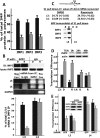
mTORC1 phosphorylates IMP2 at Ser162 and Ser164 in an amino acid-dependent manner in vivo and in a raptor-independent manner in vitro. (A) mTOR phosphorylates IMP2 and 4E-BP1 to a similar extent in vitro. Flag-mTOR wild type or N2343K (NK) were transiently expressed in 293T cells, extracted with 0.3% CHAPS, immunoprecipitated, washed, and eluted with Flag peptide. Recombinant Flag-4E-BP1 and Flag-IMP2 were extracted with CHAPS from rapamycin-treated 293 cells, immunoprecipitated with anti-Flag with (lanes 5,6 on right) or without prior treatment of the extract with DNase and RNase A, and eluted with Flag peptide. The mTOR kinase wild type was added to lanes 2, 4, and 6, and the inactive mTOR (NK) was added to lanes 1, 3, and 5; the reaction was started by addition of γ32P-ATP and was stopped after 30 min. An autoradiograph is shown. (B) Rapamycin and Torin1 abolish the concurrent phosphorylation of IMP2 Ser162 and Ser164. Phosphopeptide-specific antibodies were generated against IMP2[Ser162P], IMP2[Ser164P], and the doubly phosphorylated IMP2[Ser162P/Ser164P]; the specificity of these antibodies is shown in Supplemental Figure S5. RD cells were treated with rapamycin (200 nM, 1 h), Torin1 (100 nM, 1 h), or DMSO, and extracts were immunoblotted for the phosphorylated forms of IMP2 as well as for S6K1[Thr 389P] and 4E-BP[Thr 37P/46P]. (C) Amino acid withdrawal inhibits the concurrent phosphorylation of IMP2[Ser162/Ser164]. Rapidly growing RD cells were washed and transferred to fresh DMEM or DPBS; extracts were prepared 2 h later and immunoblotted for IMP2 polypeptide and IMP2 phosphorylation sites. (D) mTOR catalyzes the concurrent phosphorylation of IMP2[Ser162/Ser164] in vitro in a raptor-independent manner. Flag-mTOR was expressed in 293 cells and was extracted with either CHAPS (0.3%) or Triton X-100(1%). Flag-4E-BP and Flag-IMP2 were extracted from rapamycin-treated (200 nM, 1 h) 293T. The λ phosphatase was added to the extracts (10 U/mL) with incubation for 10 min at 30°C. The extracts were returned to 4°C and immobilized anti-Flag antibody was added. After 1.5 h, the beads were washed and subjected to Flag peptide elution. Kinase assay was carried out as in A, except nonradioactive ATP was employed and the reaction was analyzed by immunoblot for IMP2[Ser162P/Ser164P] and 4E-BP[Thr 37P/46P]. (E) Depletion of mTOR inhibits expression of IGF2-L3-luciferase, whereas depletion of raptor has no effect. Plasmids encoding IGF2-L3-firefly luciferase or IGF2-L4-firefly luciferase, each together with a plasmid encoding Renilla luciferase, were transfected into RD cells stably expressing shRNAs directed against GFP, mTOR, or raptor and were harvested 48 h later. Three hours prior to harvest, cells were treated with DMSO (black bars) or rapamycin (20 nM, gray bars). Translational efficiency was calculated as in Figure 1C and set to 1 for the L3-luciferase and L4-luciferase conditions in the extracts from cells treated with DMSO. Each experiment was performed in triplicate, and the combined results of three experiments are shown ±SEM. (F) Depletion of mTOR inhibits IMP2 phosphorylation, whereas comparable depletion of raptor does not. Extracts prepared from RD cells stably expressing shRNAs directed against GFP, mTOR, or raptor were immunoblotted for the polypeptides and phosphorylation sites indicated.
Figure 6.
Figure 6.
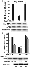
Mutation of IMP2 Ser162 and Ser164 to Ala inhibits IMP2 binding to IGF2 L3 and stimulation of L3 mRNA translation. (A) Mutation of IMP2[Ser162/164] to Ala but not Asp reduces IMP2 binding to IGF2 L3 mRNA, whereas both IMP2 mutants bind mTOR similarly. Plasmids encoding Flag-tagged IMP2 wild type (WT), IMP2[Ser162Ala/Ser164Ala] (AA), or IMP2[Ser162Asp/Ser164 Asp] (DD) were transiently expressed in RD cells. The cells were extracted 24 h later, Flag-IMP2 variants were immunoprecipitated, and matched aliquots were subjected to phenol/chloroform extraction and measurement of bound IGF2 L3 mRNA by qPCR and immunoblot for the presence of endogenous mTOR. (B) Mutation of IMP2[Ser162/164] to Ala but not Asp reduces the initiation of L3-mRNA translation. RD cells stably expressing Flag-tagged IMP2 wild type (WT), IMP2[Ser162Ala/Ser164Ala] (AA), or IMP2[Ser162Asp/Ser164Asp] (DD) at levels similar to (AA) or less than (WT, DD) endogenous IMP2 (top blot) were transiently transfected with L3-firefly luciferase together with a plasmid encoding Renilla luciferase, and the translational efficiency of L3-luciferase was measured as described in the legend for Figure 2A. The effect of IMP2[Ser162Ala/Ser164Ala] on the phosphorylation of endogenous IMP2 is shown in Supplemental Figure S6.
Figure 7.
Figure 7.
The expression of IMP2 and IMP2[Ser162P/Ser164P] in mouse embryos, adult mouse tissues, and human islets of Langerhans. (A) IMP2 exhibits concurrent phosphorylation of Ser162 and Ser164 during mouse embryonic development. Whole mouse embryos were harvested at the developmental times indicated and extracted, and aliquots were subjected immunoblot for IMP2, IMP2[Ser162P/Ser164P], and actin. (B) IMP2 uniquely is widely expressed in tissues of the adult mouse and is abundant in human islets of Langerhans. (C) Expression of IMP2 polypeptide relative to mouse brain. As in B, except normalized for total protein content and the combined results of three experiments are shown.

References

    1. Angel M, Yanik MF 2010. Innate immune suppression enables frequent transfection with RNA encoding reprogramming proteins. PLoS ONE 5: e11756 doi: 10.1371/journal.pone.0011756 - "V体育平台登录" PMC - PubMed
    1. Avruch J, Long X, Ortz-Vega S, Rapley J, Papageorgiou A, Dai N 2009. Amino acid regulation of TOR complex 1. Am J Physiol Endocrinol Metab 296: E592–E602 doi: 10.1152/ajpendo.90645.2008 - PMC - PubMed
    1. Baker J, Liu JP, Robertson EJ, Efstratiadis A 1993. Role of insulin-like growth factors in embryonic and postnatal growth. Cell 8: 73–82 - PubMed (VSports app下载)
    1. Barker DJ 2006. Adult consequences of fetal growth restriction. Clin Obstet Gynecol 49: 270–283 - "VSports在线直播" PubMed
    1. Brill LM, Salomon AR, Ficarro SB, Mukherji M, Stettler-Gill M, Peters EC 2004. Robust phosphoproteomic profiling of tyrosine phosphorylation sites from human T cells using immobilized metal affinity chromatography and tandem mass spectrometry. Anal Chem 76: 2763–2772 - PubMed (VSports手机版)

MeSH terms

Substances