<tt lang="R62r8w"></tt> Skip to main page content
U.S. flag

An official website of the United States government

Dot gov

The . gov means it’s official. Federal government websites often end in . gov or . mil VSports app下载. Before sharing sensitive information, make sure you’re on a federal government site. .

Https

The site is secure. The https:// ensures that you are connecting to the official website and that any information you provide is encrypted and transmitted securely. V体育官网.

. 2011 Jul 1;51(1):97-106.
doi: 10.1016/j.freeradbiomed.2011.04.020. Epub 2011 Apr 17.

Nrf2-dependent induction of NQO1 in mouse aortic endothelial cells overexpressing catalase (VSports注册入口)

Affiliations

Nrf2-dependent induction of NQO1 in mouse aortic endothelial cells overexpressing catalase (VSports最新版本)

Xinghua Lin et al. Free Radic Biol Med. .

Abstract (V体育2025版)

Overexpression of catalase has been shown to accelerate benzo(a)pyrene (BaP) detoxification in mouse aortic endothelial cells (MAECs). NAD(P)H:quinone oxidoreductase-1 (NQO1) is an enzyme that catalyzes BaP-quinone detoxification. Aryl hydrocarbon receptor (AhR) and nuclear factor erythroid 2-related factor-2 (Nrf2) are transcription factors that control NQO1 expression. Here, we investigated the effects of catalase overexpression on NQO1, Nrf2, and AhR expression. The levels of NQO1 mRNA and protein were comparable in MAECs isolated from wild-type and transgenic mice that overexpress human catalase (hCatTg). BaP treatment increased NQO1 mRNA and protein levels in both groups, with a significantly greater induction in hCatTg MAECs than in wild-type cells. BaP-induced NQO1 promoter activity was dramatically higher in hCatTg MAECs than in wild-type cells. Our data also showed that the basal level of AhR and the BaP-induced level of Nrf2 were significantly higher in hCatTg MAECs than in wild-type cells VSports手机版. Inhibition of specificity protein-1 (Sp1) binding to the AhR promoter region by mithramycin A reversed the enhancing effect of catalase overexpression on AhR expression. Knockdown of AhR by RNA interference diminished BaP-induced expression of Nrf2 and NQO1. Knockdown of Nrf2 significantly decreased NQO1 mRNA and protein levels in cells with or without BaP treatment. NQO1 promoter activity was abrogated by mutation of the Nrf2-binding site in this promoter. In contrast, mutation of the AhR-binding site in the NQO1 promoter did not affect the promoter activity. These results suggest that catalase overexpression upregulates BaP-induced NQO1 expression by enhancing the Sp1-AhR-Nrf2 signaling cascade. .


Figures

Fig. 1
Fig. 1
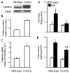
Overexpression of catalase reduces BaP-induced peroxides and lipid peroxidation. MAECs were obtained from wild-type and hCatTg mice. The catalase protein level was determined in whole cell extracts by Western blot analysis (A) and quantitated relative to β-actin (B). The catalase mRNA level was determined by quantitative real-time RT-PCR and normalized to GAPDH mRNA (C). The peroxide level (D) was measured using CDC-H2F diacetate as a probe. The F2-isoprostane level (E) was measured using GC/MS. Values represent the mean ± SEM of three separate experiments; MAECs were pooled from 4 mice. * P<0.05 vs. wild-type cells without BaP treatment, P<0.05 vs. wild-type cells with BaP treatment; and # P<0.05 vs. hCatTg cells without BaP treatment.
Fig. 2
Fig. 2
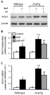
Overexpression of catalase elevates BaP-induced NQO1 expression. Wild-type and hCatTg MAECs at the 3rd or 8th-9th passages were treated with or without 1 μM BaP in the presence or absence of 10 μM aminotriazole (AT) as indicated. The level of NQO1 protein was determined in whole cell extracts by Western blot analysis (A) and quantitated relative to β-actin (B). The level of NQO1 mRNA was determined by quantitative real-time RT-PCR and normalized to GAPDH mRNA (C). Values represent the mean ± SEM of five independent experiments. * P<0.05 vs. the same genotype cells without BaP treatment; P<0.05 vs. BaP-treated wild-type cells; # P<0.05 vs. hCatTg cells without aminotriazole treatment.
Fig. 3
Fig. 3
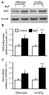
Overexpression of catalase elevates BaP-induced Nrf2 expression. Wild-type and hCatTg MAECs at the 3rd or 8th-9th passages were treated with or without 1 μM BaP as described in the Materials and Methods. The Nrf2 protein level in the whole cell was determined by Western blot analysis (A) and expressed relative to β-actin (B). The Nrf2 mRNA level was determined by quantitative real-time RT-PCR and expressed relative to GAPDH mRNA (C). Values represent the mean ± SEM of five independent experiments. * P<0.05 vs. the same genotype cells without BaP treatment; P<0.05 vs. BaP-treated wild-type cells.
Fig. 4
Fig. 4
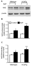
Overexpression of catalase increases BaP-induced Nrf2 accumulation in the nucleus and binding to the NQO1 promoter. Wild-type and hCatTg MAECs were treated with or without 1 μM BaP. The level of nuclear Nrf2 was determined by Western blot analysis (A) and expressed relative to β-actin (B). Nrf2 binding to the NQO1 promoter was assessed by ChIP analysis (C). Genomic DNA bound to Nrf2 was recovered and quantified by quantitative real-time PCR using primer pairs specific for the NQO1-ARE region. Data are expressed relative to the quantity of input. Values represent the mean ± SEM of five independent experiments. * P<0.05 vs. the same genotype cells without BaP treatment; P<0.05 vs. BaP-treated wild-type cells.
Fig. 5
Fig. 5
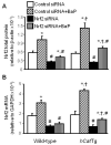
Knockdown of Nrf2 reduces NQO1 expression. Wild-type and hCatTg MAECs were transfected with Nrf2 siRNA or control siRNA, and then treated with or without 1 μM BaP as described in Materials and Methods. The level of Nrf2 (A) and NQO1 (C) proteins were determined by Western blot analysis and expressed relative to β-actin. The mRNA levels of Nrf2 (B) and NQO1 (D) were determined by quantitative real-time RT-PCR and expressed relative to GAPDH mRNA. Values represent the mean ± SEM of five independent experiments. * P<0.05 vs. the same genotype cells with same siRNA transfection and without BaP treatment; # P<0.05 vs. the same genotype cells transfected with control siRNA and the same treatment; P<0.05 vs. BaP-treated wild-type cells with same siRNA transfection.
Fig. 5
Fig. 5
Knockdown of Nrf2 reduces NQO1 expression. Wild-type and hCatTg MAECs were transfected with Nrf2 siRNA or control siRNA, and then treated with or without 1 μM BaP as described in Materials and Methods. The level of Nrf2 (A) and NQO1 (C) proteins were determined by Western blot analysis and expressed relative to β-actin. The mRNA levels of Nrf2 (B) and NQO1 (D) were determined by quantitative real-time RT-PCR and expressed relative to GAPDH mRNA. Values represent the mean ± SEM of five independent experiments. * P<0.05 vs. the same genotype cells with same siRNA transfection and without BaP treatment; # P<0.05 vs. the same genotype cells transfected with control siRNA and the same treatment; P<0.05 vs. BaP-treated wild-type cells with same siRNA transfection.
Fig. 6
Fig. 6
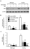
Knockdown of AhR reduces BaP-induced Nrf2 and NQO1 expression. Wild-type and hCatTg MAECs were transfected with AhR siRNA or control siRNA, and then treated with or without 1 μM BaP as described in Materials and Methods. The level of AhR protein (A) was determined by Western blot analysis and expressed relative to β-actin (B). The mRNA level of AhR was determined by quantitative real-time RT-PCR and expressed relative to GAPDH mRNA (C). The mRNA levels of Nrf2 (D) and NQO1 (E) were determined by quantitative real-time RT-PCR; BaP-induced changes were expressed as a % of those observed in cells without BaP treatment. Values represent the mean ± SEM of five independent experiments. * P<0.05 vs. the same genotype cells with same siRNA transfection and without BaP treatment; # P<0.05 vs. the same genotype cells transfected with control siRNA and the same treatment; and P<0.05 vs. BaP-treated wild-type cells with the same siRNA transfection.
Fig. 6
Fig. 6
Knockdown of AhR reduces BaP-induced Nrf2 and NQO1 expression. Wild-type and hCatTg MAECs were transfected with AhR siRNA or control siRNA, and then treated with or without 1 μM BaP as described in Materials and Methods. The level of AhR protein (A) was determined by Western blot analysis and expressed relative to β-actin (B). The mRNA level of AhR was determined by quantitative real-time RT-PCR and expressed relative to GAPDH mRNA (C). The mRNA levels of Nrf2 (D) and NQO1 (E) were determined by quantitative real-time RT-PCR; BaP-induced changes were expressed as a % of those observed in cells without BaP treatment. Values represent the mean ± SEM of five independent experiments. * P<0.05 vs. the same genotype cells with same siRNA transfection and without BaP treatment; # P<0.05 vs. the same genotype cells transfected with control siRNA and the same treatment; and P<0.05 vs. BaP-treated wild-type cells with the same siRNA transfection.
Fig. 7
Fig. 7
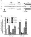
Functional analysis of the NQO1 promoter. NQO1 promoter-luciferase reporter plasmids were constructed and cotransfected into wild-type and hCatTg MAECs with a β-galactosidase expression plasmid as described in Materials and Methods. (A) Schematic diagram depicting wild-type (P1), ARE-mutated (P2), and XRE-mutated (P3) NQO1 promoter-luciferase constructs. Luciferase activity was measured using a luminescence assay and expressed relative to the luminosity of β-galactosidase assay (B). Values represent the mean ± SEM of five independent experiments. * P<0.05 vs. the same genotype cells with same plasmid transfection and without BaP treatment; # P<0.05 vs. the same genotype cells transfected with wild-type promoter (P1) and the same treatment; P<0.05 vs. wild-type cells transfected with same plasmid and treated with BaP.
Fig. 8
Fig. 8
The effect of mithramycin A on Sp1 and AhR expression, and Sp1 binding to the AhR promoter. Wild-type and hCatTg MAECs were treated with 500 nM mithramycin A (mmA) or medium alone as a control for 24 h. (A) The amount of Sp1 bound to the AhR promoter was determined using ChIP analysis, and expressed as the ratio of input controls. (B) The Sp1 protein level was determined by Western blot analysis and expressed relative to the immunoblot intensity of β-actin. (C) The AhR mRNA level was determined by quantitative real-time RT-PCR and expressed relative to GAPDH mRNA level. Values represent means ± SEM of five separate experiments; in each, MAECs were pooled from four mice. * P<0.05 vs. wild-type cells without mmA treatment; and P<0.05 vs. hCatTg cells without mmA treatment.
Fig. 9
Fig. 9
The effect of H2O2 on AhR and NQO1 expression, and Sp1 binding to the AhR promoter. Wild-type and hCatTg MAECs were treated with or without 100 μM H2O2 for 2 h, and then treated with or without 1 μM BaP for 4 h. (A) The amount of Sp1 bound to the AhR promoter was determined using ChIP analysis, and expressed as the ratio of input controls. AhR (B) and NQO1 (C) mRNA levels were determined by quantitative real-time RT-PCR and expressed relative to GAPDH mRNA. (D) The level of NQO1 protein was determined in whole cell extracts by Western blot analysis and quantitated relative to β-actin. Values represent the mean ± SEM of three independent experiments. * P<0.05 vs. the same genotype cells without H2O2 or BaP treatment (control); P<0.05 vs. the same genotype cells with BaP treatment alone; and # P<0.05 vs. wild-type cells with the same treatment.
Fig. 10
Fig. 10
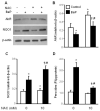
The effect of N-acetylcysteine on peroxides, and AhR and NQO1 expression. After pre-incubation with or without 10 nM N-acetylecysteine (NAC) for 1 hr, wild-type MAECs were treated with 1 μM BaP or culture medium alone (control) for 6 h (Western blot analysis) or for 1 h (peroxide measurement). The AhR and NQO1 protein levels were determined by Western blot analysis and quantitated (B, C respectively) relative to β-actin. Cellular peroxyl radicals were measured in terms of fluorescence intensity (FI) per μg protein using CDC-H2F diacetate as a probe. Values represent the mean ± SEM of five independent experiments. * P<0.05 vs. cells without NAC and BaP treatment; P<0.05 vs. cells with BaP treatment but without NAC treatment; and # P<0.05 vs. cells with NAC treatment but without BaP treatment.
Fig. 11
Fig. 11
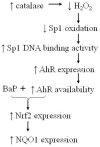
Proposed molecular mechanism for the role of catalase overexpression on BaP-induced NQO1 expression.

References

    1. Lind C. Formation of benzo[a]pyrene-3,6-quinol mono- and diglucuronides in rat liver microsomes. Arch. Biochem. Biophys. 1985;240:226–235. - "V体育安卓版" PubMed
    1. Joseph P, Jaiswal AK. NAD(P)H:quinone oxidoreductase1 (DT diaphorase) specifically prevents the formation of benzo[a]pyrene quinone-DNA adducts generated by cytochrome P4501A1 and P450 reductase. Proc. Natl. Acad. Sci. U. S. A. 1994;91:8413–8417. - V体育官网 - PMC - PubMed
    1. Kann S, Huang MY, Estes C, Reichard JF, Sartor MA, Xia Y, Puga A. Arsenite-induced aryl hydrocarbon receptor nuclear translocation results in additive induction of phase I genes and synergistic induction of phase II genes. Mol. Pharmacol. 2005;68:336–346. - PubMed
    1. Ma Q, Kinneer K, Bi Y, Chan JY, Kan YW. Induction of murine NAD(P)H:quinone oxidoreductase by 2,3,7,8-tetrachlorodibenzo-p-dioxin requires the CNC (cap ‘n’ collar) basic leucine zipper transcription factor Nrf2 (nuclear factor erythroid 2-related factor 2): cross-interaction between AhR (aryl hydrocarbon receptor) and Nrf2 signal transduction. Biochem. J. 2004;377:205–213. - PMC - PubMed
    1. Keum YS, Han YH, Liew C, Kim JH, Xu C, Yuan X, Shakarjian MP, Chong S, Kong AN. Induction of heme oxygenase-1 (HO-1) and NAD[P]H: quinone oxidoreductase 1 (NQO1) by a phenolic antioxidant, butylated hydroxyanisole (BHA) and its metabolite, tert-butylhydroquinone (tBHQ) in primary-cultured human and rat hepatocytes. Pharm. Res. 2006;23:2586–2594. - PubMed

Publication types

MeSH terms

LinkOut - more resources