<code lang="oJOExyc"></code> Skip to main page content
U.S. flag

An official website of the United States government

Dot gov

The . gov means it’s official. Federal government websites often end in . gov or . mil. Before sharing sensitive information, make sure you’re on a federal government site VSports app下载. .

Https

The site is secure V体育官网. The https:// ensures that you are connecting to the official website and that any information you provide is encrypted and transmitted securely. .

. 2011 Aug;54(2):495-508.
doi: 10.1002/hep.24396. Epub 2011 Jun 23.

"VSports注册入口" Activating transcription factor 6 plays protective and pathological roles in steatosis due to endoplasmic reticulum stress in zebrafish

Affiliations

Activating transcription factor 6 plays protective and pathological roles in steatosis due to endoplasmic reticulum stress in zebrafish

Ayca Cinaroglu et al. Hepatology. 2011 Aug.

VSports手机版 - Abstract

Many etiologies of fatty liver disease (FLD) are associated with the hyperactivation of one of the three pathways composing the unfolded protein response (UPR), which is a harbinger of endoplasmic reticulum (ER) stress. The UPR is mediated by pathways initiated by PRKR-like endoplasmic reticulum kinase, inositol-requiring 1A/X box binding protein 1, and activating transcription factor 6 (ATF6), and each of these pathways has been implicated to have a protective or pathological role in FLD. We used zebrafish with FLD and hepatic ER stress to explore the relationship between Atf6 and steatosis. A mutation of the foie gras (foigr) gene caused FLD and hepatic ER stress. The prolonged treatment of wild-type larvae with tunicamycin (TN), which caused chronic ER stress, phenocopied foigr. In contrast, acute exposure to a high dose of TN robustly activated the UPR but was less effective at inducing steatosis. The sterol regulatory element binding protein transcription factors were not required for steatosis in any of these models. Instead, depleting larvae of active Atf6 either through a membrane-bound transcription factor peptidase site 1 mutation or an atf6 morpholino injection protected them against steatosis caused by chronic ER stress, but exacerbated steatosis caused by acute TN treatment. VSports手机版.

Conclusion: ER stress causes FLD. A loss of Atf6 prevents steatosis caused by chronic ER stress but can also potentiate steatosis caused by acute ER stress V体育安卓版. This demonstrates that Atf6 can play both protective and pathological roles in FLD. .


Figures

Figure 1
Figure 1. foigr mutants develop steatosis and liver damage by 5 dpf
A. Live foigr larvae expressing dsRed in hepatocytes (Tg(fabp10:dsRed)) have large, round livers compared to their wild-type (wt) siblings. (left) Steatosis in foigr livers was detected by whole mount oil red O staining. The liver is circled. The incompletely consumed yolk in foigr mutants is labeled. Scale bar = 100 µm. (right). B. Livers dissected from oil red O stained 5 dpf wild-type and foigr larvae. C. Nearly all foigr mutants develop steatosis by 5 dpf. The percent of fish scored positive for steatosis by whole mount oil red O staining was averaged from 5 clutches (n=65 mutants; 70 wild-type; * indicates p<0.001 by a Student’s t-test). D. qPCR analysis of RNA samples isolated from whole 5 dpf larvae. Target gene expression was normalized to rpp0 to determine the ΔCt. The ΔCt in mutants was divided by ΔCt in wild-type siblings to obtain percent change in expression in each clutch and the average for at least 3 clutches is shown. All genes tested were significantly down regulated in mutants (p<0.01 for all samples using a 1 sample t-test). E. Glycogen was detected in sections of wild-type and foigr liver using the periodic acid Schiff stain. The scarce glycogen deposits in foigr hepatocytes are marked by white asterisks. Scale bar = 20 µm. F. Genes induced in response to hepatic damage were assessed by qPCR. The fold change for each clutch was calculated by dividing the ΔCt in mutants by the ΔCt in wild-type siblings and averaged (n>5 clutches; p<0.05 by a one tailed t-test). G. The percent of TUNEL positive hepatocytes on at least 15 sections of wild-type (n=1026) and foigr (n=1005) were averaged. * indicates p=0.0037 by a Student’s t-test. All error bars represent the standard deviation.
Figure 2
Figure 2. foigr larvae have hepatic ER stress
A. Electron micrographs of 5 dpf wild-type (left) and foigr (right) livers. The boxed regions in the top panels are magnified in the bottom panels. g: glycogen, m: mitochondria, L: lipid; n: nucleus; * indicates characteristic dilated ER. The cytoplasm of wild-type hepatocytes is full of grey glycogen, whereas only sparse glycogen patches are visible in mutant hepatocytes (g). B. qPCR analysis of UPR gene expression in whole 5 dpf foigr larvae normalized to expression in wild-type siblings (set to 1, indicated by the grey line). The fold change in expression of each gene in mutants compared to their wild-type siblings were averaged for at least 5 clutches and found to be significant using a 1 sample t-test (p<0.01) except where noted as not significant (ns). Genes are grouped by pathway or general function. QC: protein folding quality control. The inset is a representative Western blot of Bip on 5 dpf whole foigr mutants and their wild-type siblings. C. qPCR analysis of a subset of UPR target genes in livers dissected from 5 dpf larvae. The average fold change in 5 clutches of foigr mutant livers normalized to wild-type livers is labeled on each bar. All genes are significantly increased in mutants (p value <0.05 using a 1 sample t-test). D. In situ hybridization for bip, chop and dnajc3 on 5 dpf wild-type (top) and foigr (bottom) larvae. Images are representative of at least 20 embryos from 2 clutches. Staining is observed in the liver (arrow), jaw and exocrine pancreas. Scale bar = 200 µm. E. PCR analysis of xbp1 splicing using primers to detect both unspliced (xbp1-u) and spliced (xbp1-s) xbp1 revealed robust splicing in 5 dpf foigr livers and moderate splicing in the liver-less carcass. Data are representative of 3 experiments. F. Phosphorylated Eif2s1 was detected by Western blotting. The blot was repeated with 6 batches of 5 dpf wild-type and foigr samples and relative band intensity was normalized to α-tubulin, averaged and displayed with the standard deviation; p=0.000008 determined by a t-test. A representative blot is shown. Error bars in all graphs display the standard deviation.
Figure 3
Figure 3. Tunicamycin causes steatosis and liver damage
A. Tg(fabp10:dsRed) larvae exposed from 3–5 dpf to DMSO as a control or to 1µg/ml tunicamycin (TN) were imaged on 5 dpf live (left) or after whole mount staining with oil red O (right). The liver is circled. Scale bar = 200 µm. B. Tunicamycin treatment significantly increased the average percent of larvae with steatosis (6 clutches with n>100 for each sample; * p<0.0001 by Students t-test). Error bars represent standard deviation. Expression of genes implicated in liver function (C) and damage (D) was detected by qPCR in tunicamycin-treated embryos and normalized to controls (DMSO treated). Values represent the average fold change in 3 experiments; error bars indicate standard deviation. * indicates p<0.05 by a 1 sample t-test. E. xbp1-u and xbp1-s mRNA was detected in 5 dpf larvae treated for 48 hours with DMSO (−) or tunicamycin (+). Image is representative of 5 experiments. F. In situ hybridization of 5 dpf larvae to detect bip and chop. bip stained larvae are visualized from the ventral (top) and left lateral view (bottom) to show the jaw, pancreas and liver (arrow). Scale bar = 200 µm. Images are representative of 20 larvae per sample.
Figure 4
Figure 4. Steatosis in response to chronic ER stress does not require Srebp activation
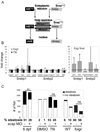
A. The activation pathways for the nSrebp and nAtf6 transcription factors utilize common components. * indicates components targeted in our studies using either a morpholino (mo) or mutant (mt). B. Srebp1 and Srebp2 target genes were assessed in at least 3 clutches of RNA isolated from whole larvae (left) and dissected livers (right). Expression in foigr mutants was normalized to wild-type siblings (grey bars) and expression in tunicamycin treated larvae was normalized to DMSO treated siblings (black bars). The level of expression in the respective controls was set to 1 (horizontal bar). * indicates p<0.01 by a 1 sampled t-test. C. The effect of scap morpholino injection on steatosis in three models (fasting, chronic tunicamycin and foigr mutation) was assessed. First, scap morphants and uninjected siblings were fasted until 6 dpf (left) or, second, they were treated with tunicamycin or DMSO from 3–5 dpf (center). Third, scap morpholino was injected into foigr mutants and wild-type siblings (right). All larvae were collected on 5 dpf, whole mount stained with oil red O and scored for steatosis. The number larvae scored positive larvae (black) and negative (white) are plotted. The percent of fish with steatosis is indicated below each bar. ** indicates a p <0.001 by Fisher’s exact test, ns: not significant.
Figure 5
Figure 5. mbtps1hi1487 mutants are protected from steatosis caused by chronic tunicamycin treatment
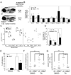
A. mbtps1hi1487 mutant larvae have a round liver, but do not develop more steatosis than their wild-type siblings. Tg(fabp10:dsRed) wild-type (top) and mbtps1hi1487 (bottom) larvae were imaged on 5 dpf and scored for steatosis by whole mount oil red O staining. B. Expression of Srebp target genes in 5 dpf wild-type andmbtps1hi1487 larvae treated with tunicamycin was analyzed by qPCR. The expression in tunicamycin treated larvae was normalized to their DMSO treated fish of the same genotype to obtain the fold change for each gene. The average fold change in wild-type and mutants from 3 clutches is plotted with error bars showing standard deviation. C. mbtps1 mutation induces UPR target genes. cDNA from 5 dpf wild-type (white dots) or mbtps1hi1487 (black dots) whole larvae (left) or dissected livers (right) was analyzed by qPCR. The ΔCt obtained for each gene is plotted as a single point for each individual clutch analyzed, with the average indicated by a horizontal bar. * indicates p<0.05, ** p<0.005 by a Student’s t-test. D. DMSO or tunicamycin treated WT and mbtps1hi1487 larvae were analyzed by qPCR for Atf6 target genes. The ΔCt in the wild-type and mutant tunicamycin treated larvae was normalized to DMSO treated wild-type larvae to obtain the fold change. The average fold change from 5 clutches is plotted with error bars indicating the standard deviation. * indicates p<0.05 by a one sampled t-test. E. mbtps1 mutation protects 5 dpf larvae from steatosis caused by chronic tunicamycin treatment. Wild-type and mbtps1hi1487 larvae treated from 3–5 dpf with 1 µg/ml tunicamycin or DMSO were stained with oil red O and scored for steatosis. Steatosis was scored in at least 8 clutches and the median percent of steatosis is plotted with bars indicating the standard deviation. ** indicates p<0.001 by Student’s t-test. In addition to whole mount oil red O staining, a subset of the samples described above were sectioned, stained with oil red O and quantified for the number of oil red O droplets (F.) and area of oil red O (G.) per liver cell. The median value for each fish is plotted as a dot, with a horizontal line to indicate the mean. * indicates p<0.05, ** indicates p<0.005 and *** indicates p<0.0005 by ANOVA. ns: not significant.
Figure 6
Figure 6. Atf6 knockdown affects UPR activation
A. atf6 morpholino injected into Tg(fabp10:dsRed) embryos does not affect development or steatosis based on whole mount oil red O staining. B. cDNA prepared from whole (left) or dissected livers (right) from 5 dpf larvae injected with the standard control (white) or atf6 morpholino (grey) was analyzed by qPCR. Each gene was normalized to rpp0 and the ΔCt from each cDNA sample is plotted as a dot with the mean indicated as a bar. p values are indicated and * indicates p<0.05. C. Uninjected embryos and atf6 morphants were treated from 24–48 hpf with 1 µg/ml tunicamycin or DMSO, collected at 48 hpf and Atf6 target gene expression was examined by qPCR. The average ΔCt value of 4 clutches is plotted with error bars indicating the standard deviation. ** indicates p<0.001 by ANOVA. The fold change for each gene is labeled below each column, and numbers with an * represent a significant difference in expression in tunicamycin treated fish compared to DMSO treated fish of the same genotype. D. atf6 morpholino was injected into embryos generated by in-crossing foigr heterozygotes. On 5 dpf, foigr mutants and phenotypically wild-type siblings with and without the atf6 morpholino were collected for qPCR analysis. The ΔCt normalized to rpp0 was calculated from 3 clutches, averaged and plotted with error bars indicating the standard deviation. * indicates p<0.01 by ANOVA. The fold change for each gene is labeled below each column, and numbers with an * represent a p<0.05 comparing expression in mutants compared to wild-type fish as by ANOVA.
Figure 7
Figure 7. atf6 depletion alleviates steatosis in foigr mutants
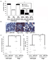
A. atf6 morpholino decreases the number of 5 dpf foigr that develop steatosis. Embryos generated by crossing foigr heterozygotes were uninjected (15 clutches) or injected with the standard control (9 clutches) or atf6 morpholino (15 clutches). Larvae were separated based on phenotype on 5 dpf, stained with oil red O, and scored for steatosis. The total number of embryos in each category is plotted and the percent fish with steatosis is labeled under each graph. * indicates p=0.01, *** indicates p< 0.0001 by chi square. B. atf6 morpholino injection decreases the amount of oil red O staining in foigr hepatocytes. Cryosections of 5 dpf either uninjected or atf6 morpholino injected wild-type and foigr mutant embryos were stained with oil red O and hematoxylin. C–D. The number of oil red O droplets per liver cell (C) and the area of each liver cell that is stained with oil red O (D) was quantified. The median value from at least 3 sections for each fish is plotted as an individual dot with the horizontal bar indicating the mean for all fish in each sample. The foigr mutants injected with atf6 morpholino are not significantly different from the wild-type controls. * indicates p<0.05, *** indicates p<0.005 by ANOVA.
Figure 8
Figure 8. Acute tunicamycin treatment causes UPR activation and steatosis that is augmented by atf6 depletion
A. Diagram of tunicamycin exposure protocols. Larvae treated with DMSO or tunicamycin for 48 (chronic; 1 µg/ml) or 12 (acute; 2 µg/ml) hours. Samples were collected at the times indicated by each letter for qPCR or oil red O staining. For protocol D, larvae were treated as in C, the tunicamycin was washed out and larvae were incubated for an additional 12 hours. B. UPR target genes were induced by all 4 protocols. The fold change for each gene in was calculated relative to DMSO (set as 1) at least 3 clutches and averaged. Error bars indicate the standard deviation, * indicates p<0.05, ** indicates p<0.005 by a ANOVA. C. Tunicamycin was administered to zebrafish according to the chronic and acute protocols outlined in panel A. The number of fish with steatosis was scored in whole mount oil red O stained larvae. The percent of steatosis in 3–5 clutches in each sample is labeled below each bar. Control fish were treated with DMSO from 3–5 dpf. The difference between tunicamycin treated and DMSO treated fish was significant for protocols A and D; *** indicates p<0.0001 by Fisher’s exact test. D. mbtps1hi1487 mutants are predisposed to steatosis caused by acute tunicamycin exposure. The number of fish with steatosis was counted in 6 clutches of mbtps1hi1487 mutants and their wild-type siblings that were treated with DMSO from 3–5 dpf or with chronic (protocol A) or acute (protocol D) tunicamycin. ** indicates p<0.002 and **** indicates p<0.0001 by Fisher’s exact test. E. atf6 morphants develop more steatosis in response to acute tunicamycin treatment. atf6 morphants, standard control morphants and uninjected embryos were treated with tunicamycin of DMSO according to protocol D. The total number of 5 dpf fish in 3 clutches that were scored for steatosis based on whole mount oil red O staining is plotted. The percent steatosis for each sample is labeled below each bar. * indicates p<0.02 by Fisher’s exact test.

References

    1. Bernales S, Papa FR, Walter P. Intracellular signaling by the unfolded protein response. Annu Rev Cell Dev Biol. 2006;22:487–508. - PubMed
    1. Rutkowski DT, Hegde RS. Regulation of basal cellular physiology by the homeostatic unfolded protein response. J Cell Biol. 2010;189:783–794. - PMC - PubMed
    1. Yoshida H, Matsui T, Yamamoto A, Okada T, Mori K. XBP1 mRNA is induced by ATF6 and spliced by IRE1 in response to ER stress to produce a highly active transcription factor. Cell. 2001;107:881–891. - "VSports app下载" PubMed
    1. Asselah T, Bieche I, Mansouri A, Laurendeau I, Cazals-Hatem D, Feldmann G, Bedossa P, et al. In vivo hepatic endoplasmic reticulum stress in patients with chronic hepatitis C. J Pathol. 2010;221:264–274. - PubMed
    1. Hotamisligil GS. Endoplasmic reticulum stress and the inflammatory basis of metabolic disease. Cell. 2010;140:900–917. - PMC - PubMed

Publication types

Substances