<noscript lang="JMJyJXR"></noscript><ins id="GfBdXX"></ins>
Skip to main page content
U.S. flag

An official website of the United States government

Dot gov

The . gov means it’s official. Federal government websites often end in VSports app下载. gov or . mil. Before sharing sensitive information, make sure you’re on a federal government site. .

Https

The site is secure V体育官网. The https:// ensures that you are connecting to the official website and that any information you provide is encrypted and transmitted securely. .

. 2011 Jun;121(6):2254-63.
doi: 10.1172/JCI44675. Epub 2011 May 2.

CD8⁺ T cells with an intraepithelial phenotype upregulate cytotoxic function upon influenza infection in human lung

Affiliations

CD8⁺ T cells with an intraepithelial phenotype upregulate cytotoxic function upon influenza infection in human lung

Berber Piet et al. J Clin Invest. 2011 Jun.

Abstract

The human lung T cell compartment contains many CD8⁺ T cells specific for respiratory viruses, suggesting that the lung is protected from recurring respiratory infections by a resident T cell pool. The entry site for respiratory viruses is the epithelium, in which a subset of lung CD8⁺ T cells expressing CD103 (αE integrin) resides. Here, we determined the specificity and function of CD103⁺CD8⁺ T cells in protecting human lung against viral infection. Mononuclear cells were isolated from human blood and lung resection samples. Variable numbers of CD103⁺CD8⁺ T cells were retrieved from the lung tissue VSports手机版. Interestingly, expression of CD103 was seen only in lung CD8⁺ T cells specific for influenza but not in those specific for EBV or CMV. CD103⁺ and influenza-reactive cells preferentially expressed NKG2A, an inhibitor of CD8⁺ T cell cytotoxic function. In contrast to CD103⁻CD8⁺ T cells, most CD103⁺CD8⁺ cells did not contain perforin or granzyme B. However, they could quickly upregulate these cytotoxic mediators when exposed to a type I IFN milieu or via contact with their specific antigen. This mechanism may provide a rapid and efficient response to influenza infection, without inducing cytotoxic damage to the delicate epithelial barrier. .


Figures

Figure 1
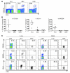
Figure 1. CD103 expression on lung CD8+ T cells is highly increased compared with that on peripheral blood CD8+ T cells.
(A) The percentages of peripheral blood CD8+ T cells and lung CD8+ T cells expressing CD103, as measured by flow cytometry, in paired peripheral blood and lung samples (n = 34). ***P < 0.0001; paired t test. (B) Representative histogram plots for the expression of CD103 on CD8+ T cells in the peripheral blood and human lung. Histogram plots show only CD3+CD8+ cells within the lymphocyte gate. The numbers in the plot represent the MFI of the CD103+ cells. (C) Preferential localization of CD103+CD8+ T cells above the basement membrane (dotted line) inside the airway epithelium (E). Original magnification, ×20. Immunohistochemistry stainings were performed on frozen human lung sections. To preserve the original tissue structure, we embedded the tissue in TissueTek prior to freezing. Representative images of airway containing tissue section. The image on the left is the original image; the image in the middle is a spectral analysis of all stained cells; and the image on the right is a spectral analysis of exclusively triple+ cells. Blue, CD3+; red, CD8+; green, CD103+; yellow, CD3/CD8/CD103 triple+ (middle and right images). n = 4 patients.
Figure 2
Figure 2. Influenza-specific lung CD8+ T cells have an intraepithelial phenotype, expressing CD103, VLA-1, and NKG2A, whereas cells specific for nonrespiratory viruses lack the expression of these epithelial markers.
(A) FACS plots showing tetramer stainings on lung CD3+ cells after gating on live cells. Numbers indicate the percentages of CD3+ cells that are tetramer+. (B) Percentages of influenza-specific, EBV-specific, and CMV-specific lung CD8+ T cells expressing CD103, NKG2A, and VLA-1, as assessed by flow cytometry (n = 5–9 per tetramer). Bars represent the median. (C) Representative FACS plots for phenotype of lung and peripheral blood CD8+ T cells. FACS plots show the expression of CD103, NKG2A, and VLA-1 on total lung CD8+ T cells (left column), on FLU- and EBV-specific lung CD8+ T cells (second and third columns, respectively), on total peripheral blood CD8+ T cells (fourth column), and on FLU- and EBV-specific peripheral blood CD8+ T cells (fifth and sixth columns, respectively). Numbers in first and fourth columns indicate the percentages of CD3+CD8+ cells that are CD103+, NKG2A+, or VLA-1+. Numbers in second, third, fifth, and sixth columns indicate the percentages of tetramer+ cells that are CD103+, NKG2A+, or VLA-1+. Plots are representative of 5 to 9 patients per tetramer (lung) or 2 to 6 patients per tetramer (peripheral blood). FLU-specific cells could often not be detected in the blood with tetramer staining, although these patients did have FLU+ cells in the lung. CD8+ T cells were all identified as CD3+CD8+ cells within the lymphocyte gate.
Figure 3
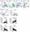
Figure 3. Lung CD103+CD8+ T cells are CD45R0+ effector or memory cells.
Differentiation stages of the CD8+ T cells within the CD103+ and CD103 cell fractions as assessed by flow cytometry. Naive cells were defined as CD27+CD45R0, memory cells were defined as CD27+CD45R0+, and effector cells were defined as CD27CD45R0±. (A) Representative FACS plots for CD103+ and CD103CD8+ T cells. Dot plots are gated on CD3+CD8+CD103+ cells among live lymphocytes or on the CD3+CD8+CD103 cells among live lymphocytes. Numbers indicate the percentages of CD3+CD8+CD103+ or CD3+CD8+CD103 cells that are located within each quadrant. (B) Naive CD8+ T cells (CD27+CD45R0), memory CD8+ T cells (CD27+CD45R0+), effector/memory (EM) CD8+ T cells (CD27CD45R0±), and CD45R0CD8+ T cells as percentages of total CD103+ and CD103CD8+ T cell fractions (n = 28). ***P < 0.0001.
Figure 4
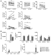
Figure 4. CD103+ and CD103 lung CD8+ T cells produce high amounts of IFN-γ and other Th1 cytokines but hardly any Th2 cytokines, IL-10, or IL-17.
CD103+CD8+ T cells produce more IFN-γ than the CD103CD8+ T cell fraction. (A) Intracellular cytokine production of lung CD8+ T cells was measured by flow cytometry after 4-hour stimulation with PMA and ionomycin in the presence of brefeldin A. Cells were stained with life/dead stain (to gate out dead cells) and with monoclonal antibodies against CD3, CD8, and CD103 and fixed, permeabilized, and stained for intracellular cytokines. Graphs show the percentages of CD103+ and CD103 CD8+ T cells that produced each indicated cytokine. n = 5–10 patients. *P ≤ 0.04, **P = 0.003. (B) Cytokine production of CD103+ and CD103 lung CD8+ T cells as measured by luminex assay. CD103+ and CD103 lung CD8+ T cells from 4 patients were sorted with flow cytometry and stimulated for 24 hours with PMA and ionomycin (PMA/iono). Culture supernatants were collected and measured by the Bio-Rad 27-plex luminex assay. The error bars show SEM. *P = 0.03.
Figure 5
Figure 5. CD8+CD103+ lung T cells have a low cytotoxic potential and a high expression of the inhibitory NK cell receptor complex CD94/NKG2A.
(A) FACS plots show the expression of perforin, granzyme B, CD94, and NKG2A plotted against CD103 expression. Plots show only CD3+CD8+ lung T cells within the lymphocyte gate and are representative of 22–30 patients. Numbers indicate the percentages of CD3+CD8+ cells that are located within each quadrant. (B) Most lung CD8+ T cells express CD94 and NKG2A to the same extent, which is suggestive of dimerization (left). Numbers indicate the percentages of CD3+CD8+ cells that are located within each quadrant. Moreover, lung CD8+ T cells hardly express the activating isoform NKG2C, and there is no difference in NKG2C expression between CD103+ and CD103 T cells (right, n = 5). (C) The percentage of CD103+CD8+ and CD103CD8+ lung T cells containing perforin or granzyme B (n = 22). (D) The percentage of CD103+CD8+ and CD103CD8+ lung T cells expressing CD94 (n = 27) and NKG2A (n = 30). All expression data were collected with flow cytometry; all FACS plots show only lung CD3+CD8+ lymphocytes within the lymphocyte gate. ***P < 0.0001.
Figure 6
Figure 6. CD8+CD103+ lung T cells have a high cytotoxic activity in vitro.
The amount of specific killing of target cells by purified T cell populations at varying effector/target (E/T) ratios is shown. A redirected killing assay was performed by incubating FACS-sorted CD103+ or CD103 lung CD8+ T cells with chromium-labeled P815 target cells at varying effector/target ratios in the presence or absence of aCD3 mAb. FACS-sorted peripheral blood naive (CD45RA+CD27bright) and effector (CD27) CD8+ T cells were included in this assay as control. Measurements were performed in triplicate (n = 3 patients). The mean ± SEM of specific killing is shown.
Figure 7
Figure 7. Lung CD8+ T cells quickly upregulate cytotoxic molecules after specific antigen contact or under influence of a type I IFN infectious milieu.
(A) LMCs were cultured for 6 days in the presence of FLU peptide, irrelevant peptide (OVA peptide), or medium. FACS plots and bar graphs show the expression of perforin and granzyme B on FLU-A2+ cells (n = 2), as measured by flow cytometry. Plots are gated on CD3+CD8+ T cells and are representative of 2 patients. Numbers indicate the percentages of CD3+CD8+ cells located in each quadrant. (B) LMCs were stimulated for 12 or 36 hours with poly I:C or different concentrations of IFN-α. Granzyme B expression was measured by flow cytometry. The percentage increase in granzyme B expression as compared with that in medium control is shown for FLU+CD3+CD8+ T cells (left) and total CD3+CD8+ T cells (right) (n = 3 patients). The error bars show SEM. *P < 0.05, **P < 0.01.

References

    1. Hogg JC, et al. The nature of small-airway obstruction in chronic obstructive pulmonary disease. . N Engl J Med. 2004;350(26):2645–2653. doi: 10.1056/NEJMoa032158. - DOI - PubMed
    1. Topham DJ, Tripp RA, Doherty PC. CD8+ T cells clear influenza virus by perforin or Fas-dependent processes. J Immunol. 1997;159(11):5197–5200. - PubMed (V体育平台登录)
    1. Russell JH, Ley TJ. Lymphocyte-mediated cyto­toxicity. Annu Rev Immunol. 2002;20:323–370. doi: 10.1146/annurev.immunol.20.100201.131730. - DOI - PubMed
    1. Trapani JA, Sutton VR, Smyth MJ. CTL granules: evolution of vesicles essential for combating virus infections. Immunol Today. 1999;20(8):351–356. doi: 10.1016/S0167-5699(99)01488-7. - DOI - PubMed
    1. Cerwenka A, Morgan TM, Dutton RW. Naive, effector, and memory CD8 T cells in protection against pulmonary influenza virus infection: homing properties rather than initial frequencies are crucial. J Immunol. 1999;163(10):5535–5543. - PubMed

Publication types

MeSH terms