Skip to main page content
U.S. flag

An official website of the United States government

Dot gov

The . gov means it’s official. Federal government websites often end in VSports app下载. gov or . mil. Before sharing sensitive information, make sure you’re on a federal government site. .

Https

The site is secure. The https:// ensures that you are connecting to the official website and that any information you provide is encrypted and transmitted securely V体育官网. .

. 2011 Apr 22;34(4):541-53.
doi: 10.1016/j.immuni.2011.04.006.

A central role for mTOR kinase in homeostatic proliferation induced CD8+ T cell memory and tumor immunity

Affiliations

A central role for mTOR kinase in homeostatic proliferation induced CD8+ T cell memory and tumor immunity

Qingsheng Li et al. Immunity. .

Abstract (VSports手机版)

The cell-intrinsic mechanisms guiding naive CD8+ T cells for clonal expansion and memory generation via homeostatic proliferation (HP) are unclear. Here, we have shown that HP of naive CD8+ T cells requires IL-7-, but not IL-15-induced mTOR kinase activation. HP-induced mTOR enhances transcription factor T-bet for functional maturation and CD122 expression, which sensitizes for an IL-15-dependent memory transition by favoring transcription factor Eomesodermin over T-bet. Inhibition of mTOR blocks T-bet and CD122 expression but preserves memory in an IL-15-independent manner by promoting Eomesodermin expression. The ability of rapamycin to augment HP-induced memory was cell-intrinsic given that silencing mTOR in CD8+ T cells generated identical outcomes. Strikingly, HP-induced CD8+ T cell memory generated by IL-15-dependent or -independent mechanisms demonstrated identical tumor efficacy VSports手机版. These results indicate a central role for mTOR in HP-induced CD8+ T cell responses and demonstrate the importance for CD8+ memory in HP-induced tumor efficacy. .


Figures

FIGURE 1
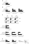
FIGURE 1. Lymphopenia induced CD8+ T cell HP and functional maturation is IL-7, but not IL-15 dependent
(A) CFSE-labeled naïve OT-1 cells (1 × 10⁶) were transferred into intact or irradiated (700 rads) syngeneic recipients on day 0. Some radiated recipients received either an isotype control (700 rad) or anti-IL-7Rα (100 µg per mouse, 700 rad+anti-IL-7Rα) on day -1 and day 2. The CD8α+Thy1.1+ cells were gated and evaluated by flow cytometric analysis of recipient spleens on day 5 for; proliferation-CFSE dilution (left), the clonal expansion-absolute number of OT-1 cells (middle) (as described in methods) and effector functions-IFN-γ and Granzyme B (Gzm B) expression detected by ICS (right). (B) The day 5 response of CFSE-labeled naive OT-1 cells in intact or irradiated syngeneic B6 or Il15−/− recipients were evaluated by flow cytometry for; proliferation-CFSE dilution (left), absolute numbers of OT-1 cells (middle), and the expression of IFN-γ and Gzm B (right). (C) In vitro CFSE-labeled naïve OT-1 cells cultured with IL-7 (10 ng/ml), IL-15 (10 ng/ml) or medium were analyzed for proliferation-CFSE dilution (left), cell recovery (middle), and the expression of IFN-γ and Gzm B (right) at 120 hr. The bars present SD of three individual samples and the results shown are representative of three independent experiments with similar outcomes (*, p<0.05; ***, p<0.001).
FIGURE 2
FIGURE 2. IL-7 induced mTOR activity is essential for HP induced functional maturation of CD8+ T cells
(A and B) OT-1 cells detected in the spleens of recipients receiving different doses of irradiation; 0, 175 or 700 rads (A), or 700 rads irradiation followed by anti-IL-7Rα (100 µg) on day -1 and day 2 (B) were evaluated for phosphorylation of S6 by ICS and flow cytomtery. (C – F), OT-1 detected in recipients treated with 700 rad or 700 rad plus daily injection of rapamycin (0.75 mg/Kg/day) from day 0 – 4 were evaluated on day 5 for phosphorylation of mTOR and S6 (C), CFSE dilution (D), absolute numbers of OT-1 cells (E), and IFN-γ and Granzyme B expression by ICS and flow cytometry (F). (G) In vivo CTL activity produced by normalized numbers of OT-1 cells, the percentage of specific lysis is indicated above histograms. The error bars are SD of three independently obtained values. A representative of three experiments with identical results is shown (**, p<0.01).
FIGURE 3
FIGURE 3. HP-IL-7 induced mTOR activity is essential for T-bet mediated functional maturation
(A – F) Naïve WT or Tbx21−/− OT-1 cells were detected by flow cytometry on day 5 after transfer into intact or irradiated (700 rads) recipients that were either untreated (A,D,E,F) or treated with anti-IL-7Rα (100 µg) on day -1 and day 2 (B), or with rapamycin (0.75 mg/Kg/day) (C) and evaluated for; (A) CFSE dilution, T-bet and CD122 expression. (B and C) T-bet and CD122 expression in recipients treated with anti-IL-7Rα (B) or in radiated recipients treated with rapamycin (C). (D) The expression of CD122 in WT or Tbx21−/− OT-1 cells. (E) CFSE dilution of WT or Tbx21−/− OT-1 cells. (F) The number of OT-1 (CD8α+Thy1.1+) cells (left), percentage of IFN-γ+ OT-1 cells (middle), and in vivo cytolysis (right). The error bars are SD of three independently obtained values. The data shown is representative of three experiments (* p<0.05; **p<0.01; n.s., not significant).
FIGURE 4
FIGURE 4. HP-induced T-bet augments IL-15 dependent CD8+ T cell memory
(A) Naïve WT or Tbx21−/− OT-1 cells transferred to radiated B6 or Il15−/−B6 recipients were evaluated for the absolute numbers of OT-1 (CD8α+Thy1.1+) cells on day 40. (B) OT-1 cell numbers (3 days post re-challenge, day 43) (left), the percentage of IFN-γ+ OT-1 cells (middle), and in vivo cytolysis (right). Numbers in parenthesis (left panel) is the OT-1 fold increase. The error bars represent SD of values from three mice/experimental group and a representative of two independent experiments is shown (**, p<0.01; ***, p<0.001).
FIGURE 5
FIGURE 5. Inhibition of mTOR promotes IL-15 independent HP-induced CD8+ T cell memory
(A – C) WT or Il15−/− irradiated OT-1 recipients that were either untreated or treated with rapamycin (0.75 mg/Kg/day) from day 0 – 7 were evaluated on day 40 for the absolute numbers of OT-1 cells (A) and antigen-recall responses measured on day 43 (B). Number of OT-1 cells (left), percentage of IFN-γ+ OT-1 cells (middle), and in vivo cytolysis (right). (C) Top, the percentage of OT-1 cells (CD8+ Thy1.1+) in WT or Il15−/− recipients on day 40 or day 43. The circles represent the OT-1 population and the numbers indicate the percent frequency. Middle, number of OT-1 cells on day 40. Bottom, number of OT-1 cells on day 43, percentage of IFN-γ+ OT-1 cells and percentage of granzyme B+ OT-1 cells on day 43. The numbers in parenthesis indicate the fold-increase in OT-1 numbers post-immunization. (D) RNAi mediated inhibition of mTOR promotes HP-induced CD8+ memory T cells. Naïve OT-1 cells activated in vivo in irradiated B6 hosts were transduced with retroviruses expressing RNAi for mTOR or empty control vector. OT-1 cells were then transferred into irradiated B6 hosts. The number of OT-1 cells was evaluated on day 40 post transduction and antigen-recall responses were measured on day 43. The numbers in parenthesis indicate the fold-increase in OT-1 numbers post-immunization (middle panel). The error bars are the SD of values obtained from three animals/experimental groups and one of two independent experiments is shown (*, p<0.05; **, p<0.01; ***, p<0.001; n.s., not significant).
FIGURE 6
FIGURE 6. IL-15 regulates the expression of T-bet and Eomes in IL-7 conditioned CD8+ T cells
(A) Naive OT-1 cells stimulated with IL-7 (10 ng/ml) for 72 hr were re-cultured with IL-15 at various concentration for additional 24 hr. The expression of T-bet and Eomes in OT-1 cells at 72 hr (left) and 72+24 hr (middle). Numbers above the lines indicate mean fluorescence intensity (MFI). The fold-increase in OT-1 cells expressing Eomes or T-bet post-treatment of IL-15 (72+24 hr/72 hr, right). The data are representative of three independent experiments. (B) Left, splenocytes from irradiated recipients or irradiated recipients followed the treatment of rapamycin (0.75 mg/Kg/day, day 0–day 7) were stained for T-bet and Eomes by ICS on day 5 or day 40. Numbers above the lines indicates mean fluorescence intensity (MFI). Right, the fold-increase in OT-1 cells expressing Eomes or T-bet (day 40/day 5). The error bars represent SD of values obtained from three animals/experimental groups. The data are representative of two independent experiments.
FIGURE 7
FIGURE 7. mTOR inhibitor enhances HP-mediated tumor protection
(A and B) B6 mice inoculated with 3 × 10⁶ EL4 or EG.7 by i.p. or s.c. injection on day -7 were irradiated (700 rad) on day -2 and received 1×10⁶ naïve OT-1 cells on day 0. Rapamycin was injected into indicated recipients from day 0 – 7 post-transfer. (A) Tumor-free survival curve. (B) Tumor size (mean ± s.e.m.) curve. Data in this figure were pooled from three independent experiments.

References

    1. Ahmed R, Gray D. Immunological memory and protective immunity: understanding their relation. Science. 1996;272:54–60. - PubMed
    1. Araki K, Turner AP, Shaffer VO, Gangappa S, Keller SA, Bachmann MF, Larsen CP, Ahmed R. mTOR regulates memory CD8 T-cell differentiation. Nature. 2009;460:108–112. - PMC - PubMed
    1. Blazar BR, Taylor PA, Panoskaltsis-Mortari A, Vallera DA. Rapamycin inhibits the generation of graft-versus-host disease- and graft-versus-leukemia-causing T cells by interfering with the production of Th1 or Th1 cytotoxic cytokines. J Immunol. 1998;160:5355–5365. - PubMed
    1. Brown VI, Fang J, Alcorn K, Barr R, Kim JM, Wasserman R, Grupp SA. Rapamycin is active against B-precursor leukemia in vitro and in vivo, an effect that is modulated by IL-7-mediated signaling. Proc Natl Acad Sci U S A. 2003;100:15113–15118. - PMC - PubMed
    1. Burnett PE, Barrow RK, Cohen NA, Snyder SH, Sabatini DM. RAFT1 phosphorylation of the translational regulators p70 S6 kinase and 4E-BP1. Proc Natl Acad Sci U S A. 1998;95:1432–1437. - PMC - PubMed

Publication types

"VSports注册入口" MeSH terms

Substances