Skip to main page content
U.S. flag

An official website of the United States government

Dot gov

The . gov means it’s official. Federal government websites often end in . gov or . mil. Before sharing sensitive information, make sure you’re on a federal government site VSports app下载. .

Https

The site is secure. The https:// ensures that you are connecting to the official website and that any information you provide is encrypted and transmitted securely. V体育官网.

. 2011 Oct;18(10):1598-607.
doi: 10.1038/cdd.2011.33. Epub 2011 Apr 8.

"VSports注册入口" Upregulation of human autophagy-initiation kinase ULK1 by tumor suppressor p53 contributes to DNA-damage-induced cell death

Affiliations

Upregulation of human autophagy-initiation kinase ULK1 by tumor suppressor p53 contributes to DNA-damage-induced cell death

W Gao (VSports注册入口) et al. Cell Death Differ. 2011 Oct.

Abstract

In yeast, activation of ATG1/ATG13 kinase complex initiates autophagy. This mechanism of autophagy initiation is conserved, as unc-51-like kinase 1 (ULK1) and unc-51-like kinase 2 (ULK2) are two mammalian functional homologues of ATG1 and form similar complex with mammalian ATG13. Here, we report that both ULK1 and ULK2 are transcriptional targets of tumor suppressor p53 VSports手机版. In response to DNA damage, ULK1 and ULK2 are upregulated by p53. The upregulation of ULK1 (ULK2)/ATG13 complex by p53 is necessary for the sustained autophagy activity induced by DNA damage. In this context, elevated autophagy contributes to subsequent cell death. These findings suggest that ULK1 and ULK2 may mediate part of tumor suppression activity in mammalian cells and contribute to the efficacy of genotoxic chemotherapeutic drugs. .


Figures

Figure 1
Figure 1
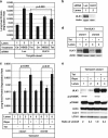
Autophagy induction by sub-lethal DNA damage. (a) U2OS cells were treated with different concentrations of camptothecin (CPT, lanes 2 and 3) or etoposide (Eto, lanes 4 and 5) for 2 days before doing the long-lived protein degradation (LLPD) assay as described in Materials and Methods. Y axis is the LLPD rate after 2 h. (b) U2OS cells were treated with either 0.5 μM CPT (lanes 3 and 4) or 5 μM Eto (lanes 5 and 6) for 2 days. On the day of measuring LLPD rate, 5 mM of 3-methyladenine (3-MA) was used to pretreat cells for 1 h (lanes 2, 4 and 6) before doing the LLPD assay. (c) U2OS cells were treated with either 0.5 μM CPT (lane 2) or 5 μM Eto (lane 3) for 2 days. The cells were collected and lysed to run western blot. The blot were probed with the antibody against unc-51-like kinase 1 (ULK1), p53, p21, T-389 phosphorylated form of p70S6K (p-p70S6K), p70S6K, AMPK-α1, -β1, -β2, -γ1, -γ2, LC3B and actin. To measure the ratio of p-p70S6K over p70S6K, the blot was developed using enhanced chemiluminescence (ECL) reagent and the band intensities were measured using Kodak Image Station 4000R pro. The relative ratio for the control is defined as 1. (d) U2OS cells were treated with 1 μM rapamycin or 0.5 μM CPT for 2, 6, 24, 48 and 72 h, after which LLPD rates were measured for each sample and plotted against treatment time. (e) U2OS cells were treated with 1 μM CPT for 1 day. The cells were then collected and lysed to do western blot. The blots were probed with the antibodies against p53, ULK1, ATG13, FIP200, ATG5, ATG7, ATG12, beclin 1 and actin. (f) MCF7 cells were untreated (E, lane 1) or treated with either Hank's buffered salt solution (HBSS) for 2 h (H, lane 2), 0.3 μM CPT for 1 day (lane 3), 1 μM CPT for 1 day (lane 4) and 1 μM rapamycin for 6 h (R, lane 5). The cells were then collected and lysed to do western blot. The blots were probed with the antibodies against ULK1, p-p70S6K and p70S6K. (g) U2OS cells were transfected with either luciferase control small interfering (si)RNA (lanes 1–3) or ATG13 siRNA (lanes 4–6) as described in Materials and Methods. After 3 days of siRNA transfection, they were treated with (lanes 2 and 5) or without (lanes 1 and 4) 0.5 μM CPT for 2 days and measured the LLPD rates. HBSS treatments (lanes 3 and 6) were used as controls. (h) The same samples as in (g) were used to do western blot to confirm the knockdown efficiency of ATG13. These data are presented as mean+S.D. and are representative of at least two independent experiments
Figure 2
Figure 2
Unc-51-like kinase 1 (ULK1) upregulation after DNA damage is dependent on p53. (a) Three U2OS clones containing tetracycline (Tet)-responsive p53shRNA (Tet-p53shRNA-clone1 and clone2) or a nonspecific shRNA (Tet-LashRNA) were treated with (lanes 3, 4, 7, 8, 11 and 12) or without (lanes 1, 2, 5, 6, 9 and 10) 1 μg/ml Tet for 2 days to knock down p53 or La, and then treated with (lanes 2, 4, 6, 8, 10 and 12) or without (lanes 1, 3, 5, 7, 9 and 11) 0.5 μM CPT for 2 days. The cells were then collected and lysed to do western blot. The blots were probed with antibodies against ULK1, p53 and p21. Actin was used as a loading control. (b) The same cells were treated as in (a) before doing the long-lived protein degradation (LLPD) assay. (c) Two U2OS clones containing Tet-responsive p53 (Tet-p53-clone1 and -clone2) were treated with (lanes 2 and 4) or without (lanes 1 and 3) 1 μg/ml Tet for 1 day to induce the expression of p53. The cells were then lysed to do western blot to test the expression level of ULK1, p53 and p21. Actin was used as a loading control. (d) One of the clone (clone1) as in (c) was transfected with either Luciferase small interfering (si)RNA (Luc, lanes 1–4) or ATG13 siRNA (ATG13i1, lanes 5 and 6) as described in Materials and Methods. After 3 days of siRNA transfection, p53 expression was induced with 1 μg/ml of Tet (lanes 2, 4 and 6) for 1 day. 3-MA (5 mM) was used to pretreat the cells for 1 h (lanes 3 and 4) before doing the LLPD assay. (e) The same clone (clone 1) as in (d) was either induced to express p53 (lane 2) or treated with 0.5 μM CPT for 2 days (lane 3), after which total RNAs were extracted from these cells. Quantitative PCR was used to test the mRNA expression levels of ULK1. These data are presented as mean±standard deviation (S.D.) and are representative of at least two independent experiments
Figure 3
Figure 3
Unc-51-like kinase 1 (ULK1) is a bona fide transcriptional target of p53. (a) Schematic representation of the putative p53 binding site of ULK1. (b). Electrophoretic mobility shift assay was carried out using biotinylated DNA oligos containing either the duplicated putative wild-type (wt) p53 binding sites (biotin-p53BS, lanes 1 and 3–5) or mutant (mt) p53 binding site (lane 2) in ULK1 gene as described in Materials and Methods. Wt (lane 4) or mt (lane 5) p53 binding sites of ULK1 were used for the competition assay. (The bands labeled with asterisk indicates nonspecific DNA complexes.) (c) Luciferase reporter assay was performed as described in Materials and Methods. Result for each condition was normalized to the firefly/Renilla luciferase activity ratio of empty control (lane 1). Ctr: empty pcDNA3.1 vector. Y axis is depicted as log scale. These data are representative of at least two independent experiments
Figure 4
Figure 4
Unc-51-like kinase 1 (ULK1) is required for p53-dependent autophagy. (a) The p53-inducible expression cell line (tetracycline (Tet)-p53-clone1) as in Figure 2 (d) was transfected with either luciferase small interfering (si)RNA (lanes 1–3) or ULK1 siRNA (lanes 4–6) as described in Materials and Methods. After 4 days of siRNA transfection, p53 was induced to be expressed (lanes 3 and 6) for 1 day with 1 μg/ml of Tet to do the long-lived protein degradation (LLPD) assay. Hank's buffered salt solution (HBSS) treatment (lanes 2 and 5) were used as positive controls. (b) The same samples in (a) were used to do western blot to test the knockdown efficiency of ULK1. Actin was used as a loading control. (c) Two U2OS clones containing Tet-responsive ULK1 (Tet-ULK1-clone1 and -clone2) were treated with 1 μg/ml of Tet for 1 day (lanes 3, 4, 7 and 8). Then, 1 μM of rapamycin was used to treat the cells for 6 h (lanes 2, 4, 6 and 8) before doing the LLPD assay as described in Materials and Methods. (d) The samples from (c) were used to do western blot to test the expression level of ULK1. Actin was used as a loading control. (e) The Tet-ULK1-clone1 cell line was treated as in (c) and the samples were collected to do western blot to test the expression level of ULK1, p-p70S6K (T-389), p70S6K, actin and LC3-I and -II. To measure the ratio of LC3-II over LC3-I, the blot was developed with enhanced chemiluminescence (ECL) reagent and the band intensities were measured with Kodak Image Station 4000R pro. The P-value was obtained using Student's t-test. These data are representative of at least two independent experiments
Figure 5
Figure 5
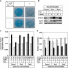
ATG13 knockdown enhances cell survival after camptothecin (CPT) treatment. (a) U2OS cells harboring tetracycline (Tet)-responsive ATG13shRNA were treated with or without 1 μg/ml of Tet for 5 days. The cells were then treated with or without 1 μM CPT for 1 day and then were washed with fresh media and allowed to grow for 3 more weeks in the presence or absence of 1 μg/ml of Tet. The cells were then stained with methylene blue as described in Materials and Methods. (b) Two ATG13 rescue cell lines (Res1 and Res2) and the parental ATG13-inducible knockdown cell line (Parent) were treated with or without Tet for 2 weeks. The cells were collected and lysed to perform western blot. The blots were probed with the antibodies against ATG13, unc-51-like kinase 1 (ULK1) and actin. (c) The same cell lines were treated as in (b) and were plated onto six-well plates to do long-lived protein degradation (LLPD) assay after being treated with or without 0.5 μM CPT for 2 days. (d) The experiments were repeated as in (a), in which parental ATG13 knockdown cell line and two rescue cell lines were used, except that the cells were left to grow for 2 more weeks after CPT treatment before counting colonies' number. Each condition was duplicated and the survival colonies were quantified. The data are presented as mean±standard deviation (S.D.) and these data are representative of at least two independent experiments
Figure 6
Figure 6
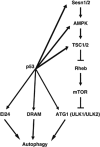
Model: p53 regulates autophagy in multiple nodes

References (VSports)

    1. Nakatogawa H, Suzuki K, Kamada Y, Ohsumi Y. Dynamics and diversity in autophagy mechanisms: lessons from yeast. Nat Rev Mol Cell Biol. 2009;10:458–467. - VSports - PubMed
    1. Kamada Y, Funakoshi T, Shintani T, Nagano K, Ohsumi M, Ohsumi Y. Tor-mediated induction of autophagy via an Apg1 protein kinase complex. J Cell Biol. 2000;150:1507–1513. - PMC - PubMed
    1. Meijer WH, van der Klei IJ, Veenhuis M, Kiel JA. ATG genes involved in non-selective autophagy are conserved from yeast to man, but the selective Cvt and pexophagy pathways also require organism-specific genes. Autophagy. 2007;3:106–116. - PubMed
    1. Kuroyanagi H, Yan J, Seki N, Yamanouchi Y, Suzuki Y, Takano T, et al. Human ULK1, a novel serine/threonine kinase related to UNC-51 kinase of Caenorhabditis elegans: cDNA cloning, expression, and chromosomal assignment. Genomics. 1998;51:76–85. - PubMed
    1. Yan J, Kuroyanagi H, Kuroiwa A, Matsuda Y, Tokumitsu H, Tomoda T, et al. Identification of mouse ULK1, a novel protein kinase structurally related to C. elegans UNC-51. Biochem Biophys Res Commun. 1998;246:222–227. - PubMed

Publication types

MeSH terms