Skip to main page content
U.S. flag

An official website of the United States government

Dot gov

The . gov means it’s official. Federal government websites often end in . gov or VSports app下载. mil. Before sharing sensitive information, make sure you’re on a federal government site. .

Https

The site is secure V体育官网. The https:// ensures that you are connecting to the official website and that any information you provide is encrypted and transmitted securely. .

. 2011 Mar 22;6(3):e18059.
doi: 10.1371/journal.pone.0018059.

The zinc transporter SLC39A14/ZIP14 controls G-protein coupled receptor-mediated signaling required for systemic growth (V体育官网入口)

Affiliations

The zinc transporter SLC39A14/ZIP14 controls G-protein coupled receptor-mediated signaling required for systemic growth

Shintaro Hojyo et al. PLoS One. .

"VSports在线直播" Abstract

Aberrant zinc (Zn) homeostasis is associated with abnormal control of mammalian growth, although the molecular mechanisms of Zn's roles in regulating systemic growth remain to be clarified. Here we report that the cell membrane-localized Zn transporter SLC39A14 controls G-protein coupled receptor (GPCR)-mediated signaling. Mice lacking Slc39a14 (Slc39a14-KO mice) exhibit growth retardation and impaired gluconeogenesis, which are attributable to disrupted GPCR signaling in the growth plate, pituitary gland, and liver. The decreased signaling is a consequence of the reduced basal level of cyclic adenosine monophosphate (cAMP) caused by increased phosphodiesterase (PDE) activity in Slc39a14-KO cells. We conclude that SLC39A14 facilitates GPCR-mediated cAMP-CREB signaling by suppressing the basal PDE activity, and that this is one mechanism for Zn's involvement in systemic growth processes. Our data highlight SLC39A14 as an important novel player in GPCR-mediated signaling VSports手机版. In addition, the Slc39a14-KO mice may be useful for studying the GPCR-associated regulation of mammalian systemic growth. .


Conflict of interest statement

Competing Interests: The authors have declared that no competing interests exist.

Figures

Figure 1
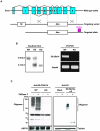
Figure 1. Generation of Slc39a14-KO mice.
(A) Schematic diagram of the construct used to generate Slc39a14-KO mice. The Slc39a14 genomic structure shows the alternatively spliced exon 4. Exon 4a encodes SLC39A14A, and exon 4b encodes SLC39A14B. (B and C) Homologous recombination by crossing heterozygotes was confirmed by southern blotting analysis (B, left) using genomic DNA from the tail, RT-PCR (B, right) using the liver mRNA, and western blotting analysis using liver whole-cell lysate (C, left), from litter mates. Cell lysates from wild-type, Slc39a14-KO liver (C, left), or Slc39a14a-v5 293T cells (C, right) were treated with or without PNGase F, and then subjected to SDS-PAGE and immunoblotting with antibodies to SLC39A14 or V5. The oligomeric bands are indicated. Non-specific bands.
Figure 2
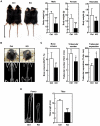
Figure 2. Dwarfism, torticollis, scoliosis, osteopenia, and shortened long bones in Slc39a14-KO mice.
(A) Left. Appearance of control (Ctrl) and Slc39a14-KO mice (4-weeks-old). Bar indicates 5 cm. Right. Body weights of 4-week-old control (Ctrl) (male, n = 9; female, n = 9), Slc39a14-KO (male, n = 10; female, n = 7), and neonatal (control, n = 22; Slc39a14-KO, n = 14) mice. Data represent the mean ± S.D. (**P<0.01, ***P<0.001). (B) Frontal views (4-week-old) and X-ray radiographs (8-week-old) of a control (Ctrl) and a Slc39a14-KO female mouse. (C) Bone histomorphometric analysis of 4-week-old control (Ctrl) and Slc39a14-KO female mice (n = 5). Data represent the mean ± S.D. (*P<0.05). (D) X-ray radiographs of the femurs (8-week-old), and the tibial length (6-week-old) of control (Ctrl) and Slc39a14-KO mice (n = 5). Data represent the mean ± S.D. (***P<0.001).
Figure 3
Figure 3. Abnormal chondrocyte differentiation in the growth plate of Slc39a14-KO mice.
(A) In situ hybridization analysis for Slc39a14 in an embryo at 16.5 dpc, and magnified images of the limb (1), spine (2), and thorax (3). Green arrowheads indicate Slc39a14 expression. (B and C) In situ hybridization analysis for Slc39a14, Col2a1, Ihh, and Col10a1 in the growth plates from 4-week-old control (Ctrl) and Slc39a14-KO mice. (D) Col10a1 levels in control (Ctrl) and Slc39a14-KO chondrocytes. Data represent the mean ± S.D. (**P<0.01). (E) H&E staining of the growth plates from control (Ctrl) and Slc39a14-KO mice (4-week-old).
Figure 4
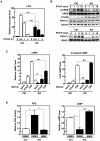
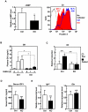
Figure 4. SLC39A14 positively regulates PTH1R-cAMP-CREB signaling in chondrocytes.
(A) PTHrP-induced c-fos levels in control (Ctrl) and Slc39a14-KO chondrocytes. Data represent the mean ± S.D. (***P<0.001). (B) CREB phosphorylation and nuclear PKA-Cα translocation in control (Ctrl) and Slc39a14-KO chondrocytes with PTHrP treatment. Whole-cell lysate (upper panels) or the nuclear fraction (lower panels) was subjected to SDS-PAGE and immunoblotting with antibodies to the indicated proteins. (C) cAMP level and the fold-increase in cAMP level in control (Ctrl) and Slc39a14-KO chondrocytes before and after PTHrP or FSK treatment. Data represent the mean ± S.D. (**P<0.01; ***P<0.001; N.S., no significance). (D) Effect of IBMX treatment on the PDE activity and cAMP level in control (Ctrl) and Slc39a14-KO chondrocytes. Data represent the mean ± S.D. (*P<0.05, ***P<0.001).
Figure 5
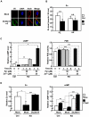
Figure 5. Zn positively regulates the cAMP level and facilitates PTH1R signaling.
(A) Localization of SLC39A14 in transfected primary chondrocytes. SLC39A14 was labeled with anti-V5 (green), nuclei with DAPI (blue), and actin with phalloidin (red). (B) Electron probe X-ray microanalysis (EPMA) in the PZ and HZ of the growth plates from control (Ctrl) and Slc39a14-KO mice. The Zn levels in 10 cells in each zone were measured. Data represent the mean ± S.D. (***P<0.001). (C) Effect of Zn plus the ionophore pyrithione on the cAMP level and the PDE activity in control (Ctrl) and Slc39a14-KO chondrocytes. The cells were treated with Zn plus pyrithione at the indicated concentrations and time points. Data represent the mean ± S.D. (*P<0.05, **P<0.01, ***P<0.001). (D) Intracellular Zn and cAMP levels in control (Ctrl) and Slc39a14-KO chondrocytes after the transduction of empty (Mock) or Slc39a14 cDNA-carrying lentivirus for 2 days. The intracellular Zn level was measured as the maximal emission at 516 nm (excitation at 494 nm) using a Varioskan after mixing FluoZin-3 with denatured cell lysates. The cAMP levels were measured after PTHrP treatment for 20 min. Data represent the mean ± S.D. (n = 3 per condition) (*P<0.05, **P<0.01, ***P<0.001).
Figure 6
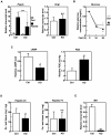
Figure 6. SLC39A14 positively regulates GH production via GHRHR signaling in the pituitary gland.
(A) cAMP and intracellular Zn levels in pituitary cells (12-week-old control (Ctrl) and Slc39a14-KO mice (n = 3)). M.F.I. represents the mean fluorescent intensity. Data represent the mean ± S.D. (*P<0.05). (B) Human GHRH (500 µg/kg) was injected intravenously into 8-week-old control (Ctrl) and Slc39a14-KO mice (n = 8). Plasma was collected at the indicated times after injection, and the GH concentration was measured. Data represent the mean ± S.E.M. (*P<0.05). (C) Gh induction in the pituitary glands from 36-hours fasted control (Ctrl) and Slc39a14-KO mice (14–17-week-old, n = 3). Data represent the mean ± S.D. (***P<0.001). (D) Serum IGF-I concentration (3-week-old control (Ctrl), n = 9; Slc39a14-KO, n = 12). Data represent the mean ± S.D. (***P<0.001). (E) Hepatic Igf-I (n = 7) and Ghr (n = 3) levels in 3-week-old control (Ctrl) and Slc39a14-KO mice. Data represent the mean ± S.D. (***P<0.001).
Figure 7
Figure 7. SLC39A14 positively regulates gluconeogenesis via GCGR signaling in the liver.
(A) Hepatic Pepck and Gcgr levels in 18-hours fed or fasted control (Ctrl) and Slc39a14-KO mice (16-week-old, n = 2). Data represent the mean ± S.D. (***P<0.001). (B) Plasma glucose level in 18–36-hours fasted control (Ctrl) and Slc39a14-KO mice (7–52-week-old, n = 8). Data represent the mean ± S.E.M. (*P<0.05). (C) cAMP level and PDE activity in the liver from control (Ctrl) and Slc39a14-KO mice (4–8-week-old, n = 3). Data represent the mean ± S.D. (*P<0.05, **P<0.01). (D) Hepatic Zn and Fe levels in control (Ctrl) and Slc39a14-KO mice measured by ICP-MS. Data represent the mean ± S.D. (*P<0.05). (E) Hepatic Mt-I level in control (Ctrl) and Slc39a14-KO mice (16-week-old, n = 2). Data represent the mean ± S.D. (**P<0.01).
Figure 8
Figure 8. Schematic model for the regulation of GPCR-mediated signaling by SLC39A14.
SLC39A14 regulates the basal cAMP level by suppressing PDE activity, through either the direct provision of Zn to PDE or the indirect provision via unidentified molecular chaperone(s) (Protein X). This system may facilitate the GPCR-cAMP-CREB pathway in endocrine-system reactions.

References

    1. Prasad AS. Zinc: an overview. Nutrition. 1995;11:93–99. - PubMed
    1. Vallee BL, Auld DS. New perspective on zinc biochemistry: cocatalytic sites in multi-zinc enzymes. Biochemistry. 1993;32:6493–6500. - PubMed
    1. Kambe T, Weaver BP, Andrews GK. The genetics of essential metal homeostasis during development. Genesis. 2008;46:214–228. - PMC - PubMed
    1. Lichten LA, Cousins RJ. Mammalian zinc transporters: nutritional and physiologic regulation. Annu Rev Nutr. 2009;29:153–176. - V体育2025版 - PubMed
    1. Vallee BL. The function of metallothionein. Neurochem Int. 1995;27:23–33. - "VSports在线直播" PubMed

Publication types

Substances (V体育安卓版)